Effects of Ultrasonic Treatment on Microstructure and Properties of Al-Based Composites Reinforced by In Situ Al2O3 Nanoparticles
-
Cunguang Chen
 ,
Ji Luo
,
Ji Luo

Abstract
An investigation on the microstructure of as-cast Al-Mg-Cu composites reinforced by in situ nano-sized Al2O3 dispersoids with ultrasonic treatment showed that ultrasonic treatment of the melt prior to casting had a significant effect on the size and sphericity of α-Al dendrites as well as on the size, continuity and sphericity of intermetallic particles (Al2CuMg) formed during cooling and solidification of the composite. More importantly, Al2O3 nanoparticles were uniformly distributed inside the grains, which were in situ produced by the displacement reaction between Al and CuO in the melt under ultrasonic treatment. The microstructural effects were mainly attributed to the cavitation and streaming phenomena which took place during ultrasonic treatment in the melt. The mechanical properties were investigated by tensile tests and hardness measurements. Ultrasonic treatment caused a significant increase in the yield strength (~43%), ultimate tensile strength (~32%) and hardness (~13%), and simultaneously slight improvement in the ductility.
Introduction
It has been well recognized that lightweight metal–matrix composites (LMMCs) (metal matrix with ceramic particles usually) can be of significance for automobile, aerospace, military industries and numerous other applications. In particularly, aluminum matrix composites (AMCs) have been widely used in the above-mentioned fields due to the interesting combination of engineering properties such as low density, high specific strength and stiffness, improved damping property, electromagnetic shield capacity, excellent machinability and good castability [1–4]. Currently, it can be carried out through many different methods for the fabrication of AMCs, which are summed up based on the literature survey in two categories including solidification processing (SP) and powder metallurgy (PM). Massive efforts have been made to develop AMCs, such as Al-SiC [5–9], Al-Al2O3 [2, 10–12], Al-B4C [13], Al-TiB2 [14], Al-CNT [15, 16], Al-Diamond [17, 18], using SP and PM in the last couple of decades. The attractive physical and mechanical properties of AMCs, including high specific modulus, superior strength, long fatigue life, and improved thermal stability, can be obtained by PM. However, it is a pity that the PM route suffers from the processing costs, the difficulty in the manufacture of large complex shaped parts and its poor ductility. On the other hand, the solidification processing, that is the melting and casting route, though cost effective, experiences problems like poor wettability between ceramic reinforcements and the melt, inhomogeneous distribution of reinforcements and unwanted interfacial reactions [19, 20].
In order to overcome the above difficulties, novel processing techniques based on in situ production of composites by displacement reactions in Al-SiO2, Al-CuO, and Al-ZnO systems have emerged in recent years [2, 21–23]. In the conventional in situ synthesis, though the in situ-formed reinforcements offer the advantages of finer size, non-contaminated surface and matched interface, a uniform distribution of in situ particles is difficult to achieve even for the mechanical stirring method due to the density difference between the liquid and solid particles. What’s more, the problem with finer reinforcement particles, particularly nanoparticles, would tend to agglomerate, especially in grain boundary, because of the high surface energy [19, 24]. With the advent of a new processing technique namely ultrasonic treatment (UST) [25], it has been possible to uniformly distribute nano-sized dispersoids in the as-cast materials resulting in a higher strength of the casting. Yang et al. [1, 26, 27] fabricated bulk aluminum alloy A356 reinforced by the SiC nanoparticles with average diameter ~30 nm by UST. It can be observed that high-power ultrasonic was effective to disperse nano-sized SiC particles in aluminum alloy A356 and high yield strength in cast Al – 7 mass% Si alloys reinforced with 2 mass% nano-sized SiC particles was obtained. Su et al. [28] produced 1 mass% nano-Al2O3/2024 aluminum matrix composites using UST. Compared with the alloy matrix, the ultimate tensile strength and yield strength of 1 mass% nano-Al2O3/2024 composite were enhanced by 37% and 81% respectively due to the uniform distribution of reinforcement and grain refinement of aluminum matrix by UST. As previously stated, much work has focused on adding nanoparticle reinforcements into the given metal melts. The available literature on aluminum matrix composites by in situ UST that utilized ultrasonic melt treatment and in situ synthesis processing was rather limited [29].
This paper presents the effects of UST on the microstructural features and mechanical properties of aluminum matrix composites fabricated by the in situ Al–CuO system. In addition, special attention is paid to the effects of UST on the second phases, including in situ nanoparticles and intermetallic phases, formed in the microstructures. This is an area less explored by other researchers.
Experimental
Materials and apparatus
In this research raw materials involved: refined aluminum ingot for remelting (99.99% purity) as the composite matrix, magnesium chips (99.5% purity) and CuO powders (99.5% purity, < 5 μm size). Magnesium addition was expected to scavenge oxygen from the surface of CuO particles and result in the elimination of the gas layer thus promoting wetting and improve the wettability between the in situ aluminum oxide and the liquid metal melt [30]. During the experimental process, an atmospheric control electrical resistance furnace with the ultimate temperature of 1,200°C and an ultrasonic processing system consisted of ultrasonic probe and ultrasonic generator and transducer were applied. The ultrasonic probe of 50 mm in diameter and 350 mm in length was made of TC4 titanium alloy specially made for aluminum melt which can withstand high processing temperature with minimum ultrasonic cavitation induced erosion. A maximum ultrasonic power of 2 KW from the transducer was found to generate adequate nonlinear effects inside the crucible.
Sample preparation
The processing temperature was controlled at approximate 200°C above the aluminum melting point (660°C). Approximately, 400 g of the pure aluminum ingot was charged into the crucible made from graphite. The required amounts of Mg and CuO were calculated according to the nominal composition Al – 2 mass% Mg – 5 mass% CuO. The mixture of Mg chips and CuO powders was wrapped in an aluminum foil by forming packets for preheated at 150°C for 2 h. After maintaining the temperature at 860°C, the preheated packets were added into molten metal of crucible while a graphite agitator was in progress to create vortex. The molten metal was continuously stirred at 300 rpm for about 10 min after addition of Mg and CuO mixture under argon gas cover. After the mixture addition through mechanical stirring, the ultrasonic probe was inserted into the melt when the processing temperature fell at 750°C. Sonication time was continued for about 5 min. The melt with the reinforced nanoparticles then poured into a preheated (200°C) Reynolds standard golf tee mold [29] made of copper. In order to identify the properties and structures of samples, the composite melts with the same components were prepared without ultrasonic treatment but in other identical conditions (including melting temperature, furnace, mechanical stirring, etc.).
Characterization of composite samples
Optical microstructures (OM) of the resulting composites were examined under the metallographic microscope (Olympus BX60M) after being etched by Keller’s reagent (1.5 ml HCl, 2.5 ml HNO3, 1 ml HF and 95 ml H2O). The morphology, size and dispersion of in situ Al2O3 particles and intermetallic phases in the matrix were observed by the scanning electron microscope (SEM, LEO 1450) and the field emission scanning electron microscope (FESEM, SUPRATM 55) equipped with an energy dispersive spectroscopy detector (EDS). The transmission electron microscope (TEM) specimen preparation process was as follows: 0.3-mm-thick slices were cut off from the specimen followed by mechanical grinding to 40–50 μm. Then the thin foils were ion milled at room temperature in a Gatan PIPS with a small incident angel till perforation. All the thin foils were examined in a Tecnai G2 F30 TEM operated at 200 kV. Phase analysis of the composite samples was carried out by X-ray diffractometer (XRD, Rigaku TTRⅢ) using monochromatic Cu Kα radiation with the wavelength of X-ray 0.154 nm, and operated at 40 kV as well as 150 mA.
According to China’s National Standard GB/T 228–2002, tensile tests were carried out at room temperature with the tensile rate of 0.5 mm · min−1 on a CMT-4105 testing machine [31]. The hardness values of the samples were determined using the Rockwell hardness tester on “B” scale (Model TH320) with 1.58 mm steel ball indenter, minor load of 10 kg, and major load of 100 kg and hardness value of 101.2 HRB as the standard block. For each test, at least three specimens were tested to obtain the average values of strength, elongation and hardness.
Results and Discussion
Microstructural characteristics
The optical micrographs of the composite samples without/with UST are shown in Figure 1(a) and (b), respectively. It indicates that the introduction of UST has reduced the grain size of aluminum matrix from 100 to 150 μm in Figure 1(a) to 40–75 μm in Figure 1(b). Besides, the primary α-Al phase dendrites formed less uniformly without UST have mostly changed into near-equiaxial or equiaxial crystals under ultrasonic vibration. Note that as it can be seen from Figure 1(b), the grains have less branches and shorter arms, which results in smaller grain perimeters and greater sphericity. Also, the uniformity of the grains in terms of their size and shape has increased by UST [32, 33].

Optical photomicrographs of as-cast composites prepared by different treatment: (a) without ultrasonic treatment and (b) with ultrasonic treatment.
In this study, UST was performed at 750°C, about 90°C above the aluminum melt point. No α-Al grain is expected to form at this temperature, and therefore the above microstructural refinement appearance could not be explained by conventional ultrasonic nonlinear effects, namely transient cavitation and acoustic streaming [26]. However, with the aid of ultrasonic transient cavitation, the in situ clustered particles such as Al2O3 could be broken up under an implosive impact, a portion of which become active and involved as the nuclei during the solidification [28, 34]. Meanwhile, distribution of the agglomerated nucleant particles existing in the melt under the effects of cavitation and streaming also increases the effective nucleation sites [35]. Thus the microstructure with fine grains as shown in Figure 1(b) can be observed.
The SEM images of as-cast composites before and after UST are presented in Figure 2(a–d) at two different magnifications. From SEM images (Figure 2), it is obvious that the grain refining effect by UST is much clearer. However, it is somewhat surprising that the porosities are not visible in the composites whether by mechanical stirring or with UST. The structural morphology and shapes of intermetallics located in the grain boundary were investigated using SEM as shown in Figure 2. The results presented by the energy dispersion spectrum (EDS) identify the intermetallics to be in the form of Aluminum Copper Magnesium phase (Al2CuMg). As can be clearly seen from Figure 2(a) and (c), under UST, the continuity and thickness of Al2CuMg particles decrease. In particular, it is observed from Figure 2(d) magnified by Figure 2(b) that the Al2CuMg particles have changed into fine axiolitic shapes with UST from coarse wormlike shapes without UST. Furthermore, the bright field TEM micrographs of the composites in Figure 3 clearly show that Al2CuMg particles of larger than 5 μm without UST are jointed together, while ones of only 1 μm with UST are separated. In other words, the size of Al2CuMg particles decreases and their sphericity increases by the applied UST.

SEM micrographs of intermetallic phases and nano-sized dispersoids observed in different samples: (a, b) without ultrasonic treatment and (c, d) with ultrasonic treatment.

Bright filed TEM micrographs of as-cast composites: (a) without ultrasonic treatment and (b) with ultrasonic treatment.
The dominant intermetallic phase in the microstructures, i.e., Al2CuMg, is formed by a eutectic reaction in the Al-Cu-Mg system at about 520°C after the displacement reaction between Al and CuO as follows:
The reaction is sought to take place leading to the formation of Al2O3 particles. Owing to its low formation temperature, Al2CuMg is the last solid phase formed during solidification, and therefore, as can be seen from Figure 2, it precipitates at the grain boundaries. As a brittle phase, Al2CuMg can have deleterious effects on the mechanical properties of the castings if it forms a continuous coarse network at the grain boundaries.
Besides, the EDS analysis was utilized to determine the composition of the composites. EDS of Particle A in Figure 2(b) magnified by the dotted circle location in Figure 2(a) shows that the precipitated particle contains some amount of O except Al, Cu and Mg as shown in Figure 4. The result of Particle B in Figure 2(d) by EDS is similar to Particle A. Magnesium addition may account for the presence of oxygen. Previous research [36, 37] demonstrated that the formation of a spinel, MgAl2O4, would occur on alumina particulate in Mg-containing aluminum alloys melt. The presence of magnesium in the melt can reduce alumina to aluminum and release the oxygen to form magnesia according to Reaction (2) or react with alumina directly, resulting in the formation of a spinel according to Reactions (3) and (4) [30].

EDS of the precipitated Particle A shown in Figure 2(b) showing peaks for Al, Cu, Mg and O.
It has been pointed out that MgAl2O4 was the stable reaction product with the concentration of Mg in the melt lower than 4 mass%, while MgO was the main reaction product with the concentration of Mg in the melt greater than 4 mass% [9, 36]. Therefore, it is believed that the main reaction product is MgAl2O4 in the present composites with 2 mass% Mg in this study, lower than 4 mass%.
As shown in Figure 2(c), within the grains, the distribution of the white dispersed particles punctuate sparsely in coarse dendrites, while a great many of nanoparticles are primarily present inside the grains in Figure 2(d). The detection zone of EDS beam is bigger than the average size of nanoparticles above mentioned, and therefore it is unclear that the ultrafine particles are consisted of in situ Al2O3 or CuAl2, which is one of the very common precipitated phases in aluminum alloys. To distinguish both of them, solution treatment was carried out at 495 ± 2°C for 8 h and then water quenched. Figure 5(a) and (b) show the microstructure of the sample with UST by solution treatment. As can be observed, after the solution treatment, nanoparticles are still clearly visible inside a grain (Figure 5(a)) and around the grain boundary (Figure 5(b)), though the quantity of nanoparticles is more or less declined compared with as observed in Figure 2(d). It should be noted that CuAl2, which usually disperses in the aluminum matrix, could dissolve into the aluminum matrix during the solution treatment. Therefore, the white nanoparticles remaining inside the grains should be Al2O3. However, a small amount of nanoparticles are found inside the grain of the sample without UST by solution treatment in Figure 5(c). This can be attributed to the pushing of nano-sized in situ Al2O3 particles by the solidification front to the regions near the grain boundaries. Hence it follows that UST can avoid nanoparticle aggregates along the grain boundary and uniformly disperse nanoparticles within the grains according to the nuclei formation mechanisms as what mentioned before.

FESEM micrographs of the distribution of nano-Al2O3 particulates after solution treatment observed in different samples: (a, b) with ultrasonic treatment and (c) without ultrasonic treatment.
Furthermore, XRD patterns of the samples with/without UST after solution treatment are shown in Figure 6. It shows that some apparent crystallographic planes of Al2O3 involve crystal diffraction with UST. However, the absence of Al2O3 peaks in the sample without UST means Al2O3 particles is extremely small or at low volume fractions. Meanwhile, MgAl2O4 peaks are clearly shown in the both patterns, confirming the formation of MgAl2O4. Unexpectedly, in the pattern of the sample with UST, Al2CuMg is not completely dissolved in the aluminum matrix during solution treatment.

XRD patterns of the samples with/without ultrasonic treatment after solution treatment.
Mechanical properties
The properties of the LMMCs depend not only on the matrix particle and the weight percent, but also on the distribution of reinforcing particles and interface bonding between the particle and matrix according to large quantities of previous studies. As mentioned above, under the applied UST, the nanoparticles Al2O3 can be uniformly dispersed in the aluminum matrix, and the size and shape of intermetallic phases formed during solidification of the as-cast composites have changed obviously. Therefore, it is predicted that the properties of the sample with UST should be better than without UST under the same other conditions.
Table 1 shows the average results of tensile and hardness testing at room temperature. As depicted in the table, the mechanical properties of as-cast composites with UST have been simultaneously enhanced as expected, the yield strength (YS) by ~43%, the ultimate tensile strength (UTS) by ~32% and hardness by ~13%, respectively. Fortunately, the elongation of the sample with UST has been slightly improved. Note that the performance improvement just results from the small amount of nano-Al2O3 particulates (~2 mass% calculated by the composition of Al – 2 mass% Mg – 5 mass% CuO) by UST. These results would be attributed to coupled effects of an increase in grain boundary area due to grain refinement and the dislocation pinning in virtue of the Orowan strengthening mechanism [28, 38]. Meanwhile, careful inspection by TEM from Figure 7 reveals a higher dislocation density close to Al2CuMg with UST (Figure 7(b)) than without UST (Figure 7(a)), which indicates that the improvement of sample properties may be related to the enhanced dislocation density. However, it is thought that discontinuity and refinement of Al2CuMg particles should play an important role in improvement of the tensile strength of the composite by UST. The results presented in the preceding sections have shown that under UST, the continuity, thickness and width of Al2CuMg particles decrease, but the sphericity increases. These would contribute to higher tensile strength and ductility improvement of the composite under UST.
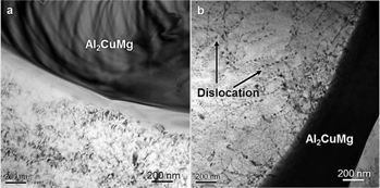
TEM micrographs of the dislocation distribution of different samples: (a) without ultrasonic treatment and (b) with ultrasonic treatment.
Above all, it seems that the collective effects of UST on these microstructural features have significantly improved the mechanical properties of the as-cast composites in Table 1.
Tensile properties and hardness of different as-cast composites.
| Condition | 0.2% YS (MPa) | UTS (MPa) | Elongation (%) | Hardness (HRB) |
| Without UST | 103 ± 4 | 186 ± 4 | 6.9 ± 0.5 | 55.6 ± 3 |
| With UST | 147 ± 6 | 245 ± 5 | 8.0 ± 0.5 | 62.8 ± 3 |
Conclusions
In this study, the effects of ultrasonic treatment on microstructural features and mechanical properties of as-cast Al-Mg-Cu composites reinforced by in situ nano-Al2O3 particulates were investigated. The results showed that ultrasonic treatment of the melt prior to casting had significant effects on the aluminum matrix and the second phases, which were mainly attributed to the cavitation and streaming phenomena taking place during ultrasonic treatment in the melt. On the one hand, grains size of the aluminum matrix decreased and the grains sphericity got greater via ultrasonic treatment. On the other hand, as to the intermetallic particles (Al2CuMg) formed during cooling and solidification of the composites, their size decreased and their sphericity increased by the applied UST. Moreover, the continuity of Al2CuMg particles was damaged by UST, improving the composites performance. Even more importantly, Al2O3 nanoparticles were uniformly distributed inside the grains, which were in situ produced by the displacement reaction between Al and CuO in the melt under ultrasonic treatment. Mechanical properties, including the yield strength, the ultimate tensile strength, the elongation and hardness, were significantly improved by ultrasonic treatment of the melt connecting microstructural features.
Funding statement: Funding: This work was financially supported by the National High Technology Research and Development Program of China (863 Program) (No. 2013AA031104) and the Research Fund for the Doctoral Program of Higher Education of China (No. 20120006110007).
Acknowledgments
The authors are also grateful to Prof. Xuexu Gao of the State Key Laboratory for Advanced Metals and Materials at the University of Science and Technology Beijing for useful discussion in the experiments.
References
[1] Y.Yang, J.Lan and X.C.Li, Mater. Sci. Eng. A, 380 (2004) 378–383.Search in Google Scholar
[2] K.D.Woo and H.B.Lee, Mater. Sci. Eng. A, 449–451 (2007) 829–832.Search in Google Scholar
[3] I.A.Ibrahim, F.A.Mohamed and E.J.Lavernia, J. Mater. Sci., 26 (1991) 1137–1156.Search in Google Scholar
[4] N.Ramakrishnan, Acta Mater., 44 (1996) 69–77.Search in Google Scholar
[5] A.El-Sabbagh, M.Soliman, M.Taha and H.Palkowski, J. Mater. Process. Tech., 212 (2012) 497–508.Search in Google Scholar
[6] S.Amirkhanlou and B.Niroumand, J. Mater. Process. Tech., 212 (2012) 841–847.Search in Google Scholar
[7] A.Mazahery and M.O.Shabani, Trans. Nonferrous Met. Soc. China, 22 (2012) 275–280.Search in Google Scholar
[8] A.McLean, H.Soda, Q.Xia, A.K.Parmanick, A.Ohno, G.Motoyasu, T.Shimizu, S.A.Gedeon and T.North, Compos. Part A, 28A (1997) 153–162.10.1016/S1359-835X(96)00107-8Search in Google Scholar
[9] M.I.Pech-Canul, M.Rodríguez-Reyes, M.A.Pech-Canul and J.C.Rendón-Angeles, Met. Mater. Int., 17 (2011) 923–929.Search in Google Scholar
[10] M.Hossein-Zadeh, M.Razavi, O.Mirzaee and R.Ghaderi, J. King Saud Univ. Eng. Sci., 25 (2013) 75–80.Search in Google Scholar
[11] I.El-Mahallawi, H.Abdelkader, L.Yousef, A.Amer, J.Mayer and A.Schwedt, J. Mater. Sci. Eng. A, 556 (2012) 76–87.Search in Google Scholar
[12] Y.B.Yang, Z.M.Zhang and X.Zhang, Mater. Sci. Eng. A, 558 (2012) 112–118.Search in Google Scholar
[13] F.Toptan, I.Kerti and L.A.Rocha, Wear, 290–291 (2012) 74–85.Search in Google Scholar
[14] Q.Guo, D.L.Sun, L.T.Jiang, G.H.Wu and X.L.Han, Micron, 43 (2012) 688–693.Search in Google Scholar
[15] Y.F.Wu, G.Y.Kim and A.M.Russell, Mater. Sci. Eng. A, 538 (2012) 164–172.Search in Google Scholar
[16] Y.F.Wu, G.Y.Kim and A.M.Russell, Mater. Sci. Eng. A, 532 (2012) 558–566.Search in Google Scholar
[17] J.P.Long, X.Li, D.D.Fang, P.Peng and Q.He, Int. J. Ref. Met. Hard Mater., 41 (2013) 85–89.Search in Google Scholar
[18] K.Mizuuchi, K.Inoue, Y.Agari, M.Sugioka, M.Tanaka, T.Takeuchi, J.Tani, M.Kawahara, Y.Makino and M.Ito, Microelectron. Reliab., 54 (2014) 2463–2470.Search in Google Scholar
[19] G.H.Zahid, T.Azhar, M.Musaddiq, S.S.Rizvi, M.Ashraf, N.Hussain and M.Iqbal, Mater. Des., 32 (2011) 1630–1635.Search in Google Scholar
[20] C.U.Atuanya and V.S.Aigbodion, J. Alloys Compd., 601 (2014) 251–259.Search in Google Scholar
[21] T.G.Durai, K.Das and S.Das, J. Alloys Compd., 457 (2008) 435–439.10.1016/j.jallcom.2007.02.142Search in Google Scholar
[22] P.C.Maity, P.N.Chakraborty and S.C.Panigrahi, Mater. Lett., 30 (1997) 147–151.10.1016/S0167-577X(96)00188-7Search in Google Scholar
[23] H.Arami, A.Simchi and S.M.Seyed Reihani, J. Alloys Compd., 465 (2008) 151–156.10.1016/j.jallcom.2007.10.099Search in Google Scholar
[24] S.Mula, P.Padhi, S.C.Panigrahi, S.K.Pabi and S.Ghosh, Mater. Res. Bull., 44 (2009) 1154–1160.10.1016/j.materresbull.2008.09.040Search in Google Scholar
[25] G.I.Eskin, Ultrason. Sonochem., 8 (2001) 319–325.10.1016/S1350-4177(00)00074-2Search in Google Scholar
[26] Y.Yang and X.C.Li, J. Manu. Sci. Eng., 129 (2007) 497–501.Search in Google Scholar
[27] X.C.Li, Y.Yang and X.D.Cheng, J. Mater. Sci., 39 (2004) 3211–3212.Search in Google Scholar
[28] H.Su, W.L.Gao, Z.H.Feng and Z.Lu, Mater. Des., 36 (2012) 590–596.Search in Google Scholar
[29] Y.F.Han, K.Li, J.Wang, D.Shu and B.D.Sun, Mater. Sci. Eng. A, 405 (2005) 306–312.Search in Google Scholar
[30] B.F.Schultz, J.B.Ferguson and P.K.Rohatgi, Mater. Sci. Eng. A, 530 (2011) 87–97.Search in Google Scholar
[31] China national standardization management committee, National standard of the People’s Republic of China – Metallic materials – Tensile testing at ambient temperature (GB/T 228–2002), Standards Press of China, Beijing, (2002).Search in Google Scholar
[32] J.W.Li and T.Momono, J. Mater. Sci. Technol., 21 (2005) 47–54.Search in Google Scholar
[33] H.B.Peng, W.Q.Chen, Y.C.Yu and H.G.Zheng, High Temp. Mater. Processes, 32 (2013) 459–465.Search in Google Scholar
[34] G.I.Eskin and D.G.Eskin, Ultrason. Sonochem., 10 (2003) 297–301.Search in Google Scholar
[35] M.Khosro Aghayani and B.Niroumand, J. Alloys Compd., 509 (2011) 114–122.Search in Google Scholar
[36] K.B.Lee, Y.S.Kim and H.Kwon, Metall. Mater. Trans. A, 29A (1998) 3087–3095.10.1007/s11661-998-0216-9Search in Google Scholar
[37] A.D.McLeod and C.M.Gabryel, Metall. Trans. A, 23A (1992) 1279–1283.10.1007/BF02665059Search in Google Scholar
[38] G.Cao, H.Choi, H.Konishi, S.Kou, R.Lakes and X.C.Li, J. Mater. Sci., 43 (2008) 5521–5526.Search in Google Scholar
©2016 by De Gruyter
This article is distributed under the terms of the Creative Commons Attribution Non-Commercial License, which permits unrestricted non-commercial use, distribution, and reproduction in any medium, provided the original work is properly cited.
Articles in the same Issue
- Frontmatter
- Numerical Simulation to Study the Effect of Arc Travelling Speed and Welding Sequences on Residual Stresses in Welded Sections of New Ferritic P92 Pipes
- Microstructural Evolution and Compressive Properties of Two-Phase Nb-Fe Alloys Containing the C14 Laves Phase NbFe2 Intermetallic Compound
- Optimization of Microwave Roasting for Dechlorination of CuCl Residue under Oxygen-Enriched Condition
- Evaluation of High Temperature Properties and Microstructural Characterization of Resistance Spot Welded Steel Lap Shear Joints
- Microstructural Changes of a Creep-Damaged Nickel-Based K002 Superalloy Containing Hf Element under Different HIP Temperatures
- Effect of Ultrasonic Treatment on the Solidification Microstructure of GCr15 Bearing Steel
- Effects of Ultrasonic Treatment on Microstructure and Properties of Al-Based Composites Reinforced by In Situ Al2O3 Nanoparticles
- High-Temperature Oxidation Behavior of Fe-Si-Ce Alloys
- Reaction Mechanism of Siderite Lump in Coal-Based Direct Reduction
- EAF Gas Waste Heat Utilization and Discussion of the Energy Conservation and CO2 Emissions Reduction
- Numerical Parametric Analysis of Bond Coat Thickness Effect on Residual Stresses in Zirconia-Based Thermal Barrier Coatings
- The Marker Conservation Law in Multiphase Systems
- Synthesis and Characterization of Strontium Carbonate Nanostructures via Simple Hydrothermal Method