Green synthesis, structural characterization, and catalytic activity of silver nanoparticles stabilized with Bridelia retusa leaf extract
-
Ramesh Vinayagam
Ramesh Vinayagam is a senior assistant professor at the Department of Biotechnology, Manipal Institute of Technology, Manipal University. He holds a Bachelor’s degree in Chemical Engineering, a Master’s degree in Industrial Biotechnology, and a Doctoral degree in the field of thermostable enzymes from Manipal University in 2016. With many research articles in reputed journals to his credit, he conducts research in the areas of nanobiotechnology and bioprocess engineering.
Thivaharan Varadavenkatesan is an associate professor at the Department of Biotechnology, Manipal Institute of Technology (MIT), Manipal University. Holding both Bachelor’s and Master’s degrees in Biotechnology, his doctoral thesis focused on biosurfactant production. He obtained his doctorate from Manipal University in 2014. His research focuses on environmental bioremediation and nanobiotechnology.
Raja Selvaraj is an associate professor at the Department of Biotechnology, Manipal Institute of Technology, Manipal University. With a Bachelor’s degree in Chemical Engineering and a Master’s degree in Biotechnology, he obtained his doctoral degree from Manipal University in 2013. He has published many research articles in reputed journals. His research areas include nano-bioremediation, environmental biotechnology, bioprocess engineering, aqueous two phase systems, and design of experiments. He is a peer-reviewer for many leading journals.


Abstract:
An environmentally benign method to synthesize silver nanoparticles (SNPs) using the leaf extract of Bridelia retusa was developed. The UV-Vis absorption spectrum of the synthesized SNPs displayed a surface plasmon peak at 420 nm. Scanning electron microscopy (SEM) revealed the irregular shaped nanoparticles, and energy dispersive X-ray (EDX) ascertained the presence of metallic silver by showing a strong signal at 3 eV. The crystalline structure of metallic silver was confirmed by X-ray diffraction (XRD). The mean size of the SNPs was calculated as 16.21 nm. Fourier infrared (FT-IR) spectroscopic studies displayed specific bands for various functional groups and affirmed the function of reduction and stabilization of SNPs. The stability was endorsed by the zeta potential value of −18.1 mV. The results evidenced that this leaf extract-mediated synthesis method is eco-friendly, rapid, and cheap. The catalytic power of the SNPs was investigated for Rhodamine B dye degradation. The SNPs completely degraded Rhodamine B within 9 min; thus, the dye degradation process was very rapid. The pseudo-first order degradation constant was found out to be 0.1323 min−1. This paves the way for the future development of novel nano-catalysts to reduce environmental pollution.
Introduction
Metallic nanoparticles (MNPs) have acquired significant momentum nowadays because of their specific features, such as simple preparation procedures, high surface-to-volume ratio, uniform particle size, surface manipulation possibility, and high stability [1]. These characteristic features have made them useful in various applications, such as catalysis [2], bio-sensing [3], [4], drug delivery [5], environmental bioremediation [6], [7], [8], and inherent biological activities like antibacterial [9], antifungal [10], anticoagulant [11], [12] and larvicidal activities [13].
Traditional routes of MNP synthesis, namely, physical and chemical methods, are energy-intensive and yield substantial quantities of toxic chemicals and wastes, which are of environmental concern. Thus, the exploration of environmentally benign methods for the synthesis of MNPs has increased in recent years [14]. These methods involve the usage of naturally available resources, such as various plant extracts [15], [16], [17], [18], bacteria, yeast, fungi [19], and algae [20]. Nevertheless, such features as being low cost, easy procedure, and the possibility of large-scale production attracted research attention to the synthesis of silver nanoparticles (SNPs) using plant extracts as compared with the microbial synthesis of nanoparticles [21]. The phytocompounds present in the plant extracts [22] and the various biomolecules, such as enzymes, amino acids, and carbohydrates, reduce and stabilize the MNPs.
SNPs are renowned nanomaterials owing to their biocompatible nature. They find applications in medicine, pharmaceutical, food, and cosmetic industries as well as environment pollutant removal. Hence, the objective of the present investigation is to establish an environmentally benign synthesis of SNPs using the leaf extract of an officinal plant, Bridelia retusa, without the addition of any separate reducing and capping chemicals. As far as we know, there are no published literature available for the nanoparticle synthesis using the B. retusa leaf extract (BRLE). It is a moderately sized tree (Figure 1) belonging to the Euphorbiaceae family, predominantly found in Bangaladesh, Nepal, southern India, and Sri Lanka. It is known as “mulluvengai” in Tamil [23].
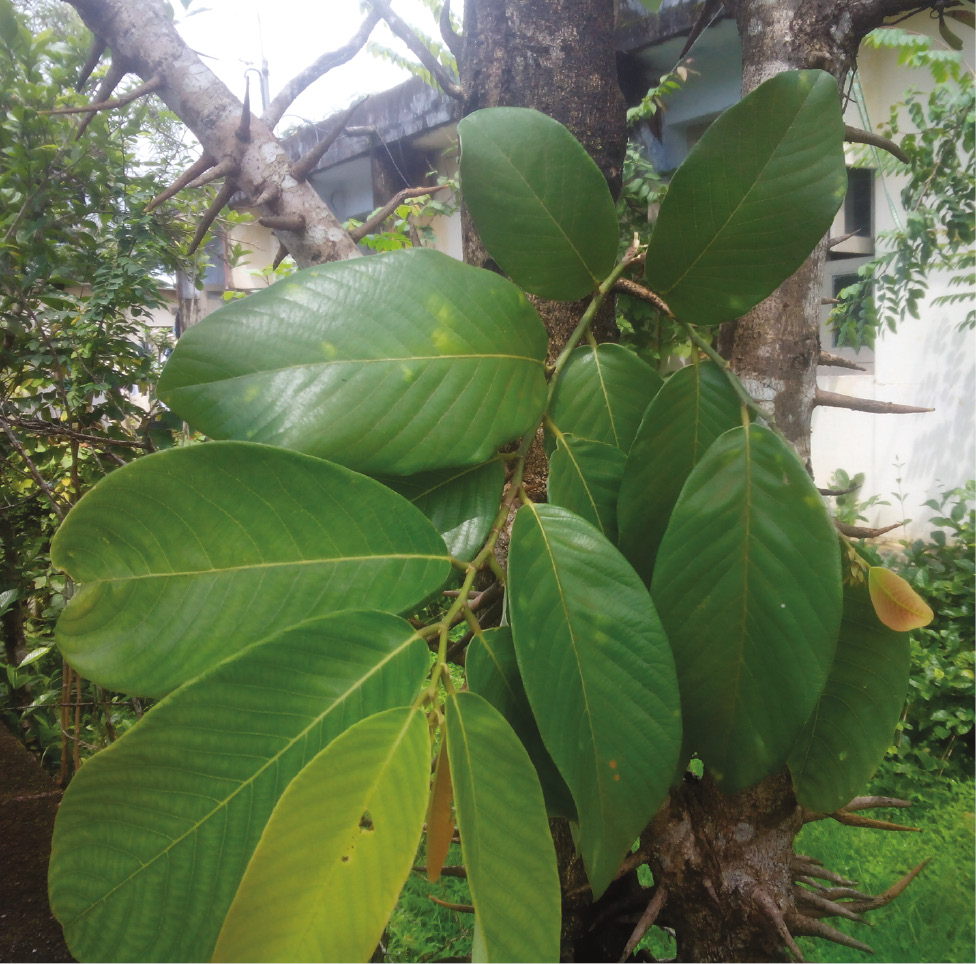
Leaves of Bridelia retusa tree used in this study.
The various parts of this tree have been widely used in folk medicine as contraceptive, as antibacterial agent, and for urinary tract infection [24], [25]. Phytocompounds like various tannins, alkaloidal compounds, glucosides, and glycosides are present in the bark, roots, and leaves of this medicinal plant [26]. Herein, we speculate that these phytocompounds could be utilized in the synthesis of MNPs.
SNPs can be used to catalyze many reduction reactions due to their high value of Fermi potential [27]. In addition, silver nanocomposites have been successfully used for the reduction of many dyes [28], [29], [30], [31], [32]. Rhodamine B (RhB) dye degradation has been considered in the present study to demonsrate the catalytic potential. It is a colorant used in various food stuff, cosmetics and textile industries. The traces of this dye in wastewater causes irritation to the eyes, skin, and respiratory tract. The conventional methods of RhB removal have many disadvantages, such as being high cost, slow pace, requiring special equipment, use of harmful chemicals and UV radiation, and so on [33], [34]. In this context, we propose an effective and rapid method for the RhB degradation using SNPs in the presence of an sodium tetrahydroborate used as a reductant.
Therefore, the present study describes the preparation of SNPs using BRLE, their characterization, and the demonstration of the catalytic potential of SNPs for the degradation of RhB.
Materials and methods
Chemical reagents and glassware
Silver nitrate and RhB dye were acquired from Merck, India. Sodium tetrahydroborate (NaBH4) was procured from SRL Pvt. Ltd., Mumbai, India. All the glassware were properly acid-cleaned and then rinsed with Millipore-MilliQ water.
Preparation of leaf extract
Fresh B. retusa leaves were collected in the month of March, in the Manipal University premises, washed initially with tap water to eliminate dirt and other stuck-on particles, and then rinsed with Millipore-MilliQ water. After allowing them to dry under the shade, the washed leaves were chopped into fine pieces and 10,000 mg of the sample was boiled in 100 ml of Millipore-MilliQ water. The resultant contents were cooled and filtered, which yielded a clear yellow filtrate. The BRLE thus collected was preserved at 4°C for future purposes.
Synthesis of SNPs
The synthesis of SNPs was initiated by mixing 10 ml of BRLE to 90 ml of 0.001 mol/l silver nitrate solution in a flask. The flask was incubated in a temperature-controlled water-bath at 80°C for 10 min. One control experiment (without BRLE) was also conducted with the same conditions. The synthesis process was monitored with respect to time to examine the plasmon resonance band pattern using UV-visible spectroscopy. The control sample containing only silver nitrate did not develop any color change. In contrast, the color of the sample containing silver nitrate and BRLE changed from yellow to deep brown, which connoted the synthesis of SNPs.
Characterization of SNPs
The optical properties of SNPs were analyzed using UV-Visible spectrophotometer (Shimadzu, Model 1700) in a range of 200–800 nm. The morphologies and sizes of the SNPs were determined by scanning electron microscope (EVO MA 18). The energy dispersive X-ray (EDX) analysis (Oxford) confirmed the elemental composition of SNPs. The X-ray diffractometer (XRD, Rigaku Miniflex 600) having Cu Kα radiation source and Ni filter was used to ascertain the crystallinity of SNPs. The scanning was done within 1–2 min at a range of 20°–80°. Fourier transform-infrared spectrometer (FT-IR, SHIMADZU-8400 S) was employed to analyze (4000–400 cm−1 range) the functional groups involved in the synthesis of MNPs. Dynamic light scattering (DLS) instrumentation (Malvern-Zetasizer nanosizer) was employed to determine the hydrodynamic size of the SNPs.
Catalytic degradation of RhB
The catalytic potential of SNPs was tested against a model dye, RhB (λmax=555 nm) in the presence of sodium tetrahydroborate as a reductant. The method suggested in our previous study [35] was carried out with few modifications. Briefly, two clean quartz cuvettes were taken, after which 2 ml of requisite concentration of RhB and newly prepared sodium tetrahydroborate (500 μl) was mixed to each of them thoroughly. To one of the cuvettes, 500 μl of SNPs was added. The extent of dye degradation process of both cuvettes was monitored with respect to time by checking the spectral details using UV-Visible spectrophotometer.
Results and discussion
Visual and UV-visible studies
The addition of the silver nitrate solution (colorless) to the BRLE (yellow color) resulted in deep brown color (Figure 2), a characteristic feature of SNPs. The change in color is due to the reduction of Ag+ to Ago by the biological components found in the leaf extract.
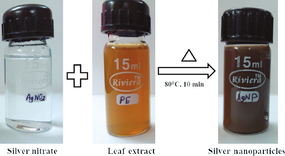
Formation of SNPs using BRLE.
In order to ascertain this reduction, UV-vis spectroscopic analysis was performed (Figure 3). In general, SNPs exhibit a UV-vis absorption maximum between 400 and 500 nm which depends on the size and shape of the SNPs formed and the biomolecules present in the medium [36]. In the present investigation, a characteristic surface plasmon resonance (SPR) peak at 420 nm was observed in the spectrum. This result is consistent with the observations made by other researchers [37], [38]. In addition, the narrow peak corroborates the fact that the synthesized SNPs are monodispersed [39].
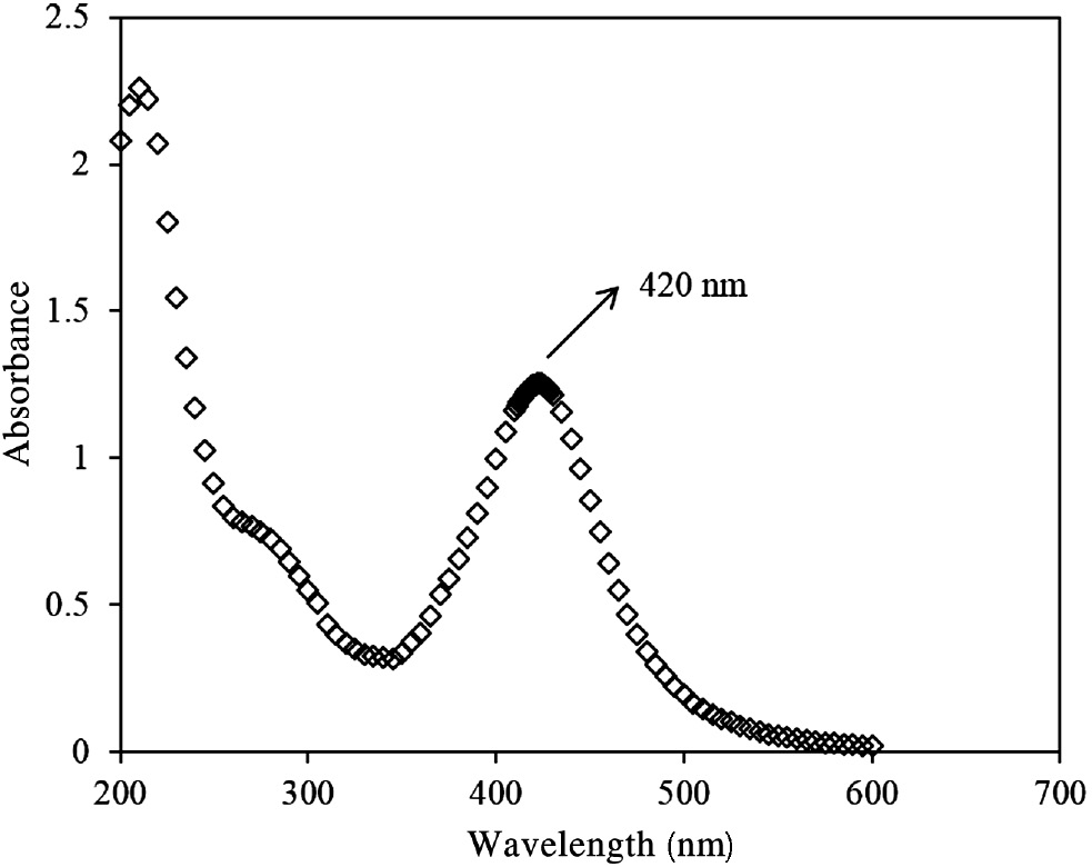
UV-vis spectra of SNPs synthesized using BRLE.
SEM with EDX analysis
SEM was used to analyze the surface morphology of the SNPs (Figure 4). The SNPs formed in this investigation were spherical in shape. Similar results were obtained by many experts for SNP synthesis using other plant extracts [40], [41]. The aggregate formation of SNPs may be due to the procedures involved during sample preparation [42] and the electrostatic interactions between the biomolecules present in the BRLE and SNPs [43].
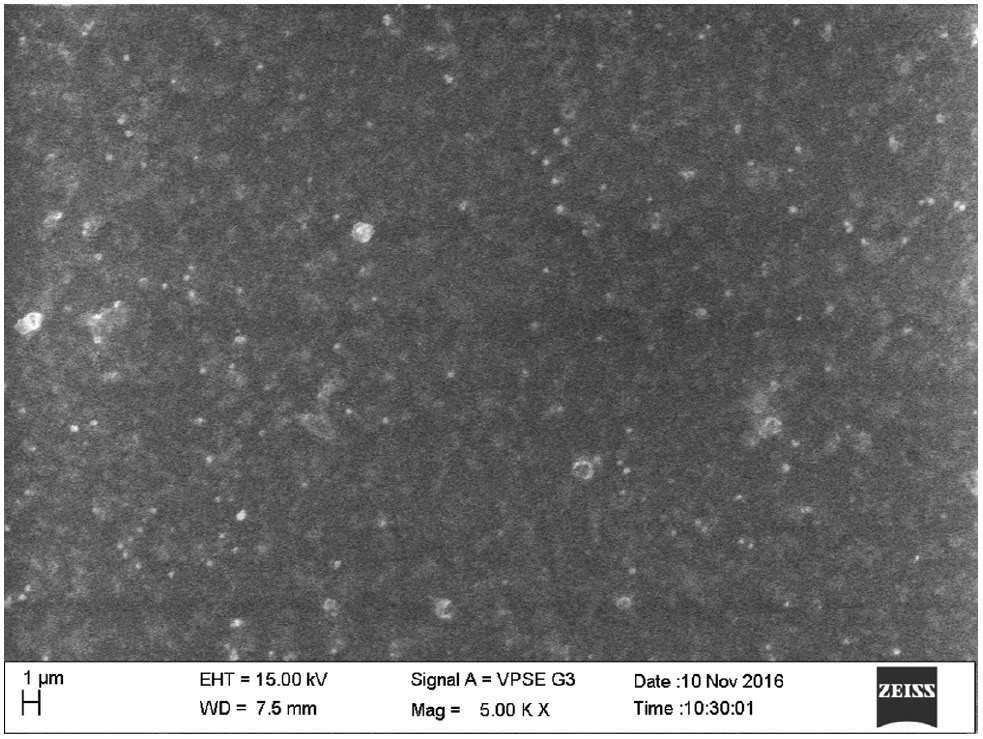
SEM image of SNPs synthesized using BRLE.
The elemental composition of the SNPs was determined by EDX analysis. Figure 5 reveals the strong signal for silver (3 keV). This finding ascertained the existence of metallic silver in the sample. The appearance of other peaks, such as Ca and Si, may be due to the glass slide that bears the sample [44].
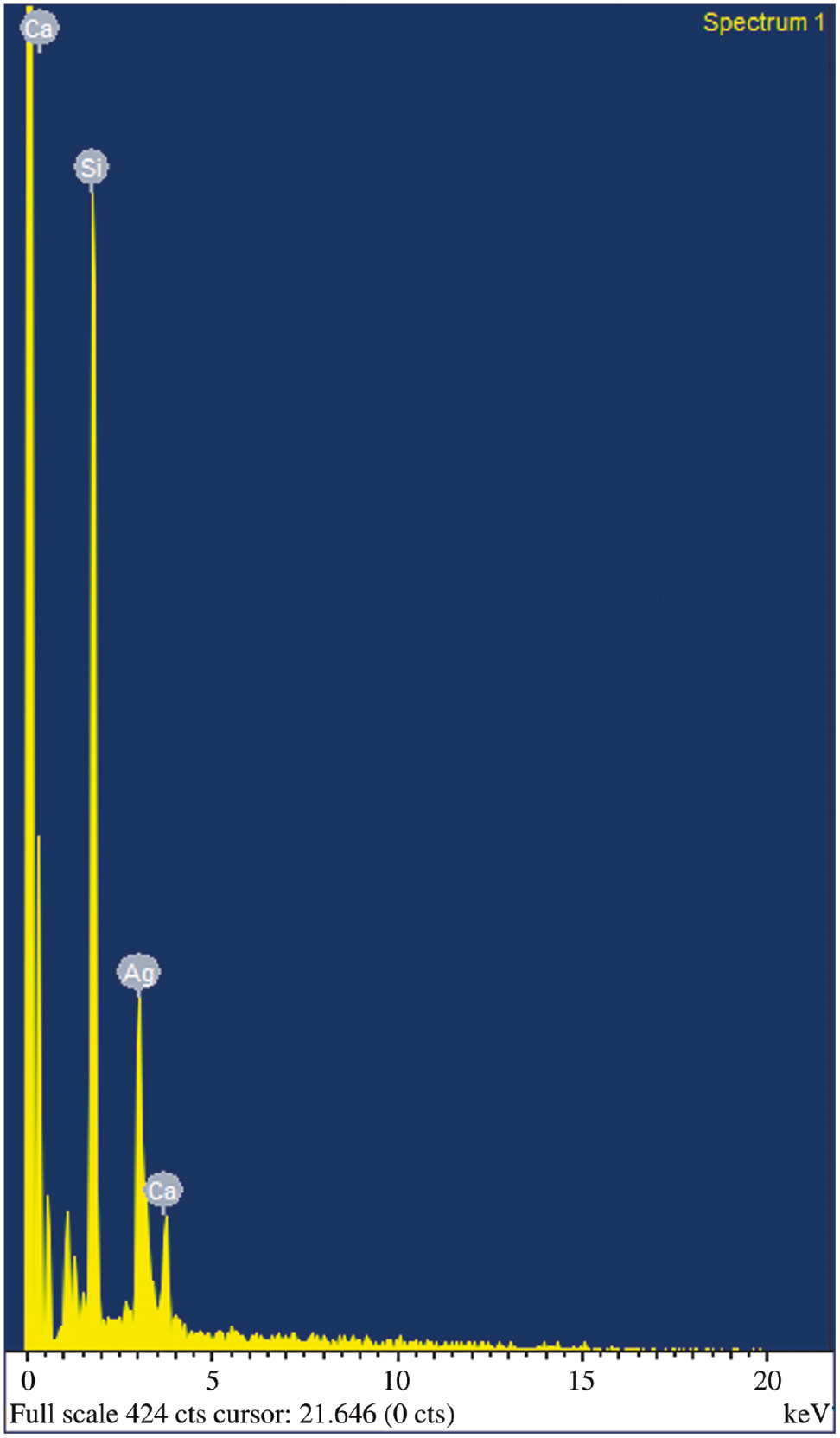
EDX spectrum of SNPs synthesized using BRLE.
XRD analysis
The X-ray diffractogram of synthesized SNPs is shown in Figure 6. The diffractogram affirms the crystallinity of the SNPs as (1 1 1) plane of the face-centered-cubic corresponding to a 2θ value of 38.65°. This result is consistent with the standard data file JCPDS No. 42-0783. This crystalline structure is comparable with the other published literature [45]. Moreover, the sharp and single peak obtained in the present study signified the purity of the synthesized SNPs. Scherrer equation (Equation 1) was used to calculate the crystallite size and is given by
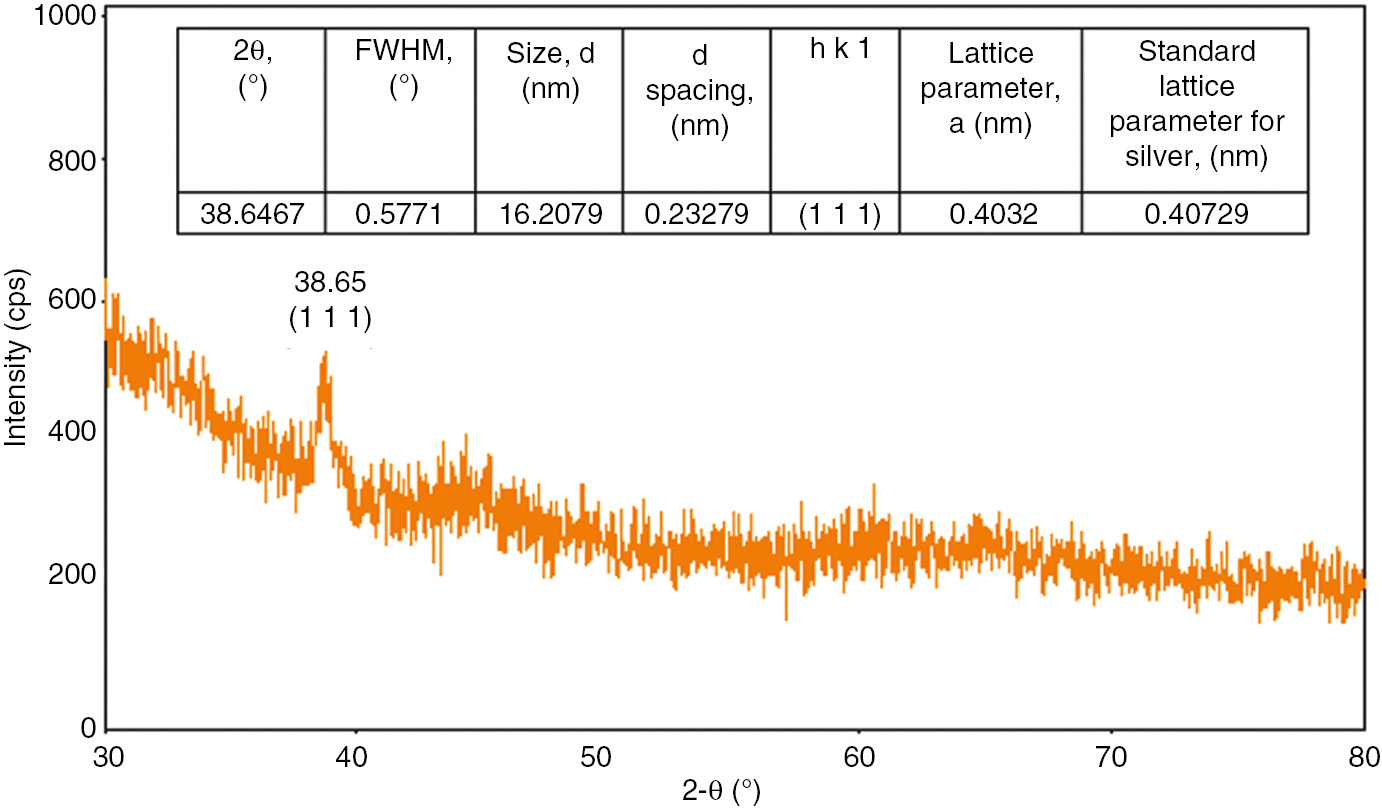
XRD pattern of SNPs synthesized using BRLE.
where d, λ, β0.5, K, and θ are the size of SNPs, wavelength (0.154 nm) of the X-ray source, full width at half maximum of the diffraction peak, Scherrer constant (0.9–1), and Bragg angle in the radian, respectively. The mean size of SNPs was calculated as 16.21 nm. The lattice parameter was 0.4032 nm, which was coherent with the standard lattice parameter of 0.40729 nm for metallic silver [46].
DLS studies
The average particle size distribution of SNPs is shown in Figure 7. The average hydrodynamic diameter of 201.68 nm obtained in the present study is acceptable because the DLS measures only the hydrodynamic diameter and not the actual size of the nanoparticle [47]. The single peak indicates the uniform distribution of nanoparticles. The extent of distribution of particle size is given by poly dispersity index, and the low value attained in this method (0.283) affirms the monodispersity of the nanoparticles [48].
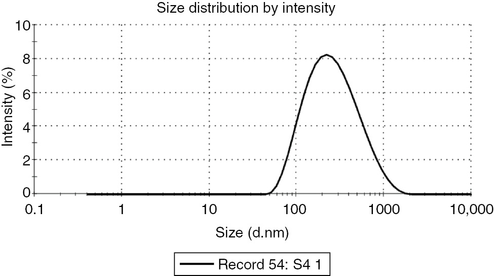
Particle size distribution of SNPs synthesized using BRLE.
The zeta potential analysis of the synthesized SNPs is shown in Figure 8. The zeta potential value obtained from the analysis is −18.1±4.46 mV, conveying good stability of SNPs [49]. It is proposed that the negative value is due to the capping effect of biomolecules in the BRLE. The repulsion among the negatively-charged particles prevents the aggregation, thereby resulting in stable nanoparticles [50].
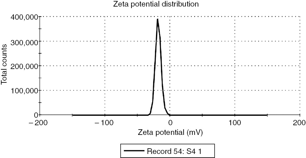
Zeta potential distribution of SNPs synthesized using BRLE.
FT-IR studies
FT-IR measurements were performed to determine the probable functional groups accountable for the reducing and capping properties of leaf extract. The BRLE is known to contain polyphenolic compounds that could act as electron donors for the conversion of Ag+ to Ag0. The major absorption bands are shown in the Figure 9.
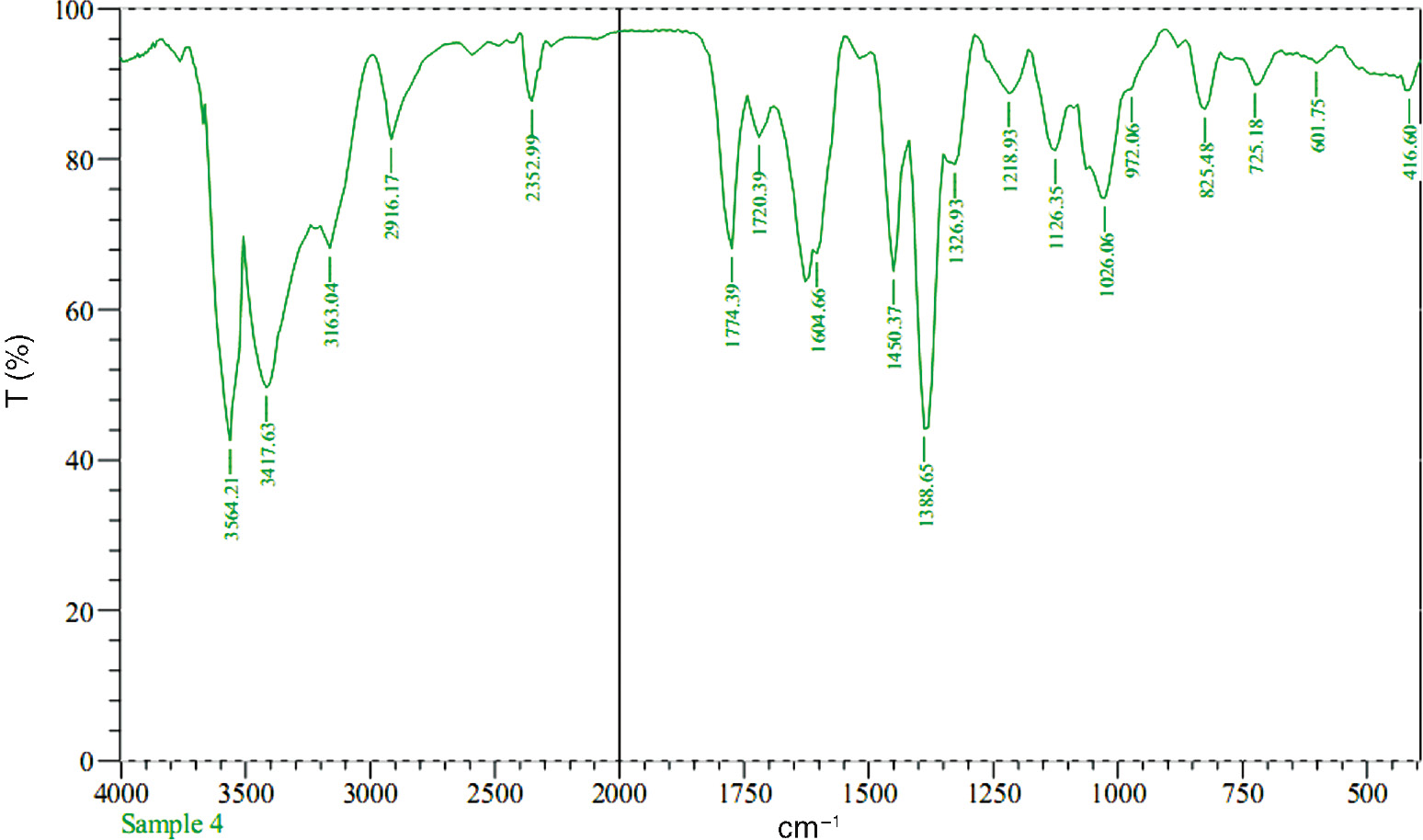
FT-IR spectra of SNPs synthesized using BRLE.
The FT-IR spectrum of the SNPs showed the presence of –OH stretching (3564.21 and 3417.63, sharp bands) owing to the existence of natural flavanols in the BRLE. This fact is further confirmed by the presence of a sharp band at 1026.06 due to the C–O stretching in alcohols [51]. The intense band at 2916.17 depicts the aliphatic –C–H stretching. The small band at 2352.99 denotes –C–NH+ stretching [52]. The presence of carbonyl group is confirmed by the band at 1774.39 (C=O stretching). Another band at 1604.66 denotes the bending vibrations of amide I group present in the BRLE which substantiate the electrostatic attraction of charged groups [53].
The presence of methyl groups (–CH3) are revealed from the bands at 1450.37 and 1388.65. The small bands at 1126.35 and 1026.06 signifies the presence of the C–O stretching of esters or C–N stretching vibrations present in the BRLE. The observed bands in this study corroborates the fact that the phytocompounds in the BRLE (as mentioned in the “Introduction” section) adsorb on the surface of SNPs [54]. Many reports have indicated that the phytocompounds in the leaf extract participate in the formation of weak interactions with SNPs and are responsible for the capping and stabilization of SNPs [55], [56], [57].
Catalytic degradation of RhB
The catalytic activity of SNPs was investigated by using them on the reduction of RhB in the presence of NaBH4. The reduction of RhB by NaBH4 in the absence of SNPs is shown in Figure 10. The UV-Vis absorption spectrum of the degradation of dye at 5-min intervals for a period of 1 h is depicted in Figure 10. At the start of the reaction, the characteristic band of RhB dye at 555 nm was observed, which originated from n→π* transitions in its conjugated structure [58]. The decrement in the intensity of absorption band at 555 nm was slow and it took 45 min to completely degrade the dye. The inset of Figure 10 indicates that the degradation process is a pseudo first order reaction (linear correlation between ln (A/A0) and the reduction time). The degradation constant was estimated as 0.0709 min−1.
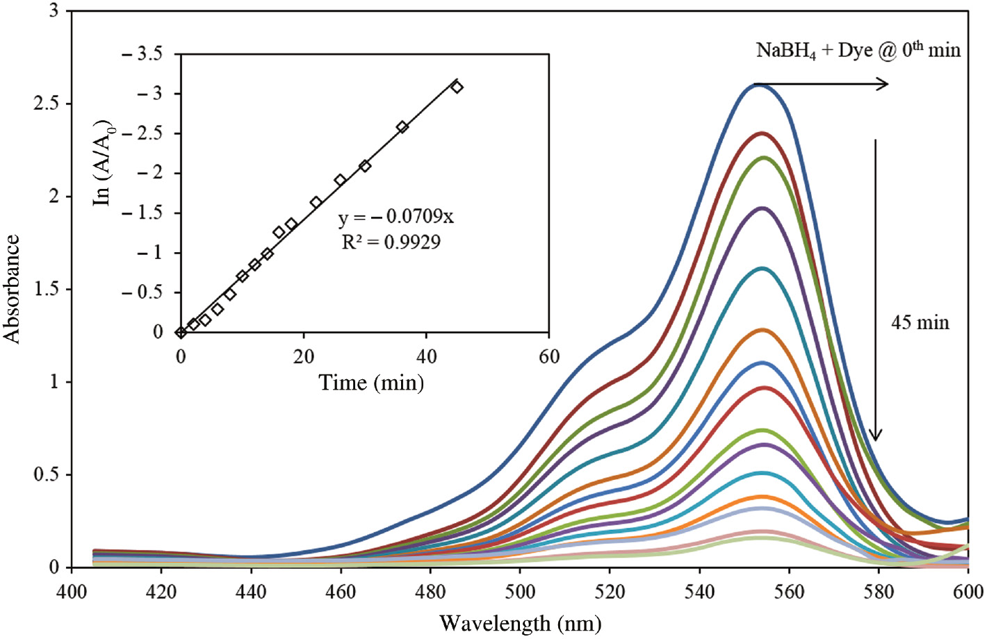
Sequential UV-vis spectra of degradation of RhB in the presence of NaBH4.
(Inset plot shows the relationship between ln (A/A0) vs. time).
Figure 11 shows the UV-Vis spectra of dye degradation in the presence of NaBH4 and SNPs catalyst for the same time period. The introduction of SNPs to the degradation of dye process accelerated the reaction rate, which can be construed from the spectra. The complete degradation of dye was accomplished within 9 min in the presence of SNPs, thus indicating a faster reaction rate as compared with the earlier case (without SNPs). Similar results were obtained by [59] for the degradation of methylene blue by Ag and Au nanoparticles. In the current study, the degradation constant in the presence of SNPs catalyst was estimated as 0.1323 min−1 (inset of Figure 11).
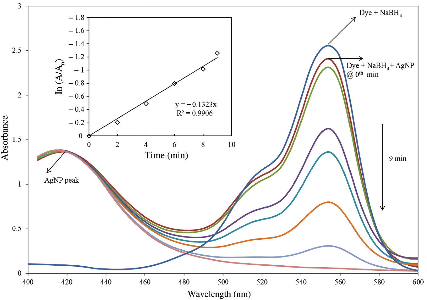
Sequential UV-vis spectra of degradation of RhB in the presence of NaBH4 and SNPs.
(Inset plot shows the relationship between ln (A/A0) vs time).
The added SNPs act as a redox catalyst (electron relay effect) by playing the role of electron transfer mediator between the RhB dye and NaBH4 [60]. To be exact, the BH4− ions donate electrons to the SNPs, and the dye molecules capture the electrons from SNPs; hence, the RhB dye is reduced to its colorless form [61]. Another noteworthy observation in Figure 11 is the appearance of SNPs peak (at 420 nm), which remains constant during the degradation process. There is neither a shift nor a change in the intensity of this peak thus demonstrate the role of catalyst in the degradation of RhB.
Conclusions
The leaf extract of Bridelia retusa was used for the synthesis of SNPs by using an environmentally benign method. The role of phytocompounds in the reduction of silver nitrate to SNPs has been studied through UV-visble and FT-IR spectroscopic studies. The sizes and morphologies were characterized by SEM studies. EDX showed the presence of metallic silver at 3 eV. The crystalline structure of metallic silver was confirmed by XRD, and the mean size of the SNPs was found to be 16.21 nm. The high negative zeta potential value (−18.1 mV) rendered stability to the synthesized SNPs. The SNPs also showed remarkable catalytic ability to degrade RhB within 9 min in the presence of NaBH4, which in turn, allowed the possibility of developing novel nano-catalysts in the future.
About the authors

Ramesh Vinayagam is a senior assistant professor at the Department of Biotechnology, Manipal Institute of Technology, Manipal University. He holds a Bachelor’s degree in Chemical Engineering, a Master’s degree in Industrial Biotechnology, and a Doctoral degree in the field of thermostable enzymes from Manipal University in 2016. With many research articles in reputed journals to his credit, he conducts research in the areas of nanobiotechnology and bioprocess engineering.

Thivaharan Varadavenkatesan is an associate professor at the Department of Biotechnology, Manipal Institute of Technology (MIT), Manipal University. Holding both Bachelor’s and Master’s degrees in Biotechnology, his doctoral thesis focused on biosurfactant production. He obtained his doctorate from Manipal University in 2014. His research focuses on environmental bioremediation and nanobiotechnology.

Raja Selvaraj is an associate professor at the Department of Biotechnology, Manipal Institute of Technology, Manipal University. With a Bachelor’s degree in Chemical Engineering and a Master’s degree in Biotechnology, he obtained his doctoral degree from Manipal University in 2013. He has published many research articles in reputed journals. His research areas include nano-bioremediation, environmental biotechnology, bioprocess engineering, aqueous two phase systems, and design of experiments. He is a peer-reviewer for many leading journals.
Acknowledgments
The authors wish to gratefully acknowledge Department of Biotechnology, MIT, Manipal University.
Conflict of interest statement: The authors declare no conflict of interest regarding the publication of this article.
References
[1] Moritz M, Geszke-Moritz M. Chem. Eng. J. 2013, 228, 596–613.10.1016/j.cej.2013.05.046Search in Google Scholar
[2] Arunachalam R, Dhanasingh S, Kalimuthu B, Uthirappan M, Rose C, Mandal AB. Colloids Surf B Biointerfaces. 2012, 94, 226–230.10.1016/j.colsurfb.2012.01.040Search in Google Scholar PubMed
[3] Ren X, Meng X, Chen D, Tang F, Jiao J. Biosens, Bioelectron. 2005, 21, 433–437.10.1016/j.bios.2004.08.052Search in Google Scholar PubMed
[4] Kumar V, Gundampati RK, Singh DK, Bano D, Jagannadham MV, Hasan SH. J. Photochem. Photobiol. B Biol. 2016, 162, 374–385.10.1016/j.jphotobiol.2016.06.037Search in Google Scholar PubMed
[5] Mudshinge SR, Deore AB, Patil S, Bhalgat CM. Saudi Pharm. J. 2011, 19, 129–141.10.1016/j.jsps.2011.04.001Search in Google Scholar PubMed PubMed Central
[6] Yan W, Lien H-L, Koel BE, Zhang W. Environ. Sci. Process Impacts. 2013, 15, 63.10.1039/C2EM30691CSearch in Google Scholar PubMed
[7] Davar F, Majedi A, Mirzaei A. J. Am. Ceram. Soc. 2015, 98, 1739–1746.10.1111/jace.13467Search in Google Scholar
[8] Hossainian H, Salavati-Niasari M, Bazarganipour M. J. Mol. Liq. 2016, 220, 747–754.10.1016/j.molliq.2016.04.129Search in Google Scholar
[9] Guzman M, Dille J, Godet S. Nanomed. Nanotechnol. Biol Med. 2012, 8, 37–45.10.1016/j.nano.2011.05.007Search in Google Scholar PubMed
[10] Narayanan KB, Park HH. Eur. J. Plant Pathol. 2014, 140, 185–192.10.1007/s10658-014-0399-4Search in Google Scholar
[11] Raja S, Ramesh V, Thivaharan V. J. Ind. Eng. Chem. 2015, 29, 257–264.10.1016/j.jiec.2015.03.033Search in Google Scholar
[12] Jeyaraj M, Varadan S, Anthony KJP, Murugan M, Raja A, Gurunathan S. J. Ind. Eng. Chem. 2013, 19, 1299–1303.10.1016/j.jiec.2012.12.031Search in Google Scholar
[13] Rajakumar G, Abdul Rahuman A. Acta Trop. 2011, 118, 196–203.10.1016/j.actatropica.2011.03.003Search in Google Scholar PubMed
[14] Nath D, Banerjee P. Environ. Toxicol. Pharmacol. 2013, 36, 997–1014.10.1016/j.etap.2013.09.002Search in Google Scholar PubMed
[15] Borase HP, Salunke BK, Salunkhe RB, Patil CD, Hallsworth JE, Kim BS, Patil SV. Appl. Biochem. Biotechnol. 2014, 173, 1–29.10.1007/s12010-014-0831-4Search in Google Scholar PubMed
[16] Issaabadi Z, Nasrollahzadeh M, Sajadi SM. J. Clean. Prod. 2017, 142, 3584–3591.10.1016/j.jclepro.2016.10.109Search in Google Scholar
[17] Atarod M, Nasrollahzadeh M, Sajadi SM. J. Colloid. Interface Sci. 2016, 465, 249–258.10.1016/j.jcis.2015.11.060Search in Google Scholar PubMed
[18] Nasrollahzadeh M, Atarod M, Jaleh B. Ceram. Int. 2016, 42, 8587–8596.10.1016/j.ceramint.2016.02.088Search in Google Scholar
[19] Narayanan KB, Sakthivel N. Adv. Colloid. Interface Sci. 2010, 156, 1–13.10.1016/j.cis.2010.02.001Search in Google Scholar PubMed
[20] Asmathunisha N, Kathiresan K. Colloids Surf B Biointerfaces. 2013, 103, 283–287.10.1016/j.colsurfb.2012.10.030Search in Google Scholar PubMed
[21] Mittal AK, Chisti Y, Banerjee UC. Biotechnol. Adv. 2013, 31, 346–356.10.1016/j.biotechadv.2013.01.003Search in Google Scholar
[22] Koupaei MH, Shareghi B, Saboury A. Saboury AA, Davar F, Semnani A, Evini M. RSC Adv. 2016, 6, 42313–42323.10.1039/C5RA24862KSearch in Google Scholar
[23] Khare CP. Indian Medicinal Plants: An Illustrated Dictionary, 1st ed., Springer-Verlag: New York, 2007.Search in Google Scholar
[24] Ngueyem TA, Brusotti G, Caccialanza G, Finzi PV. J. Ethnopharmacol. 2009, 124, 339–349.10.1016/j.jep.2009.05.019Search in Google Scholar
[25] Ayyanar M, Ignacimuthu S. J. Ethnopharmacol. 2005, 102, 246–255.10.1016/j.jep.2005.06.020Search in Google Scholar
[26] Mali S, Borges RM. Biochem. Syst. Ecol. 2003, 31, 1221–1246.10.1016/S0305-1978(03)00079-6Search in Google Scholar
[27] Sen IK, Maity K, Islam SS. Carbohydr. Polym. 2013, 91, 518–528.10.1016/j.carbpol.2012.08.058Search in Google Scholar PubMed
[28] Atarod M, Nasrollahzadeh M, Sajadi SM. J. Colloid. Interface Sci. 2016, 462, 272–279.10.1016/j.jcis.2015.09.073Search in Google Scholar PubMed
[29] Hatamifard A, Nasrollahzadeh M, Sajadi SM. New J. Chem. 2016, 40, 2501–2513.10.1039/C5NJ02909KSearch in Google Scholar
[30] Rostami-Vartooni A, Nasrollahzadeh M, Alizadeh M. J. Colloid Interface Sci. 2016, 470, 268–275.10.1016/j.jcis.2016.02.060Search in Google Scholar PubMed
[31] Tajbakhsh M, Alinezhad H, Nasrollahzadeh M, Kamali TA. J. Alloys. Compd. 2016, 685, 258–265.10.1016/j.jallcom.2016.05.278Search in Google Scholar
[32] Rostami-Vartooni A, Nasrollahzadeh M, Alizadeh M. J. Alloys Compd. 2016, 680, 309–314.10.1016/j.jallcom.2016.04.008Search in Google Scholar
[33] Nagaraja R, Kottam N, Girija CR, Nagabhushana BM. Powder Technol. 2012, 215–216, 91–97.Search in Google Scholar
[34] Torrades F, García-Montaño J. Dye Pigment. 2014, 100, 184–189.10.1016/j.dyepig.2013.09.004Search in Google Scholar
[35] Varadavenkatesan T, Selvaraj R, Vinayagam R. J. Mol. Liq. 2016, 221, 1063–1070.10.1016/j.molliq.2016.06.064Search in Google Scholar
[36] Kumar B, Smita K, Cumbal L, Debut A. Saudi J. Biol. Sci. 2014, 21, 605–609.Search in Google Scholar
[37] Raja K, Saravanakumar A, Vijayakumar R. Spectrochim. Acta A Mol. Biomol. Spectrosc. 2012, 97, 490–494.10.1016/j.saa.2012.06.038Search in Google Scholar PubMed
[38] Zahir AA, Rahuman AA. Vet. Parasitol. 2012, 187, 511–520.10.1016/j.vetpar.2012.02.001Search in Google Scholar PubMed
[39] Khalil MMH, Ismail EH, El-Baghdady KZ, Mohamed D. Arab. J. Chem. 2014, 7, 1131–1139.10.1016/j.arabjc.2013.04.007Search in Google Scholar
[40] Sathiya CK, Akilandeswari S. Spectrochim. Acta A Mol. Biomol. Spectrosc. 2014, 128, 337–341.10.1016/j.saa.2014.02.172Search in Google Scholar PubMed
[41] Kathiravan V, Ravi S, Ashokkumar S. Spectrochim. Acta A Mol. Biomol. Spectrosc. 2014, 130, 116–121.10.1016/j.saa.2014.03.107Search in Google Scholar PubMed
[42] Saravanakumar A, Ganesh M, Jayaprakash J, Jang HT. J. Ind. Eng. Chem. 2015, 28, 277–281.10.1016/j.jiec.2015.03.003Search in Google Scholar
[43] Kathiravan V, Ravi S, Ashokkumar S, Velmurugan S, Elumalai K, Khatiwada CP. Spectrochim. Acta A Mol. Biomol. Spectrosc. 2015, 139, 200–205.10.1016/j.saa.2014.12.022Search in Google Scholar PubMed
[44] Zhang W, Chen Z, Liu H, Zhang L, Gao P, Li D. Colloids Surf B Biointerfaces. 2011, 88, 196–201.10.1016/j.colsurfb.2011.06.031Search in Google Scholar PubMed
[45] Ghaedi M, Yousefinejad M, Safarpoor M, Khafri HZ, Purkait MK. J. Ind. Eng. Chem. 2015, 31, 167–172.10.1016/j.jiec.2015.06.020Search in Google Scholar
[46] Theivasanthi T, Alagar M. Nano. Biomed. Eng. 2012, 4, 1–11.Search in Google Scholar
[47] Eid M, Araby E. Appl. Biochem. Biotechnol. 2013, 171, 469–487.10.1007/s12010-013-0357-1Search in Google Scholar PubMed
[48] Kumar B, Smita K, Cumbal L, Debut A. Saudi J. Biol. Sci. 2017, 24, 45–50.Search in Google Scholar
[49] Antony JJ, Sithika MAA, Joseph TA, Suriyakalaa U, Sankarganesh A, Siva D, Kalaiselvi S, Achiraman S. Colloids Surf B Biointerfaces. 2013, 108, 185–190.10.1016/j.colsurfb.2013.02.041Search in Google Scholar PubMed
[50] Ajitha B, Reddy YAK, Shameer S, Rajesh KM, Suneetha Y, Reddy PS. J. Photochem. Photobiol. B Biol. 2015, 149, 84–92.10.1016/j.jphotobiol.2015.05.020Search in Google Scholar PubMed
[51] Swarnavalli GCJ, Dinakaran S, Raman N, Jegadeesh R, Pereira C. J. Saudi Chem. Soc. 2017, 21, 172–179.10.1016/j.jscs.2015.03.004Search in Google Scholar
[52] Raut RW, Mendhulkar VD, Kashid SB. J. Photochem. Photobiol. B Biol. 2014, 132, 45–55.10.1016/j.jphotobiol.2014.02.001Search in Google Scholar PubMed
[53] Gole A, Dash C, Ramakrishnan V, Sainkar SR, Mandale AB, Rao M, Sastry M. Langmuir 2001, 17, 1674–1679.10.1021/la001164wSearch in Google Scholar
[54] Bar H, Bhui DK, Sahoo GP, Sarkar P, Pyne S, Misra A. Colloids. Surfaces A Physicochem. Eng. Asp. 2009, 348, 212–216.10.1016/j.colsurfa.2009.07.021Search in Google Scholar
[55] Dasaratrao SB, Salimath B, Deshpande R, Dhondojirao Bedre M, Krishnamurthy Prabhakar B, Venkataraman A. Sci. Technol. Adv. Mater. 2008, 9, 35012.10.1088/1468-6996/9/3/035012Search in Google Scholar PubMed PubMed Central
[56] Balakumaran MD, Ramachandran R, Balashanmugam P, Mukeshkumar DJ, Kalaichelvan PT. Microbiol. Res. 2016, 182, 8–20.10.1016/j.micres.2015.09.009Search in Google Scholar PubMed
[57] Raja S, Ramesh V, Thivaharan V. Arab. J. Chem. 2017, 10, 253–261.10.1016/j.arabjc.2015.06.023Search in Google Scholar
[58] Khorshidi A, Mardazad N. Res. Chem. Intermed. 2016, 42, 7551–7558.10.1007/s11164-016-2552-5Search in Google Scholar
[59] Kumari MM, Philip D. Spectrochim. Acta A Mol. Biomol. Spectrosc. 2013, 111, 154–160.10.1016/j.saa.2013.03.076Search in Google Scholar PubMed
[60] Cheval N, Gindy N, Flowkes C, Fahmi A. Nanoscale Res. Lett. 2012, 7, 182.10.1186/1556-276X-7-182Search in Google Scholar PubMed PubMed Central
[61] Jeyapragasam T, Kannan RS. Russ. J. Phys. Chem. A. 2016, 90, 1334–1337.10.1134/S003602441607030XSearch in Google Scholar
©2018 Walter de Gruyter GmbH, Berlin/Boston
This article is distributed under the terms of the Creative Commons Attribution Non-Commercial License, which permits unrestricted non-commercial use, distribution, and reproduction in any medium, provided the original work is properly cited.
Articles in the same Issue
- Frontmatter
- In this issue
- Synthesis of iron oxide nanoparticles in a continuous flow spiral microreactor and Corning® advanced flow™ reactor
- Diisopropylamine as a single catalyst in the synthesis of aryl disulfides
- Processing and properties of high-purity micro-lamellate (NH4)2RuCl6 particles
- Dynamic synthesis route of zeolite Y with kaolin to improve yield
- Green synthesis, structural characterization, and catalytic activity of silver nanoparticles stabilized with Bridelia retusa leaf extract
- Hydrothermal green synthesis of gold nanoparticles using mushroom (Agaricus bisporus) extract: physico-chemical characteristics and antifungal activity studies
- Tribological behavior of a newly developed AA2014/waste eggshell/SiC hybrid green metal matrix composite at optimum parameters
- Parameters controlling the advanced oxidation degradation kinetics of nitroglycerin and pentaerythritol tetranitrate
- Kinetics of green solid-liquid extraction of useful compounds from plant materials: kinetics coefficients and modeling
- Comparison of Rhodotorula sp. and Bacillus megaterium in the removal of cadmium ions from liquid effluents