Effect of particle surface charge on drug uptake
-
Maosheng Zheng
Maosheng Zheng obtained his PhD degree in materials science & engineering in 1992 at Northwestern Polytechnic University, China. He worked in materials science & engineering for teaching and research in Xi’an Jiaotong University in 1992 till 2006 as a full time professor. Since 2006, he worked in the School of Chemical Engineering at Northwest University for materials science and nano- technology teaching and research as full time professor. His research work focuses on nano- technology, energy resource technology and materials.
 und
Jie Yu
und
Jie Yu
Jie Yu obtained her Bachelors degree in polymer materials in 1987 at Northwestern Polytechnic University, China. She worked in the polymer materials and biological engineering fields for teaching and research in Xi’an Jiaotong University in 1993 till 2007. Since 2007, she has engaged in the School of Life Science and Technology at Northwest University for biological medicine teaching and research as senior engineer. Her research work focuses on organic nano-particles, synthesis, surfaces, and their interactions with biological cells.

Abstract
In this paper, it aims to build the relationship of statically electric interaction between the surface charge of a particle drug and cellular uptake. The statically electric theory is applied to study the change of wetting between the drug particle and the cell, a factor that enhanced uptake of cells induced by particle’s surface charge is introduced, then it is formulated according to Kelvin theory for dissolving of solid particle in liquid. It is found that the change of contact angle between the surface charged particle drug and the cell can be detected if the Zeta potential reaches to 6 mV in water like solution, an increase of about 11.1% for the uptake could be obtained for a polymer particle with molar mass M∼10 kg/mol, mass density ρ∼0.9×10³ kg/m3, radius r∼15 nm at temperature 300 K and Zeta potential 10 mV condition as compared with the same particle in water-like solution but without surface charge. The effect of particle surface charge on drug uptake could be interpreted by statically electric theory.
Introduction
Due to their special physical, chemical, and biological properties, nanoparticles, with diameters ranging from 1 to 100 nm, have been used in various fields (1–4). Different nano-materials, such as carbon nanotubes, silica nanoparticles, fullerene, magnetic nanoparticles, etc. attract much more attention in the engineering and health fields (5–9). In particular, it has been realized that nanoparticles have potentials to enhance delivering drug or genetic materials to targeted cells or organs, it results in the therapeutic delivery procedures being more precise, more effective, and less harmful to healthy tissues and organs (10).
Polymer nanoparticles, such as polyethylenimine (PEI) (11), chitosan (12), and polyglycolic acid (PLGA) (13–15) have been frequently used for drug and gene delivery in the recent researches. Metallic and ceramic nanoparticles, such as Au nanoparticles (16–18), iron oxide (17, 19, 20) and silica nanoparticles (10, 21), have also been studied for drug and gene delivery. The widely used in biomedical fields such as dental, bone tissue engineering (22), orthopedic implants (23, 24) and antibacterial agents (25), hydroxyapatite (HAP), are those bone like ones due to their chemical and structural similarity with bone minerals and good biocompatibility (22). Recently, more attention has been paid to study the possibility of using HAP nanoparticles as carrier for drug and gene delivery because of their great affinity to DNA (2, 10, 26–28). It is necessary to understand the interaction between HAP nanoparticles and the cell, so as to design proper nanoparticles for gene delivery (29).
It is well known that size, shape, and surface charge are the primary parameters of colloidal particles for cellular uptake besides biomolecular approaches. Gao et al proposed a diffusive model to describe the receptor-mediated endocytosis process (30), it gives a good agreement with many cellular uptake experimental results. Other studies indicate that surface charge affects the cellular uptake and biocompatibility of nanoparticles dramatically (31–34).
In this study, the statically electric relationship between the surface charge of the particle drug and cell uptake is built, the change of wetting between drug particle and cell is studied based on statically electric theory. A factor that enhanced uptake of cells induced by surface charge effect is introduced and formulated according to Kelvin theory for dissolving of solid particle in liquid.
Action of particle surface charge with surrounding medium
The interaction between the surface charge of a particle and the surrounding medium is a statically electric one undoubtedly. Consider the particle as a charged spherical ball for the simplicity, its surrounding medium will be polarized according to the common statically electric theory in general, and there exists an opposite electric layer near the charged spherical ball due to the polarization action, thus the so-called “double charge layer” forms in between. And furthermore, it builds a spherically electric capacitor visually.
For plane infinite capacitor, its capacity can be written as (35, 36),
in which, ε0 and εr represent the dielectric constant of the vacuum and relative dielectric constant of the medium, respectively; Ssol–liq is the interface area of solid and liquid; d is the thickness of the “double charge layer”.
If the electric potential applied to the “double charge layer” capacitor is u, the increase of free energy per area due to the charge distribution in the liquid can be written as,
As to a electric charged liquid droplet, if it is placed on the solid plane, the contact angle will be changed according to the following manner by adding the electro-wetting term Δγ into Young’s equation (35, 36),
in which, γliq–vap is the free energy of liquid/vapor; θ0 is the contact angle of the liquid in absence of applied voltage and θ(u) is the contact angle of the liquid under condition of voltage u applied. Eq. [3] indicates that the contact angle decreases with u2 and independent on particle size.
Similarly, the wetting degree between electric charged drug particle and cell could be improved by Eq. [3] as well provided voltage u is applied.
Eq. [3] shows that the change of contact angle increases with electric potential (i.e. surface charge) monotonously.
As an example, lets consider water as the medium in Eq. [3]. Since the relative dielectric constant of water εr, water is about 80, and the dielectric constant of the vacuum ε0 is 8.85×10⁻¹² Coulomb2/N·m2, the free energy of water/vapor γliq–gas is about 72.8 mN/m, if the thickness of the “double charge layer” d is taken as
Ref. (31) presented the changes of contact angle with respect to preparation parameters for negatively charged chitosan hydrochloride grafted nano-particles (CMCNP), covalently labeled with rhodamine B(RhB), the size of these particles is about 150 nm. The particle size and Zeta potential were determined by using Zetasizer Nano ZS90, and the contact angle was measured by using JC 2000 A Goniometer (31). The results are cited and shown in Table 1.
Changes of contact angle with Zeta potential for CMCNP (31).
| Sample | Size, nm | Zeta potential, mV | Contact angle, ° |
|---|---|---|---|
| RhB-CMCNP (–15, 150) | 149.2±4.1 | –13.2±0.5 | 63.5±1.4 |
| RhB-CMCNP (–25, 150) | 157.3±4.7 | –23.2±1.7 | 43.1±1.0 |
| RhB-CMCNP (–40, 150) | 156.0±5.1 | –38.4±0.7 | 38.8±1.0 |
The data in Table 1 shows that the contact angle decreases with Zeta potential (i.e. surface charge) obviously, it indicates the validity of Eq. [3].
The decrease of contact angle implies the preferable wetting between the particle and the medium, which might promote the uptake of cells in principle.
The Kelvin theory for dissolving of solid particle in liquid reads (37),
in which, a is the instant activity and a0 is the original activity of solid particle dissolving in liquid at equilibrium states; M, ρ and r are the molar mass, volume density and radius of the particle, respectively; R and T are gas constant and absolute temperature, respectively, Δγ is the change of surface free energy.
The term a/a0 in Eq. [4] is in fact a ratio that reflects the enhancement of the activity of solid particle dissolving in liquid at equilibrium state.
According to Kelvin theory for dissolving of solid particle in liquid, i.e. Eq. [4], the uptake of cells induced by surface charge effect could be increased by a factor,
Under condition of polymer particle as carrier, M∼10 kg/mol, ρ∼0.9×10³ kg/m3, r=15 nm, T=300 K, u=10 mV,
Figure 1 shows the effects of particle radius and electric potential on enhanced uptake factor f, the values for M, ρ and T are taken as 10 kg/mol, 0.9×10³ kg/m3,
![Figure 1: Effect of particle radius and electric potential on enhanced factor by Eq. [5].](/document/doi/10.1515/ejnm-2015-0015/asset/graphic/j_ejnm-2015-0015_fig_001.jpg)
Effect of particle radius and electric potential on enhanced factor by Eq. [5].
He et al. studied the surface charge effect on cellular (murine macrophage) uptake for 4 types of polymer particles with the size of about 150 nm (31). They took the percentage of fluorescence associated with cells versus the amount of fluorescence present in the feed solution as the expression for the uptake (31). Here their experimental results are cited and shown in Figure 2.
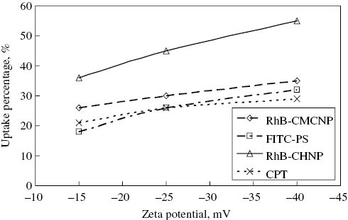
Surface charge effect on cellular (murine macrophage) uptake for 4 type of polymer particles with the size of about 150 nm (31).
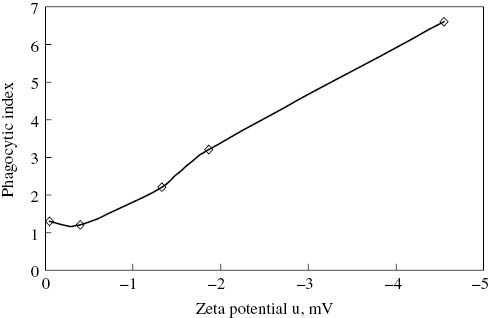
Roser et al investigated the effect of surface charges on in vitro phagocytosis and biodistribution in rats (32). The Laser Doppler Anemometry with a Malvern Zetasizer IIc was employed to determine the electrophoretic mobility, and the Smoluchowski equation was used to calculate the zeta potential from the electrophoretic mobility (32). The mouse macrophages were characterized using a 1- Naphthylesterase Diagnostik Kit (32). The correlation between zeta potential and phagocytic index is cited and shown in Figure 3, which was surface modified albumin nanoparticles using mouse macrophages for incubation time 60 min and without serum under condition of phosphate buffer, 5 mM and pH 7.2 (32).
Effect of zeta potential on phagocytic index (32).
The experimental results in Figures 2 and 3 indicate that the cellular uptake increases obviously with the value of Zeta potential for the 4 type of polymer particles with the size of about 150 nm, and for the surface modified albumin nanoparticles using mouse macrophages, their trends agree with the theoretical prediction of Eq. [5] fundamentally.
Concluding remarks
From above discussion, it could be concluded,
The correlations between the surface charged drug particle and cell, and the enhanced uptake with surface charge of particle proposed on basis of the fundamental statically electric and Kelvin theory for dissolving of solid particle in liquid in the present study are appropriate;
The correlations realize the nature of the interaction between the surface charged particle and cell quantitatively. Available experimental data verifies their validity;
The basic picture of surface charge improving cell uptake is as follows: electric interaction in statically electric double layer→wetting improving between charged drug particle and cell→solid particle dissolving in liquid improvement→cellular uptake enhancement.
About the authors

Maosheng Zheng obtained his PhD degree in materials science & engineering in 1992 at Northwestern Polytechnic University, China. He worked in materials science & engineering for teaching and research in Xi’an Jiaotong University in 1992 till 2006 as a full time professor. Since 2006, he worked in the School of Chemical Engineering at Northwest University for materials science and nano- technology teaching and research as full time professor. His research work focuses on nano- technology, energy resource technology and materials.

Jie Yu obtained her Bachelors degree in polymer materials in 1987 at Northwestern Polytechnic University, China. She worked in the polymer materials and biological engineering fields for teaching and research in Xi’an Jiaotong University in 1993 till 2007. Since 2007, she has engaged in the School of Life Science and Technology at Northwest University for biological medicine teaching and research as senior engineer. Her research work focuses on organic nano-particles, synthesis, surfaces, and their interactions with biological cells.
Acknowledgments
The support of Shaanxi provincial social development and tackling key problem program 2014-k14-03-02 is acknowledged.
Conflict of interest statement: Authors state no conflict of interest. All authors have read the journal’s Publication ethics and publication malpractice statement available at the journal’s website and hereby confirm that they comply with all parts applicable to the present scientific work.
References
1. Fayazpour F, Lucas B, Alvarez-Lorenzo C, Sanders N, Demeester J, De Smedt S. Physicochemical and transfection properties of cationic hydroxyethylcellulose / DNA nanoparticles. Bio- macromolecules 2006;7:2856–62.10.1021/bm060474bSuche in Google Scholar PubMed
2. Cai YR, Pan HH, Xu XR, Hu QH, Li L, Tang RK. Ultrasonic controlled morphology transformation of hollow calcium phosphate nanospheres: a smart and biocompatible drug release system. Chem Mater 2007;19:3081–3.10.1021/cm070298tSuche in Google Scholar
3. Yang PP, Quan ZW, Lu LL, Huang SS, Lin J. Luminescence functionalization of mesoporous silica with different morphologies and applications as drug delivery systems. Biomaterials 2008;29:692–702.10.1016/j.biomaterials.2007.10.019Suche in Google Scholar PubMed
4. Wilhelm C, Gazeau F. Universal cell labelling with anionic magnetic nanoparticles. Biomaterials 2008;29:3161–74.10.1016/j.biomaterials.2008.04.016Suche in Google Scholar PubMed
5. Chlopek J, Czajkowska B, Szaraniec B, Frackowiak E, Szostak K, Béguin F. In vitro studies of carbon nanotubes biocompatibility. Carbon 2006;44:1106–11.10.1016/j.carbon.2005.11.022Suche in Google Scholar
6. Chung TH, Wu SH, Yao M, Lu CW, Lin YS, Hung Y, et al. The effect of surface charge on the uptake and biological function of mesoporous silica nanoparticles 3T3-L1 cells and human mesenchymal stem cells. Biomaterials 2007;28:2959–66.10.1016/j.biomaterials.2007.03.006Suche in Google Scholar PubMed
7. Tang YJ, Ashcroft JM, Chen D, Min G, Kim CH, Murkhejee B, et al. Charge-associated effects of fullerene derivatives on microbial structural integrity and central metabolism. Nano Lett 2007;7:754–60.10.1021/nl063020tSuche in Google Scholar PubMed
8. Sayes CM, Gobin AM, Ausman KD. Nano-C-60 cytotoxicity is due to lipid peroxidation. Biomaterials 2005;26:7587–95.10.1016/j.biomaterials.2005.05.027Suche in Google Scholar PubMed
9. Wan SR, Huang JS, Guo M, Zhang H, Cao Y, Yan H, et al. Biocompatible superparamagnetic iron oxide nanoparticle dispersions stabilized with poly(ethylene glycol)oligo(aspartic acid) hybrids. J Biomed Mater Res A 2007;80:946–54.10.1002/jbm.a.31022Suche in Google Scholar PubMed
10. Tan K, Cheang P, Ho IA, Lam PY, Hui KM. Nanosized bioceramic particles could function as efficient gene delivery vehicles with target specificity for the spleen. Gene Ther 2007;14: 828–35.10.1038/sj.gt.3302937Suche in Google Scholar PubMed
11. Nimesh S, Chandra R. Polyethylenimine nanoparticles as an efficient in vitro siRNA delivery system. Eur J Pharm Biopharm 2009;73:43–49.10.1016/j.ejpb.2009.04.001Suche in Google Scholar PubMed
12. Woitiski CB, Neufeld RJ, Ribeiro AJ, Veiga F. Colloidal carrier integrating biomaterials for oral insulin delivery: influence of component formulation on physicochemical and biological parameters. Acta Biomater 2009;5:2475–84.10.1016/j.actbio.2009.03.007Suche in Google Scholar PubMed
13. Acharya S, Dilnawaz F, Sahoo SK. Targeted epidermal growth factor receptor nanoparticle bioconjugates for breast cancer therapy. Biomaterials 2009;30:5737–50.10.1016/j.biomaterials.2009.07.008Suche in Google Scholar PubMed
14. Thomas C, Gupta V, Ahsan F. Influence of surface charge of PLGA particles of recombinant hepatitis B surface antigen in enhancing systemic and mucosal immune responses. Int J Pharmaceut 2009;379:41–50.10.1016/j.ijpharm.2009.06.006Suche in Google Scholar PubMed
15. Zhou J, Moya S, Ma L, Gao C, Shen J. Polyelectrolyte coated PLGA nanoparticles: templation and release behavior. Macromol Biosci 2009;9:326–35.10.1002/mabi.200800188Suche in Google Scholar PubMed
16. Perrault SD, Walkey C, Jennings T, Fischer HC, Chan WC. Mediating tumor targeting efficiency of nanoparticles through design. Nano Lett 2009;9:1909–15.10.1021/nl900031ySuche in Google Scholar PubMed
17. Kamei K, Mukai Y, Kojima H, Yoshikawa T, Yoshikawa M, Kiyohara G, et al. Direct cell entry of gold/iron-oxide magnetic nanoparticles in adenovirus mediated gene delivery. Biomaterials 2009;30:1809–14.10.1016/j.biomaterials.2008.12.015Suche in Google Scholar PubMed
18. Li D, He Q, Li J. Smart core/shell nanocomposites: intelligent polymers modified gold nanoparticles. Adv Colloid Interface Sci 2009;149:28–38.10.1016/j.cis.2008.12.007Suche in Google Scholar PubMed
19. Teja AS, Koh P-Y. Synthesis, properties, and applications of magnetic iron oxide nanoparticles. Prog Cryst Growth Charact Mater 2009;55:22–45.10.1016/j.pcrysgrow.2008.08.003Suche in Google Scholar
20. Yu F, Huang Y, Cole AJ, Yang VC. The artificial peroxidase activity of magnetic iron oxide nanoparticles and its application to glucose detection. Biomaterials 2009;30:4716–22.10.1016/j.biomaterials.2009.05.005Suche in Google Scholar PubMed PubMed Central
21. Cauda V, Schlossbauer A, Kecht J, Zürner A, Bein T. Multiple core–shell functionalized colloidal mesoporous silica nanoparticles. J Am Chem Soc 2009;131:11361–70.10.1021/ja809346nSuche in Google Scholar
22. Arcis RW, Lopez-Macipe A, Toledano M, Osorio E, Rodríguez-Clemente R, Murtra J, et al. Mechanical properties of visible light-cured resins reinforced with hydroxyapatite for dental restoration. Dent Mater 2002;18:49–57.10.1016/S0109-5641(01)00019-7Suche in Google Scholar
23. Hile DD, Doherty SA, Trantolo DJ. Prediction of resorption rates for composite polylactide/hydroxylapatite internal fixation devices based on initial degradation profiles. J Biomed Mater Res B 2004;71:201–5.10.1002/jbm.b.30091Suche in Google Scholar PubMed
24. Hasegawa S, Ishii S, Tamura J, Furukawa T, Neo M, Matsusue Y, et al. A 5–7 year in vivo study of high-strength hydroxyapatite/poly(l-lactide) composite rods for the internal fixation of bone fractures. Biomaterials 2006;27:1327–32.10.1016/j.biomaterials.2005.09.003Suche in Google Scholar PubMed
25. Rameshbabu N, Kumar TS, Prabhakar TG, Sastry VS, Murty KV, Prasad Rao K. Antibacterial nanosized silver substituted hydroxyapatite: synthesis and characterization. J Biomed Mater Res A 2007;80:581–91.10.1002/jbm.a.30958Suche in Google Scholar PubMed
26. Palazzo B, Iafisco M, Laforgia M, Margiotta N, Natile G, Bianchi CL, et al. Biomimetic hydroxyapatite-drug nanocrystals as potential bone substitutes with antitumor drug delivery properties. Adv Funct Mater 2007;17:2180–8.10.1002/adfm.200600361Suche in Google Scholar
27. Olton D, Li J, Wilson M E, Rogers T, Close J, Huang L, et al. Nanostructured calcium phosphates (NanoCaPs) for non-viral gene delivery: influence of the synthesis parameters on transfection efficiency. Biomaterials 2007;28:1267–79.10.1016/j.biomaterials.2006.10.026Suche in Google Scholar PubMed
28. Zhu SH, Huang BY, Zhou KC, Huang SP, Liu F, Li YM, et al. Hydroxyapatite nanoparticles as a novel gene carrier. J Nanopart Res 2004;6:307–11.10.1023/B:NANO.0000034721.06473.23Suche in Google Scholar
29. Hu L, Mao Z, Gao C. Colloidal particles for cellular uptake and delivery. J Mater Chem 2009;19:3108–15.10.1039/b815958kSuche in Google Scholar
30. Gao H, Shi W, Freund LB. Mechanics of receptor-mediated endocytosis. Proc Natl Acad Sci 2005;102:9469–74.10.1073/pnas.0503879102Suche in Google Scholar PubMed PubMed Central
31. He C, Hu Y, Yin L, Tang C, Yin C. Effects of particle size and surface charge on cellular uptake and biodistribution of polymeric nanoparticles. Biomaterials 2010;31:3657–66.10.1016/j.biomaterials.2010.01.065Suche in Google Scholar
32. Roser M, Fischer D, Kissel T. Surface-modified biodegradable albumin nano- and microspheres. II: effect of surface charges on in vitro phagocytosis and biodistribution in rats. Eur J Pharm Biopharm 1998;46:255–63.10.1016/S0939-6411(98)00038-1Suche in Google Scholar
33. Castellanos T, Ascencio F, Bashan Y. Cell-surface hydrophobicity and cell-surface charge of Azospirillum spp. FEMS Microbiol Ecol 1997;24:159–72.10.1111/j.1574-6941.1997.tb00432.xSuche in Google Scholar
34. Yue Z, Wei W, Lv PP, Yue H, Wang LY, Su ZG, et al. Surface charge effects cellular uptake and intracellular trafficking of Chitosan-based nanoparticles. Biomacromolecules 2011;12:2440–6.10.1021/bm101482rSuche in Google Scholar PubMed
35. Verheijen HJ, Prins MW. Reversible electrowetting and trapping of charge: model and experiments. Langmuir 1999;15:6616–20.10.1021/la990548nSuche in Google Scholar
36. Vallet M, Vallade M, Berge B. Limiting phenomena for the spreading of water on polymer films by electrowetting. Eur Phys Journal B 1999;11:583–91.10.1007/s100510051186Suche in Google Scholar
37. Paul C. Hiemenz. Principles of colloid and surface chemistry. New York: Marcel Dekker Inc., 1977:270–4.Suche in Google Scholar
©2015 by De Gruyter
Artikel in diesem Heft
- Frontmatter
- In this issue
- Editorial
- Notice from the editorial office
- Reviews
- Inorganic nanoparticles for the theranostics of cancer
- Variable association of complement activation by rituximab and paclitaxel in cancer patients in vivo and in their screening serum in vitro with clinical manifestations of hypersensitivity: a pilot study
- Original Articles
- Thermotropic behavior of celecoxib-loaded beta-casein micelles: relevance to the improved bioavailability
- Effect of particle surface charge on drug uptake
- Acknowledgement to our reviewers