Nanomaterials in medicine and pharmaceuticals: nanoscale materials developed with less toxicity and more efficacy
-
Shizhu Chen
Shizhu Chen obtained his bachelor degree in Chemistry from Inner Mongolia Agricultural University (2011). Now he is a post graduate in the laboratory of Professor Jinchao Zhang at the College of Chemistry & Environmental Science from Hebei University. His current research interest focuses on the biological effects of manufactured rare earth nanomaterials.
Qun Zhang received a bachelor of chemistry from Hebei Normal University in 2010. Since 2010, she has been a post graduate in College of Chemistry & Environmental Science in Hebei University. She works under the guidance of Professor Jinchao Zhang. Her work includes the research about the effects of rare earth elements and rare earth nanoparticles on the proliferation, osteogenic differentiation, adipogenic differentiation and mineralization of primary mouse bone marrow stromal cells at cell and molecular levels.
Dr. Yingjian Hou obtained a PhD in physiology from Peking University Health Science Center in 2011. During his doctoral training, he pursued projects focusing on basic research in cardiovascular diseases. Currently serving in College of Basic Medicine, Hebei University, he proceeds to study in vascular biology. His major academic work has been published in some leading scholarly journals such as
Cir Res andArterioscler Thromb Vasc Biol .Prof. Zhang obtained his PhD in Chemistry from Zhejiang University (2001) and postdoctoral training in the Peking University (2001–2003) and City University of Hong Kong (2003–2006). He joined the Hebei University in 2007. Now he is the professor of Hebei University, the PhD supervisor, the winner of Hebei Province Science Fund for Distinguished Young Scholars, the member of National Bio-inorganic Chemistry Academic Committee, the Director of Chemical Biology Key Laboratory of Hebei Province, and the Director of Nanometer Medical Center of Hebei University. Prof. Zhang has published over 80 peer-reviewed scientific papers and 12 patents/patent applications. Now Dr. Zhang’s group is developing novel nanomaterial for biological functions and applications.
 und
Xing-Jie Liang
und
Xing-Jie Liang
Dr. Xing-Jie Liang got his PhD at National Key Laboratory of Biomacromolecules, Institute of Biophysics at CAS. He finished his postdoc at Center for Cancer Research, NCI, NIH, and worked as a Research Fellow at Surgical Neurology Branch, NINDS. Dr. Liang worked on Molecular imaging at School of Medicine, Howard University before he became deputy director of CAS Key Laboratory for Biomedical Effects of Nanomaterials and Nanosafety, National Center for Nanoscience and Technology of China. Dr. Liang is currently an editorial board member of “Acta Biophysica Sinica” and “Current Nanoscience” “Advances in Nano Research”. Developing drug delivery strategies for prevention/treatment of AIDS and cancers are current programs ongoing in Dr. Liang’s lab based on understanding of basic physio-chemical and biological processes of nanomedicine.

Abstract
Nanomaterials have unique physicochemical properties, such as large surface area to mass ratio, ultra small size and high reactivity, which are different from bulk materials with the same composition. These properties can be used to overcome some limitations found in traditional therapeutic and diagnostic agents. The application of nanomaterials in medicine and pharmaceuticals is increasing rapidly and offers excellent prospects. In this review, we will provided an overview on nanomaterial developments with less toxicity and more efficacy in the fields of imaging and diagnosis, disease therapy, drug delivery and tissue engineering. In summary, although these fields are still in their infancy, the present results have clearly demonstrated enormous potential.
Introduction
Nanomaterials have unusual mechanical, optical, electrical and chemical behaviors, they have been widely used in medicine and pharmaceuticals for the sensitive detection of key biological molecules, more precise and safer imaging of diseased tissues, and novel forms of therapeutics etc. In the last two decades, a number of nanoparticle-based therapeutic and diagnostic agents have been developed for the treatment of cancer, diabetes, pain, asthma, allergy, infections, and so on. Although these nanoscale agents may provide more effective and/or more convenient routes of administration, extend the product life cycle, and ultimately reduce health-care costs, there is growing speculation about possible nanomaterial toxicity on the basis of in vitro cell-culture and in vivo animal studies. For example, carbon nanotubes can induce asbestos-like inflammation and granulomas in female mice (1). The metabolism of CdSe Qdots leads to cadmium toxicity with adverse effects on the function, viability, and morphologic features of primary rat hepatocytes (2). To date, there is no conclusive evidence of a known human toxic response that is specifically caused by nanomaterials. However, a study has showed that high doses of nanoparticle-based therapeutic agents led to reversible kidney toxicity (3). At present, people have started to pay attention to nanomaterial developments with less toxicity and more efficacy.
Now the applications of nanomaterials in medicine and pharmaceuticals have become a large subject area including nanomaterials that act as biological mimetics, nanomachines, nanofibers and polymeric nanoconstructs as biomaterials, and nanoscale microfabrication-based devices (4). In summary, the applications of nanomaterials in medicine and pharmaceuticals are very broad. Considering that this field is currently expanding at a very fast speed, we cannot include all aspects of present nanomaterials in medicine and pharmaceuticals in details. In this review, we will provide an overview on nanomaterial developments with less toxicity and more efficacy in the fields of imaging and diagnosis, disease therapy, drug delivery and tissue engineering.
The application of nanomaterials in imaging and diagnosis
In recent years, it was found that nanomaterials possessed many advantages in bioimaging field. For example, non-invasive and deep tissue penetration by the near-infrared (NIR) excitation, sharp visible emission lines, long fluorescence lifetime, superior photo-stability, and the high signal-to-background ratio etc. This indicates that some nanomaterials may be a new generation of probes for bioimaging and have great potential utility in early-stage diagnosis of diseases. Some imaging modalities including optical imaging (OI), magnetic resonance imaging (MRI), ultrasound imaging (UI), and radionuclide imaging (RI), have been developed today (5). Figure 1 highlights the characteristics of these imaging modalities.
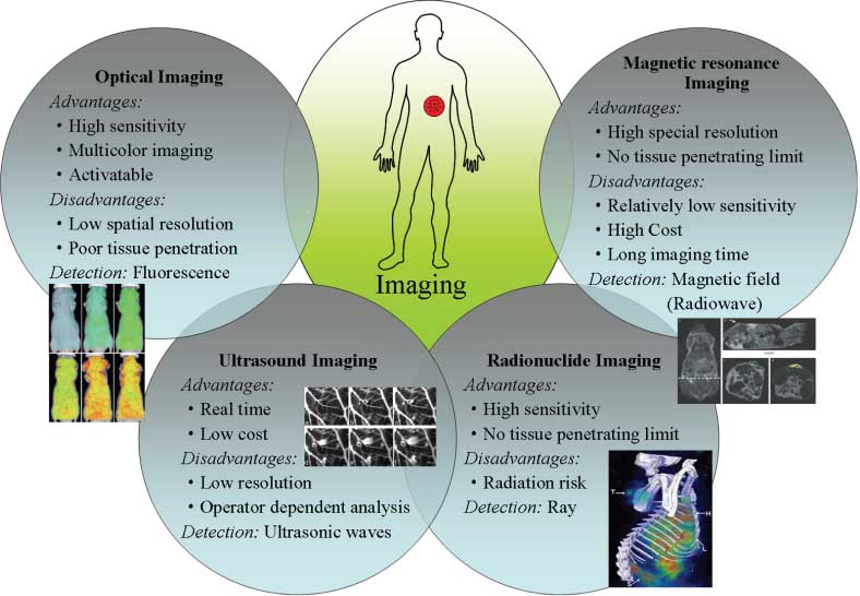
Main imaging modalities of nanomaterials used for biomedical fields.
Optical imaging (OI)
OI is an important and challenging technique in the biomedical field due to its high temporal and spatial resolutions (6–8). In living organisms, OI employs a sensitive camera to detect fluorescence emitted from fluorophores (9). Conventional fluorophores, generally including organic fluorophores, quantum dots, fluorescent proteins and luminescent transition metal complexes, have been widely used (10–15). Among them, fluorophores with long emission at the NIR region are generally preferred in living tissue for their great properties such as the lower autofluorescence and greater penetration depth (16). It was reported that lanthanide (Ln)-doped upconversion nanoparticles (UCNPs) had tremendous advantages over conventional fluorophores, such as low cytotoxicity, non-photobleaching, non-photoblinking and high spatial resolution etc (17–25). Chatterjee et al. reported that UCNPs could enhance penetration depth into tissues upon NIR excitation (26). In addition, there is a significant decrease of auto-fluorescence from surrounding tissues (25, 27). Due to the low vibration energy, fluoride-based UCNPs have also been identified as one of the most efficient upconversion fluorescent nanoparticles nowadays (28). Chatterjee et al. have further demonstrated that the luminescence from Ln-doped UCNPs could be still clearly observed upon NIR excitation when the nanoparticles were located ~10 mm beneath the skin in vivo animal imaging (26). In summary, Ln-doped UCNPs hold great potential as novel fluorophores in biomedical field (29, 30).
Another class of versatile tool for in vivo fluorescence imaging is semiconductor nanocrystals or quantum dots (QDs) (31–34). QDs typically have a core/shell structure of 2–8 nm in diameter, and are comprised of atoms of elements from group II to VI or III to V in the periodic table (35, 36). QDs are attractive for tumor imaging, which possess a broad excitation bands and narrow emission bands (37, 38). In addition, they also have other unique optical properties for in vivo optical imaging such as superior fluorescence, resistance to photo bleaching, and long blood circulation time (39, 40). After QDs conjugated with poly-N-isopropyla-crylamide (PNIPAM) were injected intravenously via the tail vein in mice bearing JHU-31 prostate cancer tumors for 3 h, tissues were cryosectioned for imaging. It was found that the QD-PNIPAM was localized at the periphery of the tissue sections while unmodified QDs were not found in the cancer tissue (41). Following by targeting of the prostate tumor cells, QDs conjugated with antibodies to the prostate-specific membrane antigen (PSMA) accumulated at the tumor xenograft in live nude mice (34). Tail vein injection showed that untargeting or passive targeting of QD probes demonstrated weak or no signals, while antibody-conjugated QDs showed intense signals inside the tumor. Because the receptor of folic acid was overexpressed on the surface of many cancer cells, including ovarian, lung, breast, endometrial, renal, and colon cancer cells, folate-QD conjugates were specifically detected in cancer cells compared to normal cells. Now CdSe QDs conjugated with folate receptors were employed for targeting mouse lymphoma cells in vivo (42). Now NIR emitting QDs were developed in order to minimize light absorption by blood and water and improve tissue penetration depth. Diagaradjane et al. conjugated QD800 (emission peak at 800 nm) with anti epidermal growth factor receptor (EGFR) antibody in order to specifically target EGFR overexpressed at tumor sites (43). Cai et al. reported the use of QD705 conjugated to the arginine-glycine-aspartic acid (RGD) tri-peptide receptors following tail vein injection in mice bearing human glioblastoma xenografts (44). Hu et al. employed NIR emitting QDs conjugated with cyclic RGD peptide for targeting in vivo tumor vasculature (45). In addition, other groups have also successfully developed procedures for multiplexed fluorescent labeling of tissue specimens by QDs (46–50).
With the increasing availability of fluorescent probes, OI is gaining momentum as an important translational tool between basic research and clinical application. Although traditional small molecule NIR dyes continue to be used, the development of fluorescent organic, biological, and inorganic nanoparticles for in vivo OI offers powerful tools for some exciting applications. The continued evolution of these new technologies will allow more molecular targets and diseases to be interrogated in vivo.
Magnetic resonance imaging (MRI)
MRI is a non-invasive imaging technique, which uses a strong magnet and radiofrequency waves to produce images of internal organs. Colloidal iron oxide nanoparticles (IONPs) generally produce enhanced proton relaxation rates at significantly lower doses than paramagnetic ions due to their larger magnetic moment, and provide negative contrast by enhancing T2 relaxivity of water protons. So, colloidal IONPs, such as superparamagnetic iron oxides (SPIOs) and ultra superparamagnetic iron oxides (USPIO), have been widely explored in MRI field.
Several SPIO agents with a variety of surface coatings have been extensively studied for the detection and diagnosis of solid tumors (51). It was found that SPIOs were mostly taken by macrophages in the liver and spleen and subsequently metabolized over several days after intravenous injections. Active targeting of SPIOs can lead to increased contrast enhancement of tumors over non-targeted tissues. SPIOs functionalized with antibodies have been used to image rectal carcinoma and breast cancer (52, 53). Although passive targeting of iron oxide NPs to tumors can be achieved through the EPR effect, a range of molecules have been attached on their surface to improve tumor targeting for MRI applications, including proteins, antibodies, peptides, and oligosaccharides (54). Researchers have recently shown preferential accumulation of SPIOs functionalized with a targeting peptide overexpressed in MMP-2 glioma tumors (55). Folic acid, a vitamin, is another ligand, which has been extensively used as targeting agent (56, 57). Iridium complexes have been loaded into magnetic NPs for dual-modal luminescent and magnetic resonance imaging.
USPIO with diameters <40 nm have also been clinically investigated as contrast agents, which can accumulate at the margins of human brain tumors, improve their delineation on MRI (56). Researchers have compared the utility of USPIOs with Gd-chelates for brain-tumor MRI and found that USPIOs could offer prolonged delineation due to lower diffusion from tumor sites and increased internalization by tumor cells (58).
In addition, it was also reported that some nanomaterials might be used in ultrasound imaging (UI) and radionuclide imaging (RI) fields (59–62). Recent advances in nanotechnology and biotechnology have contributed to the development of multifunctional nanoparticles as representative nanomedicine. Because each imaging modality has its pros and cons, the integration of several imaging agents with different properties into multifunctional nanoparticles allows precise and fast diagnosis of disease through synergetic multimodal imaging. The multifunctional probes have already appeared from iron-oxide and dendrimer-based dual MRI fluorescence imaging contrast agents to a fused X-ray CT fluorescence imaging system (63, 64). Nanoscale multimodal imaging probes which carry more than two imaging agents have the potential to overcome the limitations of a single imaging modality and provide more detailed information of the target site through targeted delivery (8, 65, 66).
In summary, this is an active area in the future because clinicians need a contrast agent that can be specifically targeted to disease tissue allowing for more accurate diagnosis of various stages of diseases.
The application of nanomaterials in disease therapy
Nanomaterials have ability to penetrate and spontaneously accumulate at biological sites by enhanced permeability and retention (EPR) effect (67), across the physiology barrier freely (68), to specifically recognize and bind to target area via surface attaching specific ligands (69). So there is great interest in developing nanomaterials for nanomedicine to overcome some limitations found in traditional therapeutic agents (70, 71). In this section, we attempt to summarize currently available information regarding nanomaterials for disease therapies including photodynamic therapy (PDT), photothermal therapy (PTT), magnetic hyperthermia (MH), sonodynamic therapy (SDT) and cryosurgery (CS).
Photodynamic therapy (PDT)
Photodynamic therapy is known as a noninvasive medical technology to treat cancer. In the process of PDT, the photosensitizer (PS) molecule transfers the photon energy to surrounding oxygen molecules, which will generate reactive oxygen species (ROS) to kill cancer cells under the irradiation of light with appropriate wavelengths (72, 73). Compared to traditional chemotherapies and radiotherapies, PDT cancer treatment shows remarkably reduced side effects and improved selectivity since only the lesion is exposed to the light, while other tissues in the dark are not affected. An important benefit of PDT is that it can be used repetitively without any immunosuppressive or myelosupressive effects. It can also be used after surgery, radiotherapy, and chemotherapy (74). Because most of the PS employed in PDT are hydrophobic and show poor water solubility, this makes them difficult to administer these drugs for in vivo applications. To increase the water solubility of PS molecules and improve their delivery into cancer cells, various nanomaterials have been developed (75).
Upconversion luminescence (UCL) refers to a non- linear optical process that converts two or more lower-energy pump photons from the NIR spectral region to a higher-energy output photon with a shorter wavelength (76, 77). In recent years, UCNPs that exhibit UCL emission have been developed. Comparing to traditional fluorescent probes, they have shown many benefits including the free of auto-fluorescence background, higher photochemical stability and tissue penetration depth (78–81). UCNPs have the ability to convert NIR light to visible photons, which can activate PS absorbed on nanoparticles to generate ROS to kill cancer cells. Wang et al. loaded Chlorin e6 (Ce6) on polymer-coated UNCPs to form an UCNP-Ce6 complex that can produce singlet oxygen and further kill cancer cells under NIR light, and excellent PDT efficiency is also achieved in tumor-bearing mice (Figure 2) (82).
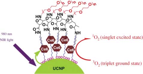
A schematic drawing showing NIR-induced PDT using UCNP-Ce6 (82).
Another biggest success in PDT is the use of Au nanoparticles. Considering that Au nanoparticles can maintain the stability and activity of hydrophobic PS in aqueous environments, there have been considerable efforts to develop Au nanoparticles as PS delivery vehicles to overcome the shortcomings of PS, for example, poor solubility and limited selectivity towards cancerous tissue (83). Gamaleia et al. have synthesized a conjugate of hematoporphrin with Au nanoparticles and evaluated its photodynamic activity. The results show that its PDT activity is much higher than that of the original PS in vitro (84).
It was reported that QD-PS complexes had better advantages over PS alone due to the high photoluminescence quantum efficiency, tunable optical properties and imaging capabilities (85, 86). The conjugation of QD to PS provides flexibility and further activates the PS molecule by utilizing variable excitation wavelength since the QDs can exhibit broad absorption spectra. Peptide-coated QDs can form extremely stable conjugates with PS, which may be used as targeted multifunctional probes for live cell targeting, imaging and PDT (73).
In addition, other kinds of PS-loaded nanoparticles, such as ceramic, liposome, and silica-based nanoparticles have been reviewed by Chatterjee et al. (87).
Photothermal therapy (PTT)
Photothermal therapy is an experimental use of electromagnetic radiation that is proposed to treat various diseases, including cancer. This activation brings the sensitizer to an excited state where it releases vibrational energy. The heat is the actual method of therapy that kills the targeted cells (88). Unlike PDT, PTT does not require oxygen to interact with the target cells or tissues (89). Current studies also show that PTT is able to use longer wavelength light, which is less energetic and therefore less harmful to other cells and tissues.
Carbon nanotubes (CNTs) are able to absorb light in the NIR region and produce heat (90). This exclusive property has been exploited as a method to kill cancer cells via thermal effects (91). A number of earlier studies have shown the promise of carbon nanotubes for photothermal ablation of cancer cells in vitro (92, 93). Recently, it was reported that intra-tumor injections of multi-walled carbon nanotubes (MWNTs) and single walled carbon nanotubes (SWNTs) followed by NIR laser irradiations at powers of 2.5 W/cm2 and 3.8 W/cm2, were able to destroy tumors grown on mice (94, 95). In a study conducted by Liu et al., SWNTs functionalized by PEGylation achieved an optimal blood circulation half-life at 12–13 h, afforded relatively low reticuloendothelial systems (RES) accumulation, high tumor uptake and low skin retention in mice, these results are ideal for cancer treatment in vivo (96).
As the sister of CNTs, nanographene can be also utilized in PTT due to the ability of NIR optical absorption (97–99). In the recent work, Yang et al. studied the in vivo behaviors of nanographene sheets (NGS) with PEG coating by a fluorescent labeling method. The results showed high tumor uptake of NGS in several xenograft tumor mouse models. Compared to PEGylated CNTs, PEGylated NGS shows some advantages in vivo behaviors including highly efficient tumor passive targeting and relatively low retention in RES (100).
Au NPs include gold nanorods, gold nanoshells and hollow Au NPs and so on, they show largely red-shifted properties which may have great potential in PTT (100–107). Similar to scatter counterpart, Au NPs absorb light millions of times stronger than the organic dye molecules. Nearly 100% absorbed light is converted to heat via the nonradiative properties (108). These features make them a new cohort photothermal contrast agents for PTT (109). It was reported that gold nanoshells demonstrated highly efficient NIR hyperthermia ability due to its strong NIR absorption (102, 110). Huang et al. reported gold nanorods as NIR hyperthermia agents for photothermal cancer therapy (111). Because the surface chemistry is a key factor for the cellular uptake and tissue penetration of nanomaterias (112), it can affect thermal therapy efficiency. Jin et al. have synthesized Au nanorods with three different polymer coatings: cetyltrimethylammonium bromide (CTAB), polystyrene sulfonate (PSS) and poly (diallyldimethylammonium chloride) (PDDAC). Then multicellular tumor spheroid (MCTS) was treated by these different nanorods, after radiated under the NIR laser for 4 min, the thermal therapy efficiency of each kind of Au nanorods is very different. In the PSS group, the compact structure of the MCTSs was broken by the Au nanorods thermal therapy, but this phenomenon in the other two groups was not observed. The result indicated that the difference in thermal therapy efficacy was due to the effective heat generated by the Au nanorods with different surface coatings, which were affected by different Au nanorods distribution patterns in the tumor (113).
Magnetic hyperthermia (MH)
Magnetic hyperthermia is based on the fact that magnetic particles, when subjected to an alternating magnetic field (AMF), produce heat. As a consequence, if magnetic particles are put inside a tumor and the whole patient is placed in an alternating magnetic field of well-chosen amplitude and frequency, the temperature in the tumor would rise and further kill cells (114).
Magnetic nanoparticles like magnetite have shown an intriguing ability to mediate a high rate of heat induction at low concentrations (115). The conversion of electromagnetic energy into heat by magnetic nanoparticles has great potential as a non-invasive and powerful therapy technique. For example, magnetic fluid hyperthermia (MFH) can be used to generate localized heating for tumor repression (116). By targeting the magnetic nanoparticles at the tumor site with an applied AMF, the temperature at tumor sites can be increased to 42–46°C, which largely reduces the viability of cancer cells (114, 117, 118). Previous studies have focused on designing and optimizing the size, composition and structure of magnetic nanoparticles to maximize the specific absorption rate (SAR) of MFH (119, 120). The size distribution of nanoparticles has a crucial influence on the SAR, where monodispersed magnetic nanoparticles show a larger SAR than polydispersed magnetite nanoparticles (121). Ma et al. found that magnetite nanoparticles with a size of 46 nm exhibited the highest SAR, whereas Hergt and Dutz reported that 30 nm magnetite nanoparticle suspensions had the highest SAR (114, 122). Other than the effect from the intrinsic nature of the inorganic component, a useful approach to improve the performance of magnetic nanoparticles is the optimization of the surface coating (117). Recently, the study on surface coating on magnetic nanoparticles has made a great progress. Liu et al. have successfully synthesized a highly monodispersed magnetite nanopaticles which are coated by mPEG. These nanoparticles show a good thermal conductivity and a high SAR, this may provide a chance for high performance hyperthermia agents (116).
Sonodynamic therapy (SDT)
Sonodynamic therapy is a recently developed anticancer treatment where ultrasound is used to activate the cytotoxic effect of chemical compounds, known as sonosensitizers. SDT diverges from PDT, as drug activation is achieved through ultrasound rather than light. Moreover, SDT is able to overcome the main drawback of PDT. Whether pulsed or continuous ultrasound is considered to be safer and more readily accepted by the patient than either radiotherapy or chemotherapy (123). In addition, ultrasound may be used as an external remote to control over time, dose, and location of drug release, provide tailor-made and localized cancer therapy (124).
Nanomaterials have some features that make them particularly suitable for SDT. For example, TiO2 NPs are useful for SDT owing to their special properties (125). Wang et al. reported that various ROS could be generated when the nanosized TiO2 powder was irradiated by ultrasound (126). Harada et al. have introduced ultrasound therapy combining with TiO2 NPs on melanoma cells. The results demonstrated that the cell viability was significantly decreased after ultrasound irradiation in the presence of TiO2 NPs (127).
Cryosurgery (CS)
Cryosurgery is the application of extreme cold to destroy abnormal or diseased tissue. It is appropriate for treating localized diseases, and solid tumors larger than 1 cm, but the tiny or diffuse metastases are usually not affected by cryosurgery. Generally, all tumors that can be reached by the cryoprobes during an operation are treatable (128–130). However, in many clinical cases, it has been found that freezing alone could not completely destroy the targeted tumor, moreover, there is always a high recurrence rate with follow-up surveys (131, 132), the major reason concerns the freezing rate, which does not produce a massive ice nucleation in tumor cells, especially at the edge of the tumor, thus the procedure cannot guarantee complete lethality to all of the tumor cells. Consequently, a major concern in conventional cryosurgery is to avoid insufficient freezing between multiple cryoprobes and maximize the freezing efficiency.
To establish an always-efficient freezing treatment for tumors, nanoparticles were introduced to significantly mediate various freezing procedures, which finally led to a generalized tumor-treatment concept called nanocryosurgery (Figure 3) (133). According to theoretical interpretation and experimental measurements, intentional loading of nanoparticles with high thermal conductivity into the target tissues can lower the final temperature appreciably, increase the maximum freezing rate, and enlarge the ice volume. Functional solutions with specific nanoparticles were injected into the targeted tissues to improve the diffusion process, regulate the freezing scale, modify iceball formation location, and prevent the surrounding healthy tissues from freeze damage. Di et al. introduced MgO NPs as a biodegradable nontoxic agent to innovate nanocryosurgery. The result shows that administering MgO NPs-mediated cryosurgery, either by direct tumor injection or intravenous delivery of nanoparticles, is possible (134). In addition, the new modality also offers many chances to ablate tumors better. For example, a minimally invasive needle with both freezing and heating capability can be designed for a combined surgery. The nanoparticles can be first injected into the targeted tumor for nanocryosurgery, which can then be followed up by a nano-hyperthermia, or vice versa.
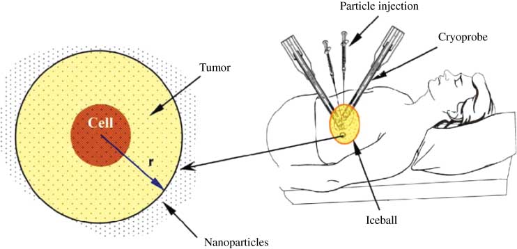
Schematic illustration of computational domain loaded with nanoparticles during cryosurgery (not to scale) (133).
In addition, nanomaterials cannot only play the role of adjuvant therapy, but also they may become a therapy agent. In 2005, Chen et al. have reported that gadolinium metallofullerenol [Gd@C82(OH)22]n nanoparticles (f-NPs) have the ability of antitumor (135). In recent years, numbers studies have indicated that f-NPs exhibit strong inhibitory activity against various tumor, such as MCF-7 cells in vivo . In addition, f-Nps have the ability of antimetastasis. The anticancer mechanism of f-NPs was also studied. The results show that the anticancer mechanism of f-NPs may be related to immune enhancement (135), G0/G1 phase arrest (138), angiogenesis inhibition (136), and scavenging of ROS (139). Liang et al. reported f-NPs could reactivate the defective endocytosis of cisplatin in the cisplatin-resisitant human prostate cancer cells and cause accumulation of intracellular cisplatin, thus tumor resistance to cisplatin was circumvented by treatment with a combination of f-NPs with cisplatin both in vitro and in vivo (140). Based on the above description about f-NPs unique properties, we believe that f-NPs may become a novel antineoplastic chemotherapy agent with high efficiency and low toxicity.
The application of nanomaterials in drug delivery
The primary goals in drug delivery system (DDS) include specific drug targeting, reduction in toxicity while maintaining therapeutic effects, and biocompatibility (141). Nano scaled carriers have revolutionalized drug delivery. Nanomaterials as drug delivery are generally <100 nm in at least one dimension, and consist of different biodegradable materials such as natural or synthetic lipids, polymers (142). At present, many nanoparticles have been studied for drug delivery. Here, we discuss the development of nanomaterial as drug delivery systems including drug targeting, controlled drug release, multi-drug resistance, and multifunctional nanocarriers.
Drug targeting
One of the challenges in developing effective drug delivery nanoparticles is to rationally design them so that they can perform two critical functions: evading the body’s normal immune response and reaching their intended targets. Based on the ability to locate and deliver a drug to the interest area in the body, NP delivery systems can be classified as passive and active target (Figure 4).
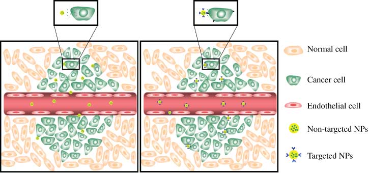
Passive (Right) vs. active targeting (Left).
The defective vascular architecture was created due to the rapid vascularization, which was necessary to serve fast-growing cancers (143, 144). Passive targeting can be achieved by the EPR effect. As a consequence of EPR effects, nanoparticles that encapsulated drugs, proteins or other therapies can accumulate in the tumor at a concentration five to ten times higher than that in normal tissue within 1–2 days (145). For instance, Abraxane®, an albumin-bound paclitaxel nanoparticles for the treatment of metastatic breast cancer and Doxil®, a poly (ethylene glycol)-coated (PEGylated) liposomal system as doxorubicin (DOX) delivery for cancer therapy have been approved by the FDA. These agents circulate in the body with a half-life about 100 times longer than that of free anticancer drugs while simultaneously reducing systemic toxicity (146–150).
Because of different degree of vascularization and porosity of tumor vessels, targeting cancer cells by the EPR effect is not feasible in all tumors, this limits the passive targeting (151, 152). Attaching target molecule to the nanoparticle surface is one approach to overcome this limitation, which is called active targeting. Active targeting, which was performed by equipping the delivery system with a “homing device” that was capable of guiding the carrier to the intended target (145). These “homing device” are special in that they can recognize and bind to complementary molecules, or receptors found on the surface of tumor cells. After such molecules are added to the drug delivery nanoparticles, they can target to the tumor cells, this will increase the efficacy of the treatment and reduce toxic effects on surrounding normal tissue. For example, Kumar et al. have synthesized a novel class of Au nanoparticles, which was functionalized with the therapeutic peptide, PMI (p12), and a targeted peptide, CRGDK for selective binding to neuropilin-1 receptors which were overexpressed on the cancer cells and regulated the process of membrane receptor-mediated internalization. In an in vitro studies, AuNPs functionalized with CRGDK resulted in maximal binding interaction between the CRGDK peptide and targeted Nrp-1 receptor overexpressed on MDA-MB-321 cell surface, which improved the delivery of therapeutic P12 peptide inside targeted cells (153).
At present, several targeted delivery systems are under clinical trials, Table 1 lists the nanoparticle-based drugs that are approved or under clinical development. Although ligand-mediated targeting drug carriers have not yet made a considerable clinical impact on human health, it will soon be feasible to develop targeted nanoparticle candidates for clinical translation.
Nanoparticle-based drugs that have been approved or under clinical development .
| Commercial name | Platform | Targeting ligand | Active pharmaceutical ingredient | Indication | Status | References |
|---|---|---|---|---|---|---|
| CALAA-01 | Cyclodextrin-containing polymeric nanoparticle | Transferrin | siRNA | Solid tumor | Phase I | (155) |
| MBP-426 | Liposome | Transferrin | Oxaliplatin | Gastric, esophageal adenocarcinoma | Phase Ib/II | (156) |
| MCC-465 | Liposome | F(ab′)2 fragment of Human Ab GAH | Doxorubicin | Metastatic stomach cancer | Phase I | (157) |
| BIND-014 | PLGA-PEG nanoparticle | PSMA-specific peptide | Doxetaxel | Solid tumor | Phase I | (158) |
| SGT53-01 | Liposome | Transferring receptor specific-scAb | P53 Gene | Solid tumor | Phase I | (159) |
Controlled drug release
An ideal drug carrier should not only have the nature with high load of drug, but also be able to have the ability of controlled drug release. When some drugs are early released in the blood circulation, they will damage normal tissues and produce serious side effects. Nowadays, with the developing of nanotechnology, nanomaterials as a drug delivery can overcome these side effects.
Several biodegradable materials such as liposomes, polymeric nanoparticles and dendrimers have been used as “smart” carriers that can controllably release pharmaceutical drugs in aqueous solution upon the structural degradation of the carriers triggered by various chemical factors, such as pH. Several exciting DDS have been prepared following this approach (160). Though structurally unstable “soft” materials are beneficial for the drug delivery, it is difficult to achieve “zero” premature release of drugs. In many cases, the matrix-entrapped drug molecules would start to release from the biodegradable carrier as soon as the system was introduced in vivo. The premature release problem not only limits the usage of these biodegradable DDS materials for effective cancer treatment, but also presents a major challenge for the site-selective delivery of protein and nucleotide-based drugs via oral administration. The precious pharmaceutical or nutriceutical cargo of enzymes, DNAs, and RNAs would decompose in the highly acidic environment of stomach if the carriers could not offer the necessary protection (161).
Recent research has focused on developing structurally stable drug delivery systems that are able to deliver a relatively large amount of drug molecules without any premature release problem to targeted tissues or even intracellular organelles. In 2003, Lai et al. first designed a mesoporous silica nanosphere (MSN)-based stimuli-responsive system using a concept of gatekeeping (162). This system has the advantage of a variety of chemical entities as “gatekeepers” to regulate the encapsulation and release of drug molecules (Figure 5). Kim et al. reported a unique methodology for controlled-release delivery systems (163, 164). Cyclodextrin gatekeepers on the surface of MSNs can be hydrolyzed by amylase to release guest molecules from the porous reservoir. The ester linkage in the stalk part of the cyclodextrin gatekeeper can also be cleaved by lipase to release guests from the channel. Other groups also explored some control release systems that employed different gate based on MSNs, such as Au nanoparticles, silver nanoparticles, dendrimer and other gatekeepers (165–168). Among structurally stable materials that have been investigated for drug delivery, Bio-MSN becomes the best candidate, because biodegradable MSN have many advantages for intracellular delivery, such as large surface area, tunable pore sizes and volumes, and encapsulation of drugs, proteins and biogenic molecules (169).
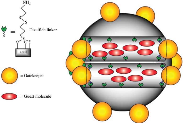
Representation of an MSN loaded with guest molecule and end-capped with a general gatekeeper (162).
In addition, to achieve the goal of drug controlled release, nanocarriers have been designed as an environment sensitive delivery. Recently, by self-assembly approach, Hu et al. fabricated a pH/thermo-responsive nanocarriers with dually responsive poly(N-isopropylacrylamide) (PNiPAM)-co-acrylic acid (AA) hydrogel core enclosed in silica shell. Comparing with common MSNs, these core-shell particles have a more uniform size and better dispersity. In vitro cell culture studies, it is more biocompatible and less cytotxic than MSNs. In addition, the drug release results indicate that PNiPAM/AA@SiO2 particles have pH responsive characteristics and higher releasing efficiency than MSN particles, which are very attractive for cancer treatment (170).
Multi-drug resistance
In the treatment of disease, especially cancer, drug-sensitive cells were killed, but a proportion of drug-resistant cells are left. This is so called multi-drug resistance (MDR) (171). One mechanism of MDR is increasing drug efflux (172). The increasing drug efflux is caused by the integral membrane proteins, known as MDR transporters. The transporters such as p-glycoprotein (MDR1), multidrug resistance associated proteins (MRPs) can actively pump chemotherapeutic drugs out of the cell and reduce the intracellular drug dose below lethal threshold level (173). The onset of nanomaterials has endowed the ability to provide solution to this limitation by bypassing the MDR transporters (174, 175). For example, SP1049C, a novel P-glycoprotein targeting micellar formulation of DOX exhibits greater efficacy than DOX against a variety of drug-resistance tumor cell lines due to the ability of circumventing p-glycoprotein-mediated drug resistance (176). Other studies have addressed the challenge of MDR using polymeric nanoparticles (177), lipid nanocapsules (178), and micelles (179) within cell lines or in mouse tumor models. Although those nanocarriers can bypass the transporters to an extent, it is not sufficient for overcoming drug resistance. Taking into account the mechanism of drug resistance, the overexpression of transporters is one of the major mechanisms of drug resistance in cancer cells, knockdown of those gene expressions by siRNA could help to restore the intracellular drug levels to the concentrations requiring for induction of apoptosis and cytotxicity. So, the nanobased drug co-delivery systems have been designed for targeting the transporters concurrently delivering drugs for increasing intracellular drug concentration (175). Meng et al. have reported a new approach to overcome MDR by utilizing siRNA to silence the expression of efflux transporter. In their studies, MSNs were functionalized to effectively deliver DOX as well as p-glycoprotein siRNA to a drug-resistant cancer cell line in an additive or synergistic fashion (180).
Another problem in MDR is activation of anti-apoptotic pathways. Cells have developed strategies to avoid death by the anti-apoptotic pathways (181). It often involves the genetic and epigenetic alterations of cancer cells including overexpression of B-cell lymphoma 2 (Bcl-2), an anti-apoptosis regulator protein and mutations in tumor suppressor p53 gene. The nanocarrier is also possible to co-delivery cytotoxic drugs and gene targeting apoptosis regulator protein including Bcl-2 (182), transcription factor NF-κB and hypoxia-inducible factor alpha (HIF-1α) (146). With the synergistic effect, the intracellular drug accumulation can increase therapy efficacy. Several groups have designed DDS based on nanoparticles or liposomes for co-delivery of anticancer drugs and siRNA. For example, Chen et al. obtained CaCO3/DNA/DOX nanoparticles by CaCO3 co-precipitation technique for co-delivery of p53 gene and DOX (183). These nanoparticles could induce a higher cell inhibition and promote tumor cell apoptosis more effectively.
Multifunctional nanocarrier
By a synergistic effect, multifunctional nanocarriers as delivery systems are capable of delivering therapeutic payloads and/or image contrast enhancement agents to targeted sites in the body. Kim et al. designed a tumor-homing chitosan-based nanoparticles (CNPs) containing a near-infrared fluorescent (Cy5.5) and anticancer drug (paclitaxel) for cancer treatment. The CNPs have many advantages such as stability in serum, deformability, and rapid uptake by tumor cells. It presents a new strategy in cancer treatment, in which early stage cancer diagnosis, drug delivery, and real-time non-invasive monitoring for therapeutic efficacy are carried out simultaneously (184). Yang et al. have developed a multifunctional nanosystem combining magnetic nanocrystals with therapeutic antibodies and the chemotherapeutic drug DOX, this nanosystem has the ability of imaging, targeting and treating (185). Multifunctional nanomaterials have also been used for in vivo imaging, siRNA delivery and silencing in tumors. Medarova et al. have obtained a nanosystem, which is consisted of magnetic nanoparticles labeled with a near-infrared dye and covalently linked to siRNA molecules specific for model target. This nanosystem is easily translocated to cytosol and has high gene transduction efficiency (186). Guo et al. designed and prepared a kind of multifunctional nanoparticles by graft-degradable amphiphilic cationic copolymer, poly(ɛ-caprolactone)-graft-poly(2-(N,N-dimethylamino)ethyl methacrylate) (PCL-g-PDMAEMA). It was found that PCL-g-PDMAEMA nanoparticles were able to simultaneously entrap hydrophobic paclitaxel and load DNA. These nanoparticles had pH dependent sensitive ability, drug could be released faster in an acidic environment than in a neutral environment. In addition, the PCL-g-PDMAEMA nanoparticles showed high gene transfection efficiency than Lipofectamine™ 2000. The most interesting thing is that these nanoparticles can escape from the endosome and release the payloads effectively in cytoplasm. All of those advantages suggest that PCL-g-PDMAEMA has great potential for achieving the synergistic effect of drug and gene therapies in vivo (187).
The application of nanomaterials as drug delivery has made a great success. Current pharmaceutical nanocarriers, such as liposomes, micelles, biodegradable MSN, demonstrate a broad variety of useful properties, such as longevity in the blood, enhanced intracellular penetration, specific targeting. With continuous advances in new biomarkers and targeting ligands, it will be increasingly feasible to develop optimized nanocarriers as promising candidates for clinical translation.
Tissue engineering
The application of nanomaterials in tissue engineering has been paid more attention in recent years because of their special biology properties. With the rapid development of nanotechnology, a great deal of progress has been made in nanomaterials for tissue engineering including bone, cartilage, nerve, blood vessel, bladder tissue engineering.
Bone
As a type of dense connective tissue, bone provides a frame to support and protect internal organs. Due to the crucial roles of the skeleton for the human body, rehabilitation of bone tissue is certain to be imperative when it is damaged by trauma, severe infection and other disorders. Recently, several nanomaterials have been applied in tissue engineering.
As a major component and an essential ingredient of normal bone and teeth, the size of natural hydroxyapatite(HAp) particles is demonstrated to be nano-scale shaped like fibers, 70% of HAp particles have 2–5 nm in width and 20–80 nm in length (188). Besides their mechanical functions, natural HAp particles can modulate activity of bone cells. Hence, artificial nanomaterials substituted for HAp are required not only to possess good mechanical behavior, but also to benefit bone cells. Nowadays, the novel ways to modify nano-HAp are found continuously to better fulfill the mechanical and biological requirements. Kong et al. synthesized nano-HAp/chitosan scaffolds with high porosity, the scaffolds showed better biocompatibility than pure chitosan scaffolds (189). Zandi et al. prepared rod-like nano-HAp/gelatin scaffolds coated with nano-HAp particles. The HAp particles on the scaffold wall easily enter the microscopic fractures, and increase the mechanical strength. The incorporation of rod-like nano-HAp and scaffolds coated with nano-HAp particles make the scaffolds possess desirable biocompatibility, high bioactivity, and sufficient mechanical strength in comparison with noncoated HAp samples (190). Liao et al. developed a nano-HAp/collagen/PLA three-dimensional porous scaffold by biomimetic synthesis. This scaffold mimics the microstructure of cancellous bone and shows some features of natural bone both in main composition and hierarchical microstructure. Cell culture and animal model tests showed that the scaffold was bioactive (191). Another three-dimensional scaffold consisting of porous nonwoven silk fibroin net/nano-apatite composite was prepared by Zhao et al. The nano-sized HAp crystals were needle like, with a length of approximately 100–300 nm and a diameter of approximately 20–60 nm. The results demonstrated that the nonwoven silk fibroin net/nano-apatite composite showed excellent cytocompatibility for the growth of osteoblasts and had the capability to improve the viability of osteoblasts (192).
Titanium and its alloys have been widely applied to implantation materials in orthopedic and dental surgery. In contrast to pure Ti metal, TiO2 imparts stronger bioactivity and chemical bonding to bone (193). Great progress in nanotechnology makes further improvement of TiO2 as a bioactive material for tissue engineering. Nanophase titania/PLGA composites were designed and synthesized by combining the advantages of degradable polymers with nano-size ceramic grains to optimize physical and biological properties for bone regeneration. Liu et al. found that the PLGA composites with nanophase titania could enhance osteoblast functions. The study also revealed that the dispersion of titania in PLGA was enhanced by increasing the intensity of sonication and osteoblast adhesion was correlated with the dispersion of nanophase titania (194). To obtain more excellent biomechanical compatibility and bioactivity, Li et al. prepared bioactive nano-titania ceramics. The data showed that the mechanical properties of nano-titania ceramics were analogous to that of the human bone. Moreover, HAp additive plays an important role in adjusting the grain/particle size of nano-titania ceramics, which has a great effect on the osteoblast proliferation (195). Wang et al. prepared PLGA/nano-TiO2-particle composite microspheres. The introduction of TiO2 component has been proven to be capable of largely enhancing mechanical properties of PLGA/TiO2 microsphere-sintered scaffold. In addition, nano-TiO2 additives are also capable of increasing the proliferation, calcium secretion and alkaline phosphatase (ALP) activity of osteoblasts (196).
In addition, we previously reported the effects of AuNPs on the differentiation of MSC and the associated molecular mechanisms (197). Cellular assay results demonstrated that AuNPs promoted the differentiation of MSCs toward osteoblasts over adipocytes with increasing ALP activity and mineralization of the extra-cellular matrix. The results at the molecular level showed that the interaction of MSCs with AuNPs may lead to the activation of the p38 mitogen-activated protein kinase pathway (MAPK) signaling pathway and the up-regulation of osteogenic genes and the down-regulation of adipogenesis specific genes.
Cartilage
Cartilage is a flexible connective tissue composed of chondroblasts and a large amount of extracellular matrix (ECM). Several diseases are correlated closely with cartilage damage, which is difficult to heal because of disability of chondrocytes to migrate and shortage of blood supply in chondral tissue. Nanomaterials for tissue engineering have emerged as an interesting therapeutic option for the treatment of articular chondral defects with good and potentially durable results. To mimic the native ECM of cartilage for tissue repair and regeneration, many types of 3D tissue engineering scaffolds were fabricated as a collagen-like nano-fiber. Wimpenny et al. improved the nano-scaffolds using poly(L, D-lactide) (PLDLA) nano-fiber coating on microfibers or films. The nanostructure promoted the expression of chondrogenic markers, such as collagen type IIaI and aggrecan. The lower wettability of polymeric nanofibers favored the maintenance of rounded chondrocyte morphology (198). Compared to the above-mentioned nano-scaffolds, some bioactive peptide-linked nanomaterials may exhibit a more potent therapeutic effect for cartilage damage. Sethi et al. prepared a nanostructure combining vasoactive intestinal peptide (VIP) with sterically stabilized micelles (SSMs). It was found that the nano-sized SSM-VIP protected the bioactive peptide from degradation or inactivation in biological fluids, and had superior actions to traditional medications. Kon et al. reported that a 46-year-old athletic patient was successfully treated with a closing-wedge high tibial osteotomy and the implant of a newly developed biomimetic nanostructured osteochondral bioactive scaffold. After 1 year of follow-up, the patient was pain-free, had full knee range of motion, and returned to his pre-operation level of athletic activity (199).
Nerve
The repair of damaged nerves is believed to be a tough clinical problem. For example, one of the biggest challenges in peripheral nerve tissue engineering is to create an artificial nerve graft that can mimic the ECM and assist in nerve regeneration. It was reported that nanotopography or orientation of the fibers within the scaffolds greatly influenced the nerve cell morphology and outgrowth, and the alignment of the fibers ensured better contact guidance of the cells (200).
The initial experimental results indicated that nanostructures might lead to suppress nerve regeneration because of hindering nutrient and oxygen supply, which are essential for nerve growth. In order to avoid to this problem, Oh et al. developed the nanomaterials for repairment of nerve. Nerve guide conduits (NGCs) with selective permeability and hydrophilicity were fabricated using PLGA and pluronic F127. The inner surface of the tube has 50 nm nano-size pores, which can effectively prevent from fibrous tissue infiltration but permeate nutrients and retain neurotrophic factors, while the outer surface has micro-size pores, which can allow vascular ingrowth for effective supply of nutrients and oxygen into the tube. From the investigations of mechanical property, water absorbability, and model nutrient permeability of the tubes, the hydrophilized PLGA/F127 (3 wt%) tube seems to be a good candidate as a NGC for the effective permeation of nutrients as well as the good mechanical strength to maintain a stable support structure for the nerve regeneration (201).
The chitosan nanofiber mesh tube may be also a promising substitute for autogenous nerve graft. Wang et al. developed bilayered, deacetylation rate and oriented chitosan tube (202–204). Bilayered chitosan tube that comprises an outer layer of chitosan film and an inner layer of chitosan nonwoven nano/microfiber mesh. Nerve regeneration in chitosan tubes, on which the CGGGGGGYIGSR peptide was immobilized, exhibited efficacy similar to that of the isograft and represented a promising candidate for promoting peripheral nerve repair (202). It was reported that the chitosan nano/microfiber mesh tubes with a deacetylation rate of 93% had sufficient mechanical properties to preserve tube space, provided a better scaffold for cell migration and attachment, and facilitated humoral permeation to enhance nerve regeneration. As a result of fiber orientation, the tensile strength along the axis of the sheet increased (203). Because Schwann cells aligned along the nanofibers, oriented fibrous sheets could exhibited a Schwann cell column, thus functional and electrophysiological recovery occurred in time in the oriented group as well as in the bilayered group. Furthermore, histological analysis revealed that the sprouting of myelinated axons occurred vigorously followed by axonal maturation in the isograft, oriented, and bilayered group in the order (204).
In addition, the applications of nanomaterials in bladder and blood vessel tissue engineering have been reported (205, 206). In summary, at present, although there are many problems, we believe that nanomaterials are very important in tissue engineering fields in the future.
Summary and perspective
Over the last ten years, there have been substantial developments in nanomedicine. Building upon these advances, much work needs to be done over the next 5 to 10 years to more fully realize the promise of nanomedicine. First, the future of nanomedicine will depend on rational design of nanomaterials and tools based around a detailed and thorough understanding of biological and medical processes. For example, drug carrier design and targeting strategies may vary in relation to the type, developmental stage, and location of the disease. Second, more complex systems such as multifunctional nanoparticles that are concurrently capable of targeting, imaging, and therapy are subjects of future research. Third, toxicity issues are of particular concern, it is essential that fundamental research should be carried out to address these issues if successful efficient application of these technologies is going to be achieved. In addition, a major challenge to the field remains its highly interdisciplinary nature, the best course will be to train a new generation of truly interdisciplinary investigators who have a strong interdisciplinary grounding. In summary, although nanomedicine is still in its infancy, these practical applications clearly demonstrate its enormous potential.
About the authors
Shizhu Chen obtained his bachelor degree in Chemistry from Inner Mongolia Agricultural University (2011). Now he is a post graduate in the laboratory of Professor Jinchao Zhang at the College of Chemistry & Environmental Science from Hebei University. His current research interest focuses on the biological effects of manufactured rare earth nanomaterials.
Qun Zhang received a bachelor of chemistry from Hebei Normal University in 2010. Since 2010, she has been a post graduate in College of Chemistry & Environmental Science in Hebei University. She works under the guidance of Professor Jinchao Zhang. Her work includes the research about the effects of rare earth elements and rare earth nanoparticles on the proliferation, osteogenic differentiation, adipogenic differentiation and mineralization of primary mouse bone marrow stromal cells at cell and molecular levels.
Dr. Yingjian Hou obtained a PhD in physiology from Peking University Health Science Center in 2011. During his doctoral training, he pursued projects focusing on basic research in cardiovascular diseases. Currently serving in College of Basic Medicine, Hebei University, he proceeds to study in vascular biology. His major academic work has been published in some leading scholarly journals such as Cir Res and Arterioscler Thromb Vasc Biol.
Prof. Zhang obtained his PhD in Chemistry from Zhejiang University (2001) and postdoctoral training in the Peking University (2001–2003) and City University of Hong Kong (2003–2006). He joined the Hebei University in 2007. Now he is the professor of Hebei University, the PhD supervisor, the winner of Hebei Province Science Fund for Distinguished Young Scholars, the member of National Bio-inorganic Chemistry Academic Committee, the Director of Chemical Biology Key Laboratory of Hebei Province, and the Director of Nanometer Medical Center of Hebei University. Prof. Zhang has published over 80 peer-reviewed scientific papers and 12 patents/patent applications. Now Dr. Zhang’s group is developing novel nanomaterial for biological functions and applications.
Dr. Xing-Jie Liang got his PhD at National Key Laboratory of Biomacromolecules, Institute of Biophysics at CAS. He finished his postdoc at Center for Cancer Research, NCI, NIH, and worked as a Research Fellow at Surgical Neurology Branch, NINDS. Dr. Liang worked on Molecular imaging at School of Medicine, Howard University before he became deputy director of CAS Key Laboratory for Biomedical Effects of Nanomaterials and Nanosafety, National Center for Nanoscience and Technology of China. Dr. Liang is currently an editorial board member of “Acta Biophysica Sinica” and “Current Nanoscience” “Advances in Nano Research”. Developing drug delivery strategies for prevention/treatment of AIDS and cancers are current programs ongoing in Dr. Liang’s lab based on understanding of basic physio-chemical and biological processes of nanomedicine.
This work was supported in part by Chinese Natural Science Foundation project (No. 30970784, No. 81171455, No. 21271059, No. 20971034 and No. 81200078), National Key Basic Research Program of China (2009CB930200), a National Distinguished Young Scholars grant (31225009) from the National Natural Science Foundation of China, the State High-Tech Development Plan (2012AA020804) and Chinese Academy of Sciences (CAS) “Hundred Talents Program” (07165111ZX), CAS Knowledge Innovation Program and Research Fund for the Doctoral Program of Higher Education of China (No. 20111301110004).
References
1. Poland CA, Duffin R, Kinloch I, Maynard A, Wallace WA, Seaton A, et al. Carbon nanotubes introduced into the abdominal cavity of mice show asbestos-like pathogenicity in a pilot study. Nat Nanotech 2008;3:423–8.10.1038/nnano.2008.111Suche in Google Scholar PubMed
2. Shiohara A, Hoshino A, Hanaki K, Suzuki K, Yamamoto K. On the cyto-toxicity caused by quantum dots. Microbiol Immunol 2004;48:669–75.10.1111/j.1348-0421.2004.tb03478.xSuche in Google Scholar PubMed
3. Zhang L, Gu FX, Chan JM, Wang AZ, Langer RS, Farokhzad OC. Nanoparticles in medicine: therapeutic applications and developments. Clin Pharmacol Ther 2008;83: 761–9.10.1038/sj.clpt.6100400Suche in Google Scholar PubMed
4. Juliano RL. The future of nanomedicine: promises and limitations. Sci Public Policy 2012;39:99–104.10.3152/030234212X13214603531969Suche in Google Scholar
5. Priyanka P, Vandana P. The upcoming field of theranostic nanomedicine: an overview. J Biomed Nanotechnol 2012;8:859–82.10.1166/jbn.2012.1459Suche in Google Scholar PubMed
6. Berezin M, Achilefu S. Fluorescence lifetime measurements and biological imaging. Chem Rev 2010;110:2641–84.10.1021/cr900343zSuche in Google Scholar PubMed PubMed Central
7. Kobayashi H, Ogawa M, Alford R, Choyke PL, Urano Y. New strategies for flourescent probe design in medical diagmpstic imaging. Chem Rev 2011;110:2620–40.10.1021/cr900263jSuche in Google Scholar PubMed PubMed Central
8. Louie A. Multimodality imaging probes: design and challenges. Chem Rev 2010;110:3146–95.10.1021/cr9003538Suche in Google Scholar PubMed PubMed Central
9. Rao J, Dragulescu-Andrasi A, Yao H. Fluorescence imaging in vivo: recent advances. Curr Opin Biotechnol 2007;18:17–25.10.1016/j.copbio.2007.01.003Suche in Google Scholar PubMed
10. Lau JS, Lee P, Tsang KH, Ng CH, Lam Y, Cheng S, et al. Luminescent cyclometalated iridium (III) polypyridine indole properties, cytotoxicity, and cellular uptake. Chart 2009;48:708–18.Suche in Google Scholar
11. Malkani N, Schmid JA. Some secrets of fluorescent proteins: distinct bleaching in various mouting fluids and photoactivation of cyan flourescent proteins at YFP-excitation. Nat Preceding 2011;6:e18586.10.1371/journal.pone.0018586Suche in Google Scholar PubMed PubMed Central
12. Medintz IL, Uyeda HT, Goldman ER, Mattoussi H. Quantum dot bioconjugates for imaging, labelling and sensing. Nat Mater 2005;4:435–46.10.1038/nmat1390Suche in Google Scholar PubMed
13. Wang C, Ma Q, Dou W, Kanwal S, Wang G, Yuan P, et al. Synthesis of aqueous CdTe quantum dots embedded silica nanoparticles and their applications as fluorescence probes. Talanta 2009;15:1358–64.10.1016/j.talanta.2008.09.018Suche in Google Scholar PubMed
14. Yu M, Zhao Q, Shi L, Li F, Zhou Z, Yang H, et al. Cationic iridium(III) complexes for phosphorescence staining in the cytoplasm of living cells. Chem Commun 2008;14:2115–7.10.1039/b800939bSuche in Google Scholar PubMed
15. Zhao Q, Yu M, Shi L, Liu S, Li C, Shi M, et al. Cationic iridium(III) complexes with tunable emission color as phosphorescent dyes for live cell imaging. Organometallics 2010;29:1085–91.10.1021/om900691rSuche in Google Scholar
16. Janib SM, Moses AS, MacKay JA. Imaging and drug delivery using theranostic nanoparticles. Adv Drug Deliver Rev 2010;30:1052–63.10.1016/j.addr.2010.08.004Suche in Google Scholar PubMed PubMed Central
17. Chatterjee DK, Yong Z. Upconverting nanoparticles as nanotransducers for photodynamic therapy in cancer cells. Nanomedicine 2008;3:373–82.Suche in Google Scholar
18. Abdul Jalil R, Zhang Y. Biocompatibility of silica coated NaYF4 upconversion fluorescent nanocrystals. Biomaterials 2008;29:4122–8.10.1016/j.biomaterials.2008.07.012Suche in Google Scholar PubMed
19. Tsien RY. The green fluorescent protein. Ann Rev Biochem 1998;67:509–44.10.1146/annurev.biochem.67.1.509Suche in Google Scholar PubMed
20. Wang L, Li Y. Green upconversion nanocrystals for DNA detection. Chem Commun 2006;28:2557–9.10.1039/b604871dSuche in Google Scholar PubMed
21. Xiong LQ, Chen ZG, Yu MX, Li FY, Liu C, Huang CH. Synthesis, characterization, and in vivo targeted imaging of amine-functionalized rare-earth up-converting nanophosphors. Biomaterials 2009;30:5592–600.10.1016/j.biomaterials.2009.06.015Suche in Google Scholar PubMed
22. Xiong L, Yang T, Yang Y, Xu C, Li F. Long-term in vivo biodistribution imaging and toxicity of polyacrylic acid-coated upconversion nanophosphors. Biomaterials 2011;31:7078–85.10.1016/j.biomaterials.2010.05.065Suche in Google Scholar PubMed
23. Idris NM, Li Z, Ye L, Sim EK, Mahendran R, Ho PC-L, et al. Tracking transplanted cells in live animal using upconversion fluorescent nanoparticles. Biomaterials 2009;30:5104–13.10.1016/j.biomaterials.2009.05.062Suche in Google Scholar PubMed
24. Park YI, Kim JH, Lee KT, Jeon KS, Na HB, Yu JH, et al. Nonblinking and nonbleaching upconverting nanoparticles as an optical imaging nanoprobe and T1 magnetic resonance imaging contrast agent. Adv Mater 2009;26:4467–71.10.1002/adma.200901356Suche in Google Scholar
25. Sudhagar S, Sathya S, Pandian K, Lakshmi BS. Targeting and sensing cancer cells with ZnO nanoprobes in vitro. Biotechnol Lett 2011;33:1891–6.10.1007/s10529-011-0641-5Suche in Google Scholar PubMed
26. Chatterjee DK, Rufaihah AJ, Zhang Y. Upconversion fluorescence imaging of cells and small animals using lanthanide doped nanocrystals. Biomaterials 2008;29:937–43.10.1016/j.biomaterials.2007.10.051Suche in Google Scholar PubMed
27. Wu X, Zhang Q, Wang X, Yang H, Zhu Y. One-Pot synthesis of carboxyl-functionalized rare earth fluoride nanocrystals with monodispersity, ultrasmall size and very bright luminescence. Eur J Inorg Chem 2011;16:2158–63.10.1002/ejic.201001149Suche in Google Scholar
28. Boyer J, Cuccia LA, Capobianco JA. Synthesis of colloidal upconverting monodisperse nanocrystals. Nano Lett 2007;7:847–52.10.1021/nl070235+Suche in Google Scholar PubMed
29. Heer S, Kömpe K, Güdel H-U, Haase M. Highly efficient multicolour upconversion emission in transparent colloids of lanthanide-doped NaYF4 nanocrystals. Adv Mater 2004;17:2102–5.10.1002/adma.200400772Suche in Google Scholar
30. Yi G, Chow G. Core/shell/shell nanoparticles with significant enhancement of upconversion fluorescence. Chem Mater 2007;19:341–3.10.1021/cm062447ySuche in Google Scholar
31. Yezhelyev MV, Qi L, O’Regan RM, Nie S, Gao X. Proton-sponge coated quantum dots for siRNA delivery and intracellular imaging. J Am Chem Soc 2008;16:9006–12.10.1021/ja800086uSuche in Google Scholar PubMed PubMed Central
32. Qi L, Gao X. Quantum dot-amphipol nanocomplex time imaging of siRNA. ACS Nano 2008;2:1403–10.10.1021/nn800280rSuche in Google Scholar PubMed PubMed Central
33. Derfus AM, Chen A, Min DH, Ruoslahti E, Bhatia SN. Targeted quantum dot conjugates for siRNA delivery. Bioconjug Chem 2007;18:1391–6.10.1021/bc060367eSuche in Google Scholar PubMed
34. Gao X, Yang L, Petros J, Marshall FF, Simons JW, Nie S. In vivo molecular and cellular imaging with quantum dots. Curr Opin Biotechnol 2005;16:63–72.10.1016/j.copbio.2004.11.003Suche in Google Scholar PubMed
35. Alivisatos AP. Semiconductor clusters, nanocrystals, and quantum dots. Science 1996;271:933–6.10.1126/science.271.5251.933Suche in Google Scholar
36. Aldana J, Wang Y, Peng X. Photochemical instability of CdSe nanocrystals coated by hydrophilic thiols. J Am Chem Soc 2001;123:8844–50.10.1021/ja016424qSuche in Google Scholar PubMed
37. Yong KT, Qian J, Roy I, Lee HH, Bergey EJ, Tramposch KM, et al. Quantum rod bioconjugates as targeted probes for confocal and two-photon fluorescence imaging of cancer cells. Nanolett 2007;7:761–5.10.1021/nl063031mSuche in Google Scholar PubMed
38. Dubertret B, Skourides P, Norris DJ, Noireaux V, Brivanlou AH, Libchaber A. In vivo imaging of quantum dots encapsulated in phospholipid micelles. Science 2002;298:1759–62.10.1126/science.1077194Suche in Google Scholar PubMed
39. Ho YP, Leong KW. Quantum dot-based theranostics. Nanoscale 2010;2:60–8.10.1039/B9NR00178FSuche in Google Scholar
40. Sajja HH, East MP, Mao H, Wang AY, Nie S, Yang L. Development of multifunctional nanoparticles for targeted drug delivery and non invasive imaging of therapeutic effect. Curr Drug Discov Technol 2009;6:43–51.10.2174/157016309787581066Suche in Google Scholar PubMed PubMed Central
41. Nair A, Shen J, Thevenot P, Zou L, Cai T, Hu Z, et al. Enhanced intratumoral uptake of quantum dots concealed within hydrogel nanoparticles. Nanotechnology 2008;19:485102.10.1088/0957-4484/19/48/485102Suche in Google Scholar PubMed
42. Schroeder JE, Shweky I, Shmeeda H, Banin U, Gabizon A. Folate-mediated tumor cell uptake of quantum dots entrapped in lipid nanoparticles. J Control Release 2007;4:28–34.10.1016/j.jconrel.2007.08.028Suche in Google Scholar PubMed
43. Diagaradjane P, Orenstein-Cardona JM, Colón-Casasnovas NE, Deorukhkar A, Shentu S, Kuno N, et al. Imaging epidermal growth factor receptor expression in vivo: pharmacokinetic and biodistribution characterization of a bioconjugated quantum dot nanoprobe. Clin Cancer Res 2008;14:731–41.10.1158/1078-0432.CCR-07-1958Suche in Google Scholar PubMed
44. Cai W, Shin DW, Chen K, Gheysens O, Cao Q, Wang SX, et al. Peptide-labeled near-infrared quantum dots for imaging tumor vasculature in living subjects. Nanolett 2006;6:669–76.10.1021/nl052405tSuche in Google Scholar PubMed
45. Hu R, Yong KT, Roy I, Ding H, Law WC, Cai H, et al. Functionalized near-infrared quantum dots for in vivo tumor vasculature imaging. Nanotechnology 2010;21:145105.10.1088/0957-4484/21/14/145105Suche in Google Scholar PubMed
46. Xing Y, Chaudry Q, Shen C, Kong KY, Zhau HE, Chung LW, et al. Bioconjugated quantum dots for multiplexed and quantitative immunohistochemistry. Nat Protoc 2007;2:1152–65.10.1038/nprot.2007.107Suche in Google Scholar PubMed
47. Yezhelyev MV, Al-Hajj A, Morris C, Marcus I, Liu T, Lewis M, et al. In situ molecular profiling of breast cancer biomarkers with multicolor quantum dots. Adv Mater 2007;17:3146–51.10.1002/adma.200701983Suche in Google Scholar
48. Ghazani A, Lee J, Klostranec J, Xiang Q, Dacosta RS, Wilson BC, et al. High throughput quantification of protein expression of cancer antigens in tissue microarray using quantum dot nanocrystals. Nanolett 2006;6:2881–6.10.1021/nl062111nSuche in Google Scholar PubMed
49. Tholouli E, Hoyland J, Di Vizio D, O’Connell F, Macdermott S, Twomey D, et al. Imaging of multiple mRNA targets using quantum dot based in situ hybridization and spectral deconvolution in clinical biopsies. Biochem Biophys Res Commun 2006;22:628–36.10.1016/j.bbrc.2006.07.122Suche in Google Scholar PubMed
50. Byers RJ, Di Vizio D, O’connell F, Tholouli E, Levenson RM, Gossage K, et al. Semiautomated multiplexed quantum dot-based in situ hybridization and spectral deconvolution. J Mol Diagn 2007;9:20–9.10.2353/jmoldx.2007.060119Suche in Google Scholar PubMed PubMed Central
51. Reimer P, Tombach B. Abdominal radiology review article Hepatic MRI with SPIO: detection and characterization of focal liver lesions. Eur Radiol 1998;1204:1198–204.10.1007/s003300050535Suche in Google Scholar PubMed
52. Toma A, Otsuji E, Kuriu Y, Okamoto K, Ichikawa D, Hagiwara A, et al. Monoclonal antibody A7-superparamagnetic iron oxide as contrast agent of MR imaging of rectal carcinoma. Br J Cancer 2005;93:131–6.10.1038/sj.bjc.6602668Suche in Google Scholar PubMed PubMed Central
53. Funovics M, Kapeller B, Hoeller C, Su HS, Kunstfeld R, Puig S, et al. MR imaging of the her2/neu and 9.2.27 tumor antigens using immunospecific contrast agents. Magn Reson Imaging 2004;22:843–50.10.1016/j.mri.2004.01.050Suche in Google Scholar PubMed
54. Chang S, Dai Y, Kang B, Han W, Mao L, Chen D. UV-enhanced cytotoxicity of thiol-capped CdTe quantum dots in human pancreatic carcinoma cells. Toxicology Letters 2009;188:104–11.10.1016/j.toxlet.2009.03.013Suche in Google Scholar PubMed
55. Sun C, Veiseh O, Gunn J, Fang C, Hansen S, Lee D, et al. In vivo MRI detection of gliomas by chlorotoxin-conjugated superparamagnetic nanoprobes. Small 2008;4:372–9.10.1002/smll.200700784Suche in Google Scholar PubMed PubMed Central
56. Weitman SD, Lark RH, Coney LR, Fort DW, Frasca V, Zurawski VR, et al. Distribution of the folate receptor GP38 in normal and malignant cell lines and tissues. Cancer Res 1992;52:3396–401.Suche in Google Scholar
57. Lines C, Ross JF, Chaudhuri PK, Ratnam M. Differential regulation of folate receptor isoforms in normal and malignant tissues in vivo and in established cell lines. Cancer 1993;73:2432–43.Suche in Google Scholar
58. Harisinghani MG, Weissleder R. Sensitive, noninvasive detection of lymph node metastases. PLoS Medicine 2004;1:e66.10.1371/journal.pmed.0010066Suche in Google Scholar PubMed PubMed Central
59. Liu YL, Wu YH, Tsai WB, Tsai CC, Chen WS, Wu CS. Core-shell silica@chitosan nanoparticles and hollow chitosan nanospheres using silica nanoparticles as templates: preparation and ultrasound bubble application. Carbohydr Polym 2011;84:770–4.10.1016/j.carbpol.2010.03.033Suche in Google Scholar
60. Xie J, Jon S. Magnetic nanoparticle-based theranostics. Theranostics 2012;2:122–4.10.7150/thno.4051Suche in Google Scholar PubMed PubMed Central
61. Kang E, Min HS, Lee J, Han MH, Ahn HJ, Yoon IC, et al. Nanobubbles from gas-generating polymeric nanoparticles: ultrasound imaging of living subjects. Angew Chem Int Ed Engl 2010;49:524–8.10.1002/anie.200903841Suche in Google Scholar PubMed
62. Ma H, Wang PC, Qian F, Liang XJ. Biological effects of nanomaterials and drugs measured by the small-animal SPECT/CT imaging system in vivo. Acta Bioph Sin 2010;16:691–701.Suche in Google Scholar
63. Kircher MF, Mahmood U, King RS, Weissleder R, Josephson L. A multimodal nanoparticle for preoperative magnetic resonance imaging and intraoperative optical brain tumor delineation advances in brief. Cancer Res 2003;63:8122–5.Suche in Google Scholar
64. Talanov VS, Regino CS, Kobayashi H, Bernardo M, Choyke PL, Brechbiel MW. Dendrimer-based nanoprobe for dual modality magnetic resonance and fluorescence imaging. Nanolett 2006;6:1459–63.10.1021/nl060765qSuche in Google Scholar PubMed
65. Jennings LE, Long NJ. “Two is better than one” – probes for dual-modality molecular imaging. Chem Commun 2009;12:3511–24.Suche in Google Scholar
66. Sun C, Yang H, Yuan Y, Tian X, Wang L, Guo Y, et al. Controlling assembly of paired gold clusters within apoferritin nanoreactor for in vivo kidney targeting and biomedical imaging. J Am Chem Soc 2011;133:8617–24.10.1021/ja200746pSuche in Google Scholar PubMed
67. Ruoslahti E, Bhatia SN, Sailor MJ. Targeting of drugs and nanoparticles to tumors. J Cell Biol 2010;188:759–68.10.1083/jcb.200910104Suche in Google Scholar PubMed PubMed Central
68. Bhaskar S, Tian F, Stoeger T, Kreyling W, De la Fuente JM, Grazú V, et al. Multifunctional nanocarriers for diagnostics, drug delivery and targeted treatment across blood-brain barrier: perspectives on tracking and neuroimaging. Part Fibre Toxicol 2010;7:3.10.1186/1743-8977-7-3Suche in Google Scholar
69. Torchilin VP. Targeted pharmaceutical nanocarriers for cancer therapy and imaging. AAPS J 2007;9:E128–47.10.1208/aapsj0902015Suche in Google Scholar
70. Sumer B, Gao J. Theranostic nanomedicine for cancer. Nanomedicine 2008;3:137–40.10.2217/17435889.3.2.137Suche in Google Scholar
71. Bae KH, Chung HJ, Park TG. Nanomaterials for cancer therapy and imaging. Mol Cells 2011;31:295–302.10.1007/s10059-011-0051-5Suche in Google Scholar
72. De Rosa A, Naviglio D, Di Luccia A. Advances in photodynamic therapy of cancer. Curr Cancer Ther Rev 2011;7:234–47.10.2174/157339411796234889Suche in Google Scholar
73. Olivo M, Bhuvaneswari R, Lucky SS, Dendukuri N, Soo-Ping Thong P. Targeted therapy of cancer using photodynamic therapy in combination with multi-faceted anti-tumor modalities. Pharmaceuticals 2010;3:1507–29.10.3390/ph3051507Suche in Google Scholar
74. Misra R, Acharya S, Sahoo SK. Cancer nanotechnology: application of nanotechnology in cancer therapy. Drug Discov Today 2010;15:842–50.10.1016/j.drudis.2010.08.006Suche in Google Scholar
75. Konan YN, Gurny R, Allémann E. State of the art in the delivery of photosensitizers for photodynamic therapy. J Photochem Photobiol B 2002;66:89–106.10.1016/S1011-1344(01)00267-6Suche in Google Scholar
76. Mai H, Zhang Y. Highly efficient multicolor up-conversion emissions and their mechanisms of monodisperse NaYF4: Yb, Er core and core/shell-structured nanocrystals. J Phys Chem C 2007;111:13721–9.10.1021/jp073920dSuche in Google Scholar
77. Auzel F. Upconversion and anti-Stokes processes with f and d ions in solids. Chem Rev 2009;104:139–73.10.1021/cr020357gSuche in Google Scholar PubMed
78. Zijlmans HJ, Bonnet J, Burton J, Kardos K, Vail T, Niedbala RS, et al. Detection of cell and tissue surface antigens using up-converting phosphors: a new reporter technology. Anal Biochem 1999;267:30–6.10.1006/abio.1998.2965Suche in Google Scholar PubMed
79. Cheng L, Yang K, Zhang S, Shao M, Lee S, Liu Z. Highly-sensitive multiplexed in vivo imaging using pegylated upconversion nanoparticles. Nano Res 2010;3:722–32.10.1007/s12274-010-0036-2Suche in Google Scholar
80. Yu M, Li F, Chen Z, Hu H, Zhan C, Yang H, et al. Laser scanning up-conversion luminescence microscopy for imaging cells labeled with rare-earth nanophosphors. Anal Chem 2009;81:930–5.10.1021/ac802072dSuche in Google Scholar PubMed
81. Wu S, Han G, Milliron DJ, Aloni S, Altoe V, Talapin DV, et al. Non-blinking and photostable upconverted luminescence from single lanthanide-doped nanocrystals. Proc Natl Acad Sci USA 2009;106:10917–21.10.1073/pnas.0904792106Suche in Google Scholar PubMed PubMed Central
82. Wang C, Tao H, Cheng L, Liu Z. Near-infrared light induced in vivo photodynamic therapy of cancer based on upconversion nanoparticles. Biomaterials 2011;32:6145–54.10.1016/j.biomaterials.2011.05.007Suche in Google Scholar PubMed
83. Bechet D, Couleaud P, Frochot C, Viriot M-L, Guillemin F, Barberi-Heyob M. Nanoparticles as vehicles for delivery of photodynamic therapy agents. Trends Biotechnol 2008;26:612–21.10.1016/j.tibtech.2008.07.007Suche in Google Scholar PubMed
84. Gamaleia NF, Shishko ED, Dolinsky GA, Shcherbakov AB, Usatenko AV, Kholin VV. Photodynamic activity of hematoporphyrin conjugates with gold nanoparticles: experiments in vitro. Exp Oncol 2010;32:44–7.Suche in Google Scholar
85. Ma J, Chen J-Y, Idowu M, Nyokong T. Generation of singlet oxygen via the composites of water-soluble thiol-capped CdTe quantum dots-sulfonated aluminum phthalocyanines. J Phys Chem B 2008;112:4465–9.10.1021/jp711537jSuche in Google Scholar PubMed
86. Samia AC, Chen X, Burda C. Semiconductor quantum dots for photodynamic therapy. J Am Chem Soc 2003;125:15736–7.10.1021/ja0386905Suche in Google Scholar PubMed
87. Chatterjee DK, Fong LS, Zhang Y. Nanoparticles in photodynamic therapy: an emerging paradigm. Adv Drug Deliv Rev 2008;60:1627–37.10.1016/j.addr.2008.08.003Suche in Google Scholar PubMed
88. Neal DP, Hirsch LR, Halas NJ, Payne JD, West JL. Photo-thermal tumor ablation in mice using near infrared-absorbing nanoparticles. Cancer Lett 2004;209:171–6.10.1016/j.canlet.2004.02.004Suche in Google Scholar PubMed
89. Liao X, Zhang X. Preparation, characterization and cytotoxicity of carbon nanotube-chitosan-phycocyanin complex. Nanotechnology 2012;23:035101.10.1088/0957-4484/23/3/035101Suche in Google Scholar PubMed
90. Zhou F, Resasco DE, Chen WR, Xing D, Ou Z, Wu B. Cancer photothermal therapy in the near-infrared region by using single-walled carbon nanotubes. J Biomed Opt 2009;14:021009.10.1117/1.3078803Suche in Google Scholar PubMed PubMed Central
91. Gannon CJ, Cherukuri P, Yakobson BI, Cognet L, Kanzius JS, Kittrell C, et al. Carbon nanotube-enhanced thermal destruction of cancer cells in a noninvasive radiofrequency field. Cancer 2007;110:2654–65.10.1002/cncr.23155Suche in Google Scholar PubMed
92. Chakravarty P, Marches R, Zimmerman NS, Swafford AD, Bajaj P, Musselman IH, et al. Thermal ablation of tumor cells with carbon nanotubes. Proc Natl Acad Sci USA 2008;105:8697–702.10.1073/pnas.0803557105Suche in Google Scholar PubMed PubMed Central
93. Prencipe G, Tabakman SM, Welsher K, Liu Z, Goodwin AP, Zhang L, et al. PEG branched polymer for functionalization of nanomaterials with ultralong blood circulation. J Am Chem Soc 2009;131:4783–7.10.1021/ja809086qSuche in Google Scholar PubMed PubMed Central
94. Ghosh S, Dutta S, Gomes E, Carroll D, D’Agostino R, Olson J, et al. Increased heating efficiency and selective thermal ablation of malignant tissue with DNA-encased multiwalled carbon nanotubes. ACS Nano 2009;3:2667–73.10.1021/nn900368bSuche in Google Scholar PubMed PubMed Central
95. Moon HK, Lee SH, Choi HC. In vivo near-infrared mediated tumor destruction by photothermal effect of. ACS Nano 2009;3:3707–13.10.1021/nn900904hSuche in Google Scholar PubMed
96. Liu X, Tao H, Yang K, Zhang S, Lee S-T, Liu Z. Optimization of surface chemistry on single-walled carbon nanotubes for in vivo photothermal ablation of tumors. Biomaterials 2011;32:144–51.10.1016/j.biomaterials.2010.08.096Suche in Google Scholar PubMed
97. Kam NW, O’Connell M, Wisdom J, Dai H. Carbon nanotubes as multifunctional biological transporters and near-infrared agents for selective cancer cell destruction. Proc Natl Acad Sci USA 2005;102:11600–5.10.1073/pnas.0502680102Suche in Google Scholar PubMed PubMed Central
98. Liu Z, Li X, Tabakman SM, Jiang K, Fan S, Dai H. Multiplexed multicolor Raman imaging of live cells with isotopically modified single walled carbon nanotubes. J Am Chem Soc 2008;130:13540–1.10.1021/ja806242tSuche in Google Scholar PubMed PubMed Central
99. Welsher K, Liu Z, Daranciang D, Dai H. Selective probing and imaging of cells with single walled carbon nanotubes as near-infrared fluorescent molecules. Nanolett 2008;8:586–90.10.1021/nl072949qSuche in Google Scholar
100. Yang K, Zhang S, Zhang G, Sun X, Lee S-T, Liu Z. Graphene in mice: ultrahigh in vivo tumor uptake and efficient photothermal therapy. Nanolett 2010;10:3318–23.10.1021/nl100996uSuche in Google Scholar
101. Chang SS, Lee CL, Wang CR. Gold nanorods: electrochemical synthesis and optical properties. J Phys Chem B 1997;101:6661–4.10.1021/jp971656qSuche in Google Scholar
102. Hirsch LR, Stafford RJ, Bankson JA, Sershen SR, Rivera B, Price RE, et al. Nanoshell-mediated near-infrared thermal therapy of tumors under magnetic resonance guidance. Proc Natl Acad Sci USA 2003;100:13549–54.10.1073/pnas.2232479100Suche in Google Scholar
103. Huang X, Jain PK, El-Sayed IH, El-Sayed MA. Gold nanoparticles: interesting optical properties and recent applications in cancer diagnostics and therapy. Nanomedicine 2007;2:681–93.10.2217/17435889.2.5.681Suche in Google Scholar
104. Murphy CJ, Sau TK, Gole AM, Orendorff CJ, Gao J, Gou L, et al. Anisotropic metal nanoparticles: synthesis, assembly, and optical applications. J Phys Chem B 2005;109:13857–70.10.1021/jp0516846Suche in Google Scholar
105. Nikoobakht B, El-Sayed M. Preparation and growth mechanism of gold nanorods (NRs) using seed-mediated growth method. Chem Mater 2003;15:1957–62.10.1021/cm020732lSuche in Google Scholar
106. Oldenburg S, Averitt R, Westcott S, Halas N. Nanoengineering of optical resonances. Chem Phys Lett 1998;288:243–7.10.1016/S0009-2614(98)00277-2Suche in Google Scholar
107. Sun Y, Mayers BT, Xia Y. Template-engaged replacement reaction: a one-step approach to the large-scale synthesis of metal nanostructures with hollow interiors. Nano Lett 2002;2:481–5.10.1021/nl025531vSuche in Google Scholar
108. Huang X, El-Sayed M. Gold nanoparticles: optical properties and implementations in cancer diagnosis and photothermal therapy. J Adv Res 2010;1:13–28.10.1016/j.jare.2010.02.002Suche in Google Scholar
109. Svaasand LO, Gomer CJ, Morinelli E. On the physical rationale of laser induced hyperthermia. Lasers Med Sci 1990;5:121–8.10.1007/BF02031373Suche in Google Scholar
110. Loo C, Lowery A, Halas N, West J, Drezek R. Immunotargeted nanoshells for integrated cancer imaging and therapy. Nano Lett 2005;5:709–11.10.1021/nl050127sSuche in Google Scholar
111. Huang X, El-Sayed IH, Qian W, El-Sayed M. Cancer cell imaging and photothermal therapy in the near-infrared region by using gold nanorods. J Am Chem Soc 2006;128:2115–20.10.1021/ja057254aSuche in Google Scholar
112. Verma A, Stellacci F. Effect of surface properties on nanoparticle-cell interactions. Small 2010;6:12–21.10.1002/smll.200901158Suche in Google Scholar
113. Jin S, Ma X, Ma H, Zheng K, Liu J, Hou S, et al. Surface chemistry-mediated penetration and gold nanorod thermotherapy in multicellular tumor spheroids. Nanoscale 2013;5:143–6.10.1039/C2NR31877FSuche in Google Scholar
114. Hergt R, Dutz S. Magnetic particle hyperthermia: nanoparticle magnetism and materials development for cancer therapy. J Phys Condens Matter 2006;18:2919–34.10.1088/0953-8984/18/38/S26Suche in Google Scholar
115. Jordan A, Scholz R, Wust P, Schirra H, Schiestel T, Schmidt H, et al. Endocytosis of dextran and silan-coated magnetite nanoparticles and the effect of intracellular hyperthermia on human mammary carcinoma cells in vitro. J Magn Mater 1999;194:185–96.10.1016/S0304-8853(98)00558-7Suche in Google Scholar
116. Liu X, Fan H, Yi J, Yang Y. Optimization of surface coating on Fe3O4 nanoparticles for high performance magnetic hyperthermia agents. J Mater Chem 2012;22:8235–44.10.1039/c2jm30472dSuche in Google Scholar
117. Rosensweig R. Heating magnetic fluid with alternating magnetic field. J Magn Magn Mater 2002;252:370–4.10.1016/S0304-8853(02)00706-0Suche in Google Scholar
118. Fortin J, Wilhelm C. Size-sorted anionic iron oxide nanomagnets as colloidal mediators for magnetic hyperthermia. J Am Chem Soc 2007;129:2628–35.10.1021/ja067457eSuche in Google Scholar PubMed
119. Ibrahim EM, Khavrus VO, Krupskaya Y, Hampel S, Leonhardt A, Bu B, et al. The synthesis of carbon coated Fe, Co and Ni nanoparticles and an examination of their magnetic properties. Carbon 2009;47:2821–8.10.1016/j.carbon.2009.06.025Suche in Google Scholar
120. Nanoscale C, Panissod P, Pichon BP, Pourroy G, Guillon D, Donnio B. Nanoscale Size-dependent properties of magnetic iron oxide nanocrystals. Nanoscale 2011;3:225–32.10.1039/C0NR00521ESuche in Google Scholar
121. Khandhar AP, Ferguson RM, Krishnan KM. Monodispersed magnetite nanoparticles optimized for magnetic fluid hyperthermia: implications in biological systems. J Appl Phys 2011;310:109.10.1063/1.3556948Suche in Google Scholar
122. Ma M, Wu Y, Zhou J, Sun Y, Zhang Y, Gu N. Size dependence of specific power absorption of Fe3O4 particles in AC magnetic field. J Magn Mater 2004;268:33–9.10.1016/S0304-8853(03)00426-8Suche in Google Scholar
123. Tachibana K Jr, Feril LB, Ikeda-dantsuji Y. Sonodynamic therapy. Ultrasonics 2008;48:253–9.10.1016/j.ultras.2008.02.003Suche in Google Scholar
124. Umemura S, Kawabata K, Sasaki K, Yumita N, Umemura K, Nishigaki R. Recent advances in sonodynamic approach to cancer therapy. Ultrason Sonochem 1996;3:S187–91.10.1016/S1350-4177(96)00024-7Suche in Google Scholar
125. Ghosh P, Han G, De M, Kim CK, Rotello VM. Gold nanoparticles in delivery applications. Adv Drug Deliv Rev 2008;60:1307–15.10.1016/j.addr.2008.03.016Suche in Google Scholar PubMed
126. Wang J, Guo Y, Liu B, Jin X, Liu L, Xu R, et al. Ultrasonics sonochemistry detection and analysis of reactive oxygen species (ROS) generated by nano-sized TiO2 powder under ultrasonic irradiation and application in sonocatalytic degradation of organic dyes. Ultrason Sonochem 2011;18:177–83.10.1016/j.ultsonch.2010.05.002Suche in Google Scholar PubMed
127. Harada Y, Ogawa K, Irie Y, Endo H, Feril LB, Uemura T, et al. Ultrasound activation of TiO2 in melanoma tumors. J Clin Oncol 2011;149:190–5.10.1016/j.jconrel.2010.10.012Suche in Google Scholar PubMed
128. Baust JG, Gage A. The molecular basis of cryosurgery. BJU Int 2005;95:1187–91.10.1111/j.1464-410X.2005.05502.xSuche in Google Scholar PubMed
129. Rubinsky B, Onik G. Cryosurgery: advances in the application of low temperatures to medicine. Int J Refrig 1991;14:190–9.10.1016/0140-7007(91)90003-YSuche in Google Scholar
130. Rubinsky B. Cryosurgery. Annu Rev Biomed Eng 2000;2:157–87.10.1146/annurev.bioeng.2.1.157Suche in Google Scholar PubMed
131. Izawa BJ, Madsen LT, Scott SM, Tran J, Mcguire EJ, Eschenbach AC Von, et al. Salvage cryotherapy for recurrent prostate cancer after radiotherapy: variables affecting patient outcome. J Clin Oncol 2002;20:2664–71.10.1200/JCO.2002.06.086Suche in Google Scholar PubMed
132. Seifert JK, Springer A, Baier P, Junginger T. Liver resection or cryotherapy for colorectal liver metastases a prospective case control study. Int J Colorectal Dis 2005;20:507–20.10.1007/s00384-004-0723-0Suche in Google Scholar PubMed
133. Yan JF, Liu J. Nanocryosurgery and its mechanisms for enhancing freezing efficiency of tumor tissues. Nanomedicine 2008;4:79–87.10.1016/j.nano.2007.11.002Suche in Google Scholar PubMed
134. Di DR, He ZZ, Sun ZQ, Liu J. A new nano-cryosurgical modality for tumor treatment using biodegradable MgO nanoparticles. Nanomedicine 2012;8:1233–41.10.1016/j.nano.2012.02.010Suche in Google Scholar PubMed
135. Chen C, Xing G, Wang J, Zhao Y, Li B, Tang J, et al. Multihydroxylated [Gd@C82(OH)22]n nanoparticles: antineoplastic activity of high efficiency and low toxicity. Nano Lett 2005;5:2050–7.10.1021/nl051624bSuche in Google Scholar PubMed
136. Meng H, Xing G, Sun B, Zhao F, Lei H, Li W, et al. Potent angiogenesis inhibition by the particulate form of fullerene derivatives. ACS Nano 2010;4:2773–83.10.1021/nn100448zSuche in Google Scholar PubMed
137. Meng J, Xing J, Wang Y, Lu J, Zhao Y, Gao X, et al. Epigenetic modulation of human breast cancer by metallofullerenol nanoparticles: in vivo treatment and in vitro analysis. Nanoscale 2011;3:4713–9.10.1039/c1nr10898kSuche in Google Scholar PubMed
138. Meng J, Xing J, Ma X, Cao W, Lu J, Wang Y. Metallofullerol nanoparticles with low toxicity inhibit tumor growth by induction of a G0/G1 arrest. Nanomedicine 2013;8:203–13.10.2217/nnm.12.95Suche in Google Scholar PubMed
139. Yin JJ, Lao F, Fu PP, Wamer WG, Zhao Y, Wang PC, et al. The scavenging of reactive oxygen species and the potential for cell protection by functionalized fullerene materials. Biomaterials 2009;30:611–21.10.1016/j.biomaterials.2008.09.061Suche in Google Scholar
140. Liang XJ, Meng H, Wang Y, He H, Meng J, Lu J, et al. Metallofullerene nanoparticles circumvent tumor resistance to cisplatin by reactivating endocytosis. Proc Natl Acad Sci USA 2010;107:7449–54.10.1073/pnas.0909707107Suche in Google Scholar
141. De Jong WH, Borm PJ. Drug delivery and nanoparticles: applications and hazards. Int J Nanomed 2008;3:133–49.10.2147/IJN.S596Suche in Google Scholar
142. Suri SS, Fenniri H, Singh B. Nanotechnology-based drug delivery systems. J Occup Med Toxicol 2007;2:16.10.1186/1745-6673-2-16Suche in Google Scholar
143. Sledge G, Miller K. Exploiting the hallmarks of cancer: the future conquest of breast cancer. Eur J Cancer 2003;39:1668–75.10.1016/S0959-8049(03)00273-9Suche in Google Scholar
144. Van Vlerken LE, Vyas TK, Amiji MM. Poly(ethylene glycol)-modified nanocarriers for tumor-targeted and intracellular delivery. Pharm Res 2007;24:1405–14.10.1007/s11095-007-9284-6Suche in Google Scholar PubMed
145. Barreto JA, O’Malley W, Kubeil M, Graham B, Stephan H, Spiccia L. Nanomaterials: applications in cancer imaging and therapy. Adv Mater 2011;23:H18–40.10.1002/adma.201100140Suche in Google Scholar PubMed
146. Abdollahi A, Folkman J. Evading tumor evasion: current concepts and perspectives of anti-angiogenic cancer therapy. Drug Resist Updat 2010;13:16–28.10.1016/j.drup.2009.12.001Suche in Google Scholar PubMed
147. Desai N, Trieu V, Yao Z, Louie L, Ci S, Yang A, et al. Increased antitumor activity, intratumor paclitaxel concentrations, and endothelial cell transport of cremophor-free, albumin-bound paclitaxel, ABI-007, compared with cremophor-based paclitaxel. Clin Cancer Res 2006;12:1317–24.10.1158/1078-0432.CCR-05-1634Suche in Google Scholar PubMed
148. Lu W, Qi X, Zhang Q, Li R, Wang G, Zhang R. A pegylated liposomal platform: pharmacokinetics, pharmacodynamics, and toxicity in mice using doxorubicin as a model drug. J Pharmacol Sci 2004;389:381–9.10.1254/jphs.FPJ04001XSuche in Google Scholar PubMed
149. Miele E, Spinelli GP, Miele E, Tomao F, Tomao S. Albumin-bound formulation of paclitaxel (Abraxane ABI-007) in the treatment of breast cancer. Int J Nanomed 2009;4:99–105.Suche in Google Scholar
150. Park JW. Liposome-based drug delivery in breast cancer treatment. Breast Cancer Res 2002;4:95–9.10.1186/bcr432Suche in Google Scholar PubMed PubMed Central
151. Bae YH. Drug targeting and tumor heterogeneity. J Control Release 2009;133:2–3.10.1016/j.jconrel.2008.09.074Suche in Google Scholar PubMed PubMed Central
152. Hobbs SK, Monsky WL, Yuan F, Roberts WG, Griffith L, Torchilin VP, et al. Regulation of transport pathways in tumor vessels: role of tumor type and microenvironment. Proc Natl Acad Sci USA 1998;95:4607–12.10.1073/pnas.95.8.4607Suche in Google Scholar PubMed PubMed Central
153. Kumar A, Ma H, Zhang X, Huang K, Jin S, Liu J, et al. Gold nanoparticles functionalized with therapeutic and targeted peptides for cancer treatment. Biomaterials 2012;33:1180–9.10.1016/j.biomaterials.2011.10.058Suche in Google Scholar PubMed
154. Yu MK, Park J, Jon S. Targeting strategies for multifunctional nanoparticles in cancer imaging and therapy. Theranostics 2012;2:3–44.10.7150/thno.3463Suche in Google Scholar PubMed PubMed Central
155. Sarris AH, Hagemeister F, Romaguera J, Rodriguez MA, Mclaughlin P, Tsimberidou AM, et al. Liposomal vincristine in relapsed non-Hodgkin’s lymphomas: early results of an ongoing phase II trial. Ann Oncol 2000;11:69–72.10.1023/A:1008348010437Suche in Google Scholar
156. Di Costanzo F, Gasperoni S, Rotella V, Di Costanzo F. Targeted delivery of albumin bound paclitaxel in the treatment of advanced breast cancer. Onco Targets Ther 2009;2:179–88.10.2147/OTT.S3863Suche in Google Scholar
157. Lim WT, Tan EH, Toh CK, Hee SW, Leong SS, Ang PC, et al. Phase I pharmacokinetic study of a weekly liposomal paclitaxel formulation (Genexol-PM) in patients with solid tumors. Ann Oncol 2010;21:382–8.10.1093/annonc/mdp315Suche in Google Scholar PubMed
158. Davis ME. The first targeted delivery of siRNA in humans via a nanoparticle: from concept to clinic. Mol Pharm 2009;6: 659–68.10.1021/mp900015ySuche in Google Scholar PubMed
159. Sankhala K, Mita A, Adinin R, Wood L, Beeram M, Yamagata SB, et al. A phase I, pharmacokinetic (PK) study of MBP-426, a novel liposome encapsulated oxaliplatin. J Clin Oncol 2009;27:S2535.10.1200/jco.2009.27.15_suppl.2535Suche in Google Scholar
160. Davis M. The first targeted delivery of siRNA in humans via a self-assembling, cyclodextrin polymer-based nanoparticle: from concept to clinic. Mol Pharm 2009;6:659–98.10.1021/mp900015ySuche in Google Scholar PubMed
161. Slowing II, Vivero-Escoto JL, Wu C-W, Lin VS-Y. Mesoporous silica nanoparticles as controlled release drug delivery and gene transfection carriers. Adv Drug Deliv Rev 2008;60: 1278–88.10.1016/j.addr.2008.03.012Suche in Google Scholar PubMed
162. Lai CY, Trewyn BG, Jeftinija DM, Jeftinija K, Xu S, Jeftinija S, et al. A mesoporous silica nanosphere-based carrier system with chemically removable CdS nanoparticle caps for stimuli-responsive controlled release of neurotransmitters and drug molecules. J Am Chem Soc 2003;125:4451–9.10.1021/ja028650lSuche in Google Scholar PubMed
163. Park C, Kim H, Kim S, Kim C. Enzyme responsive nanocontainers with cyclodextrin gatekeepers and synergistic effects in release of guests. J Am Chem Soc 2009;131:16614–5.10.1021/ja9061085Suche in Google Scholar PubMed
164. Park C, Oh K, Lee SC, Kim C. Controlled release of guest molecules from mesoporous silica particles based on a pH-responsive polypseudorotaxane motif. Angew Chem Int Ed Engl 2007;46:1455–67.10.1002/anie.200603404Suche in Google Scholar PubMed
165. Aznar E, Marcos MD, Martínez-Máñez R, Sancenón F, Soto J, Amorós P, et al. pH- and Photo-switched release of guest molecules from mesoporous silica supports. J Am Chem Soc 2009;131:6833–43.10.1021/ja810011pSuche in Google Scholar PubMed
166. Hernandez R, Tseng H-R, Wong JW, Stoddart JF, Zink JI. An operational supramolecular nanovalve. J Am Chem Soc 2004;126:3370–1.10.1021/ja039424uSuche in Google Scholar PubMed
167. Leung KC, Nguyen TD, Stoddart JF, Zink JI. Supramolecular nanovalves controlled by proton abstraction and competitive binding. Chem Mater 2006;18:5919–28.10.1021/cm061682dSuche in Google Scholar
168. Radu DR, Lai C, Jeftinija K, Rowe EW, Jeftinija S, Lin VS. A Polyamidoamine dendrimer-capped mesoporous silica nanosphere-based gene transfection reagent. J Am Chem Soc 2004;126:13216–7.10.1021/ja046275mSuche in Google Scholar PubMed
169. Tang F, Li L, Chen D. Mesoporous silica nanoparticles: synthesis, biocompatibility and drug delivery. Adv Mater 2012;24:1504–34.10.1002/adma.201104763Suche in Google Scholar PubMed
170. Hu X, Hao X, Wu Y, Zhang J, Zhang X, Wang PC, et al. Multifunctional hybrid silica nanoparticles for controlled doxorubicin loading and release with thermal and pH dually response. J Mater Chem B 2013;1:1109–18.10.1039/c2tb00223jSuche in Google Scholar
171. Persidis A. Cancer multidrug resistance. Nat Biotechnol 1999;17:94–5.10.1038/5289Suche in Google Scholar PubMed
172. Gottesman MM. Mechanisms of cancer drug resistance. Annu Rev Med 2002;53:615–27.10.1146/annurev.med.53.082901.103929Suche in Google Scholar PubMed
173. Peer D, Margalit R. Fluoxetine and reversal of multidrug resistance. Cancer Lett 2006;237:180–7.10.1016/j.canlet.2005.06.003Suche in Google Scholar PubMed
174. Iversen T-G, Skotland T, Sandvig K. Endocytosis and intracellular transport of nanoparticles: present knowledge and need for future studies. Nano Today 2011;6:176–85.10.1016/j.nantod.2011.02.003Suche in Google Scholar
175. Shapira A, Livney YD, Broxterman HJ, Assaraf YG. Nanomedicine for targeted cancer therapy: towards the overcoming of drug resistance. Drug Resist Updat 2011;14:150–63.10.1016/j.drup.2011.01.003Suche in Google Scholar PubMed
176. Danson S, Ferry D, Alakhov V, Margison J, Kerr D, Jowle D, et al. Phase I dose escalation and pharmacokinetic study of pluronic polymer-bound doxorubicin (SP1049C) in patients with advanced cancer. Br J Cancer 2004;90:2085–91.10.1038/sj.bjc.6601856Suche in Google Scholar PubMed PubMed Central
177. Wong HL, Rauth AM, Bendayan R, Manias JL, Ramaswamy M, Liu Z, et al. A new polymer-lipid hybrid nanoparticle system increases cytotoxicity of doxorubicin against multidrug-resistant human breast cancer cells. Pharm Res 2006;23:1574–85.10.1007/s11095-006-0282-xSuche in Google Scholar PubMed
178. Garcion E, Lamprecht A, Heurtault B, Paillard A, Aubert-Pouessel A, Denizot B, et al. A new generation of anticancer, drug-loaded, colloidal vectors reverses multidrug resistance in glioma and reduces tumor progression in rats. Mol Cancer Ther 2006;5:1710–22.10.1158/1535-7163.MCT-06-0289Suche in Google Scholar PubMed
179. Lee ES, Na K, Bae YH. Doxorubicin loaded pH-sensitive polymeric micelles for reversal of resistant MCF-7 tumor. J Control Release 2005;103:405–18.10.1016/j.jconrel.2004.12.018Suche in Google Scholar PubMed
180. Meng H, Liong M, Xia T, Li Z, Ji Z, Zink JI, et al. Engineered design of mesoporous silica nanoparticles to deliver doxorubicin and P-glycoprotein siRNA to overcome drug resistance in a cancer cell line. ACS Nano 2010;4:4539–50.10.1021/nn100690mSuche in Google Scholar PubMed PubMed Central
181. Adams JM, Cory S. The Bcl-2-regulated apoptosis switch: mechanism and therapeutic potential. Curr Opin Immunol 2009;19:488–96.Suche in Google Scholar
182. AlexM C, Min Z, Dongguang W, Dirk S, Oleh T. Co-delivery of Doxorubicin and Bcl-2 siRNA by mesoporous silica nanoparticles enhances the efficacy of chemotherapy in multidrug resistant cancer cells. Small 2009;5:2673–7.10.1002/smll.200900621Suche in Google Scholar PubMed PubMed Central
183. Chen S, Zhao D, Li F, Zhuo R-X, Cheng S-X. Co-delivery of genes and drugs with nanostructured calcium carbonate for cancer therapy. RSC Adv 2012;2:1820–6.10.1039/c1ra00527hSuche in Google Scholar
184. Kim K, Kim JH, Park H, Kim Y-S, Park K, Nam H, et al. Tumor-homing multifunctional nanoparticles for cancer theragnosis: simultaneous diagnosis, drug delivery, and therapeutic monitoring. J Control Release 2010;146:219–27.10.1016/j.jconrel.2010.04.004Suche in Google Scholar PubMed
185. Yang J, Lee C-H, Ko H-J, Suh J-S, Yoon H-G, Lee K, et al. Multifunctional magneto-polymeric nanohybrids for targeted detection and synergistic therapeutic effects on breast cancer. Angew Chem Int Ed Engl 2007;46:8836–9.10.1002/anie.200703554Suche in Google Scholar PubMed
186. Medarova Z, Pham W, Farrar C, Petkova V, Moore A. In vivo imaging of siRNA delivery and silencing in tumors. Nat Med 2007;13:372–7.10.1038/nm1486Suche in Google Scholar PubMed
187. Guo S, Qiao Y, Wang W, He H, Deng L, Xing J, et al. Poly(ε-caprolactone)-graft-poly(2-(N, N-dimethylamino) ethyl methacrylate) nanoparticles: pH dependent thermo-sensitive multifunctional carriers for gene and drug delivery. J Mater Chem 2010;20:6935–41.10.1039/c0jm00506aSuche in Google Scholar
188. Taton TA. Boning up on biology. Nature 2001;412:491–2.10.1038/35087687Suche in Google Scholar PubMed
189. Kong L, Gao Y, Cao W, Gong Y, Zhao N, Zhang X. Preparation and characterization of nano-hydroxyapatite/chitosan composite scaffolds. J Biomed Mater Res A 2005;75:275–82.10.1002/jbm.a.30414Suche in Google Scholar PubMed
190. Zandi M, Mirzadeh H, Mayer C, Urch H, Eslaminejad MB, Bagheri F, et al. Biocompatibility evaluation of nano-rod hydroxyapatite/gelatin coated with nano-HAp as a novel scaffold using mesenchymal stem cells. J Biomed Mater Res A 2010;92:1244–55.Suche in Google Scholar
191. Liao SS, Cui FZ, Zhang W, Feng QL. Hierarchically biomimetic bone scaffold materials: nano-HA/collagen/PLA composite. J Biomed Mater Res B 2004;69:158–65.10.1002/jbm.b.20035Suche in Google Scholar PubMed
192. Zhao Y, Chen J, Chou AH, Li G, LeGeros RZ. Nonwoven silk fibroin net/nano-hydroxyapatite scaffold: preparation and characterization. J Biomed Mater Res A 2009;91:1140–9.10.1002/jbm.a.32272Suche in Google Scholar PubMed
193. Oh S, Daraio C, Chen L, Pisanic TR, Fin RR, Jin S. Significantly accelerated osteoblast cell growth on aligned TiO2 nanotubes. J Biomed Mater Res A 2006;78:97–103.10.1002/jbm.a.30722Suche in Google Scholar PubMed
194. Liu H, Slamovich EB, Webster TJ. Increased osteoblast functions among nanophase titania/poly (lactide-co-glycolide) composites of the highest nanometer surface roughness. J Biomed Mater Res A 2006;78:798–807.10.1002/jbm.a.30734Suche in Google Scholar PubMed
195. Li Z, Qu Y, Yang B, Zhang B, Kim H-M, Zhao H, et al. Effects of hydroxyapatite additive content on the bioactivity and biomechanical compatibility of bioactive nano-titania ceramics. J Biomed Mater Res A 2008;86:333–8.10.1002/jbm.a.31591Suche in Google Scholar PubMed
196. Wang Y, Shi X, Ren L, Yao Y, Zhang F, Wang DA. Poly(lactide-co-glycolide)/titania composite microsphere-sintered scaffolds for bone tissue engineering applications. J Biomed Mater Res B 2010;93:84–92.10.1002/jbm.b.31561Suche in Google Scholar PubMed
197. Yi C, Liu D, Fong CC, Zhang J, Yang M. Gold nanoparticles promote osteogenic differentiation of mesenchymal stem cells through p38 MAPK pathway. ACS Nano 2010;4:6439–48.10.1021/nn101373rSuche in Google Scholar PubMed
198. Wimpenny I, Ashammakhi N, Yang Y. Chondrogenic potential of electrospun nanofibres for cartilage tissue engineering. J Tissue Eng Regen Med 2012;6:536–49.10.1002/term.459Suche in Google Scholar PubMed
199. Kon E, Delcogliano M, Filardo G, Altadonna G, Marcacci M. Novel nano-composite multi-layered biomaterial for the treatment of multifocal degenerative cartilage lesions. Knee Surg Sports Traumatol Arthrosc 2009;17:1312–5.10.1007/s00167-009-0819-8Suche in Google Scholar PubMed PubMed Central
200. Kijeńska E, Prabhakaran MP, Swieszkowski W, Kurzydlowski KJ, Ramakrishna S. Electrospun bio-composite P(LLA-CL)/collagen I/collagen III scaffolds for nerve tissue engineering. J Biomed Mater Res B 2012;100:1093–102.10.1002/jbm.b.32676Suche in Google Scholar PubMed
201. Oh SH, Lee JH. Fabrication and characterization of hydrophilized porous PLGA nerve guide conduits by a modified immersion precipitation method. J Biomed Mater Res A 2007;80:530–8.Suche in Google Scholar
202. Wang W, Itoh S, Matsuda A, Aizawa T, Demura M, Ichinose S, et al. Enhanced nerve regeneration through a bilayered chitosan tube: the effect of introduction of glycine spacer into the CYIGSR sequence. J Biomed Mater Res A 2008;85:919–28.10.1002/jbm.a.31522Suche in Google Scholar PubMed
203. Wang W, Itoh S, Matsuda A, Ichinose S, Shinomiya K, Hata Y, et al. Influences of mechanical properties and permeability on chitosan nano/microfiber mesh tubes as a scaffold for nerve regeneration. J Biomed Mater Res A 2008;84:557–66.Suche in Google Scholar
204. Wang W, Itoh S, Konno K, Kikkawa T, Ichinose S, Sakai K, et al. Effects of Schwann cell alignment along the oriented electrospun chitosan nanofibers on nerve regeneration. J Biomed Mater Res A 2009;91:994–1005.10.1002/jbm.a.32329Suche in Google Scholar PubMed
205. Alpaslan E, Ercan B, Webster TJ. Anodized 20 nm diameter nanotubular titanium for improved bladder stent applications. Int J Nanomedicine 2011;6:219–25.Suche in Google Scholar
206. Miralles P, Church TL, Harris AT. Toxicity, uptake, and translocation of engineered nanomaterials in vascular plants. Environ Sci Technol 2012;46:9224–39.10.1021/es202995dSuche in Google Scholar PubMed
©2013 by Walter de Gruyter Berlin Boston
Artikel in diesem Heft
- Masthead
- Masthead
- What’s up in Nanomedicine?
- Reviews
- Nanomaterials in medicine and pharmaceuticals: nanoscale materials developed with less toxicity and more efficacy
- A contemporary review on – polymer stereocomplexes and its biomedical application
- Nanoemulsions: increasing possibilities in drug delivery