Elaboration of targeted nanodelivery systems based on colloidal polyelectrolyte complexes (PEC) of chitosan (CH)-dextran sulphate (DS)
-
Ramona C. Polexe
Ramona Cristina Polexe was born in Romania in 1983, she successfully defended her PhD in Physico-Chemistry of Materials Science, in 2010 at the Institut Charles Gerhardt Montpellier, France. She received a post-doctoral fellowship at University Lyon1 and under the mentorship of Thierry Delair. Recently, she became R&D Project Manager at Cytosial Biomedic, France. During her PhD, Ramona studied and developed chitosan/layered double hydroxide/phospholipids, new hybrids materials for the formulation and the delivery of drugs. In her post-doc elaborated colloidal polyelectrolyte complexes based on chitosan used for drug-targeting delivery systems. Ramona has published two articles in international journals and participated at international conferences.
Céline Terrat was born in France in 1976, and graduated in Biochemistry and Biotechnology. She has been an Engineer Assistant since 1999, in Bernard Verrier’s team in the department of “tissue biology and therapeutic engineering” in Lyon, France. She has mastered all process of formulation of proteins antigens onto various colloidal vectors and has elaborated more than 20 vaccine formulations, all of them being evaluated in various animal models (mice, rabbits or non human primates). Céline has co-authored five articles in international journals and trained more than 10 masters student and technicians to share her unique expertise.
Bernard Verrier was born in France 1957 and received a PhD in Molecular Virollogy in 1985 and a PhD from EMBL, Germany. He returned to France to study human retrovirology, designing HIV vaccine candidates, through a permanent position at CNRS in Lyon. He has been Director since 2004 and is currently Head of the Department of Tissue Biology and Therapeutic Engineering at IBCP (
www.ibcp.fr ) where his team is elaborating biodegradable particulate vaccines for human infectious diseases (HIV, TB, HSV). This expertise allows him to coordinate and participate in various national and European research programmes involving particulate vaccine and adjuvants, such as Cuthivac (www.cuthivac.eu ), Aditec (www.aditecproject.eu ) or iNanoDCs (www.euronanomed.net ).Armelle Cuvillier, PhD, was born in France in 1973 and graduated in Biology from Bordeaux University in 2000. She acquired solid expertise in cellular biology and immunology during her 1-year post-doctoral training at the Oswaldo Cruz National Institute in Brazil and her 2 and a half years training at Harvard Medical School in US. After a 1-year business training at H.E.C (French business school), she created B CELL DESIGN, a biotechnology company, with three other founders, in 2007. She is Chief Scientific Officer at B CELL DESIGN where she is in charge of the scientific developments and business strategies of the company. She also continues research programs in immunology and cancer therapies. She has published 10 articles in international journals.
Gaël Champier was born in France in 1977, he obtained a Master’s degree in Biotechnology in 2002 and a PhD in Biology Science Health in 2006. He initially worked on cellular and molecular studies of antiviral resistances, particularly concerning Herpes viruses and supported the development of investigation and detection methods of hCMV resistant strains; his results and those of other teams allowed the creation of the French hCMV National Reference Center in Limoges. He is is a co-founder of B Cell Design Company, producing human chimeric antibodies for both diagnostic and therapeutic approaches. He supervised the development of the industrial processes of chimeric antibodies production and purification of IgA and secretory IgA. He published nine articles in international journals (eight concerning hCMV antiviral resistance and one on anti-gliadin IgA in celiac disease).
Thierry Delair was born in France in 1958, and received his PhD in Organic Chemistry in 1986 and post-doctorated for 1 year at the Standford Research Institute (California). He has been Professor at University Lyon 1, since November 2008. Previously, he spent 4 years as a research scientist at Rhône-Poulenc Agrochimie (now Bayer Crops Science) and 20 years in the R&D Department at bio-Mérieux a medical diagnostics company. He developed polymeric materials for
in vitro diagnostics applications and for vaccine delivery. He has supervised 21 PhD students, numerous trainees and postdoctoral students. He has published 121 articles in international peered-reviewed journals (h-index 27), filed 16 patents, participated in eight book chapters, and has given 60 oral conferences (12 as an invited speaker). His results allowed the creation of two companies: Ademtech (magnetic particles) and Anabior (particles-based adjuvants for vaccines).

Abstract
This work reports the first drug nanodelivery system based on polyelectrolyte complexes (PECs) between chitosan (CH) and dextran sulphate (DS), stable in physiological media surface and functionalised by antibodies for drug targeting. The formation of colloidal PECs occurred at room temperature, under moderate stirring, by mixing two water solutions of the polysaccharides, which makes this process attractive for safety and environmental reasons and also for the incorporation of labile molecules. The polymer interactions were characterized by thermal analysis and the morphology was observed by scanning electron microscopy. The resulting particles had a spherical shape with a controlled size distribution (350–590 nm, PI=0.1) and displayed a positive surface charge. They remained stable for 1 month in physiological conditions (PBS pH 7.4), 150 mM NaCl or acidic conditions: 0.1 M citric acid, pH 5. In PBS, the model drug (tenofovir) was quantitatively encapsulated. The antibody sorption onto the PECs was carried out in the buffers used for the particles formulation. We reported that the sorption capacity increased with particles at lower concentration (<0.1 wt%). Furthermore, the adsorbed antibodies remained bioactivities, as shown by ELISA, confirming that this formulation is very promising for the development of new delivery systems.
Introduction
Bionanoparticles consisting of protein molecules immobilised on/or trapped within, colloids offer a great potentiality of applications in biotechnology and biomedicine as drug delivery systems [1, 2]. Colloids obtained by polyelectrolyte complexation (PEC) are quite attractive because they result from the mixing of two aqueous solutions of oppositely charged polymers without any potentially toxic organic solvents nor chemical cross-linkers. At low ionic strength, the PEC process is entropy driven, thanks to the release of small counterions initially bound to the polyelectrolytes. But other types of interactions can favour the ion-pairing process such as hydrogen bonding or van der Waals interaction [3].
Over the last years we developed PECs nanoparticles made from natural polymers, like chitosan (CH) and dextran sulphate (DS) [4]. Chitosan is a polysaccharide bearing primary amine groups (–NH2) that can be protonated in weak acidic environment. Chitosan is particularly attractive for medical applications because of its biocompatibility [5], biodegradability [6], nontoxicity [7]. Moreover, it is rather inexpensive [8] as it is considered as a valorisation product of biomass. For PECs formation with CH, a great variety of polyanions were used: DNA [9], poly (γ-L-glutamic acid) [10], carboxymethyl cellulose [11], alginates [12] and dextran sulphate [13]. Dextran sulphate is a biocompatible and bioresorbable polysaccharide with a good safety profile, widely used for pharmaceutical applications [14]. Also, it is cheap, easily available and the presence of the sulphate groups ensures strong electrostatic interactions with the ammonium groups of chitosan [15].
The formation of PECs at the colloidal scale requires high dilution of polymer solutions and nonstoichiometric systems, as described by Dautzenberg [16]. Schatz et al. [17] developed CH-DS PECs by a one-shot addition method and showed that the particle formulation could be controlled by the charge ratio mixture and that the particle size of the PECs depended on the degree of acetylation (DA i.e., the molar fraction of residual N-acetyl moieties in the polymer chain) and Mw (weight-average molar mass) of chitosan [17]. The importance of salt concentration and pH was demonstrated by Etrych [18]. Drogoz et al. [19] provided a complexation mechanism for the formation of cationic or anionic PECs as a function of the charge ratio and the nature of the polymer in excess. Furthermore, they showed that, according to the global surface charge of the colloidal PECs, the adsorption kinetics of a model antigen, were different: <2 h for negative PECs and around 20 h for positive PECs [20]. The colloidal stability of cationic PECs in the presence of physiological salt concentration was investigated by us in a recent paper and we showed that it was related to the use of chitosan samples of high DAs [21]. The obtained colloidal PECs were successfully associated to a model antigen for vaccine delivery.
Tenofovir (TF) is a water-soluble, molecular drug, containing a phosphate group, negatively charged in a NaOH solution, which can interact with the ammonium moieties of chitosan through electrostatic forces. Meng et al. [22] loaded TF in chitosan-triphenylphosphine nanoparticles with an encapsulation efficiently of 20% by ionic gelation. The in vitro release, cytotoxicity and mucoadhesiveness suggested that the nanoparticles have the potential to be microbicide delivery system. The TF effect in the prevention HIV/AIDS infection was established by previous report [23].
Recently, Sharma et al. [24] investigated the entrapment of immunoglobulin (IgA) and pertussis toxoid within CH-DS nanoparticles. The entrapment efficacy was 90% for both proteins in positive charged PECs of diameter around 300–350 nm and a PI between 0.39 and 0.45. But the authors observed that the carriers were not colloidal stable in physiological conditions.
In this paper, we report for the first time the elaboration of multifunctional targeted nanodelivery systems based on positively charged PECs of CH-DS, stable in physiological media. We will establish that the encapsulation of tenofovir (TF) and the sorption of immunoglobulin A (IgA) are feasible and afford stable colloids with active recognition properties and capability to deliver a biologically active molecule. The final objective of this work is to target the latently infected reservoirs of the HIV virus, which are the intestinal lymphoid cells [25]. The challenge was of finding a cure for HIV infection using IgA as a targeting agent and the oral/enteric route of administration. These results open the door towards new tools for the efficient and safe delivery of bioactive molecules.
Materials and methods
Materials
Chitosan was from Mahtani chitosan PVT, Veraval, India, batch 113, Mw 430,000 g/mol and Degree of Acetylation of 5% extracted from squid pen chitin. The sample was first purified by filtration through Millipore membranes (Millipore, Molsheim, France). Purified high molar mass chitosans were N-acetylated with acetic anhydride in a hydro-alcoholic mixture according to the procedure previously described by Vachoud [26]. After re-acetylation, chitosans were neutralised, rinsed with deionised water, and then freeze-dried. In addition, controlled nitrous deaminations [27] were carried out to produce low molar mass polymers. Chitosans were dissolved at 0.5% (w/v) in a 0.2 M acetic acid/0.1 M sodium acetate buffer. A 0.15 M sodium nitrite solution was added to chitosan solutions to obtain a nitrite/glucosamine unit molar ratio of 0.5. The reaction was run under moderate magnetic stirring for 1 h to obtain Mw lower then 150,000 g/mol. Dextran sulphate with a Mw of 740,000 g/mol (DS 500 k) were provided by Sigma (Saint Quentin Fallavier, France) and used without further purification. The antiretroviral drug model tenofovir (99% purity) was purchased from R&D Systems (Lille, France). The antibodies Immunoglobulin A (IgA) anti-hCEA and anti-α4β7 were provided by B-Cell Design (Limoges, France). Their molecular weights and isoelectric points were ca 170 kg/mol and 6.8, respectively, as provided by the producer. Their concentrations were confirmed by BCA Assay according to the procedure provided by Pierce (Thermo Fischer Scientific, Courtabœuf, France), phosphate buffer solution (PBS) and water from Invitrogen® and citric acid from Sigma (Saint Quentin Fallavier, France).
Polymer characterization
Degrees of acetylation were determined on purified chitosans by 1H NMR spectroscopy (Varian, 500 MHz, Palo Alto, USA), according to the method developed by Hirai [28]. The water content was determined by Thermogravimetric analysis (TGA) (SETARAM, Caluire, France). The weight-average molecular weight and the polydispersity index (PI) were measured by size exclusion chromatography (SEC) (3000 and 6000 PW TSK gel columns, 7.8 mm inner diameter and 300 mm length) coupled on line with a differential refractometer (Waters 410) and a multi-angle-laser-light-scattering (Waters, Guyancourt, France) spectrophotometer equipped with a 5 mW He/Ne laser operating at k=632.8 nm. Analyses were performed in micro-batch mode using the K5 flow cell. A degassed 0.2 M acetic acid/0.15 M ammonium acetate buffer (pH 4.5) was used as eluent. The flow rate was maintained at 0.5 mL/min. Refractive index increments (dn/dc) were determined from a master curve previously established under identical conditions, in the same solvent and with an interferometer (NFT ScanRef, Gottingen, Germany). The chitosan used in this investigation had a DA of 48% and an Mw of 130,000 g/mol. The molecular characteristic of DS was also determined by SEC using the same system as described above with a PL aquagel-OH mixed column. A 0.1N NaNO3 solution adjusted at pH 7.0 was used as solvent and eluent.
Polyelectrolyte complex formation
Chitosan was dispersed at 0.1% (w/w) in Versol® water (Aguettant, France) containing 50 mM NaCl [17], taking into account the residual water. Dissolution was achieved by adding a stoichiometric amount of acetic acid, with respect to the free amino functions. Then, solutions were adjusted to pH 4.0 with 0.1 M sodium hydroxide or hydrochloric acid. Dextran sulphate solutions, at 0.1% (w/w), were prepared directly in Versol® water, 50 mM sodium chloride was added to obtain the required ionic strength and pH was adjusted at 4.0. Both solutions were filtered through 0.22 μm Millipore membranes before use.
Colloidal PECs were formed in non-stoichiometric conditions at a molar charge ratio R (n+/n–)=2, at room temperature using chitosan as starting solution. The solution containing DS was added in one shot to the starting solution, under a constant magnetic stirring of 750 rpm. The final volume of the particles dispersion was 37.4 mL (30 mL CH and 7.4 mL DS) with a solid content of 0.1% w/w. In all experiments, the initial amino and sulphate concentrations in the starting solutions of chitosan and dextran sulphate, respectively, were set at 6×10⁻³ M corresponding to a weight concentration close to 0.1% for both polymer solutions. Because of non-stoichiometric conditions, the polymer in excess was not completely consumed, thus, a low amount of free polymer still remained in solution. To remove it, particles were separated from the continuous phase by centrifugation at 7000 g for 30 min at 20°C. The supernatant was discarded and the particles were re-dispersed in three different buffers: PBS pH 7.4, 150 mM NaCl or 0.1 M citric acid, pH 5 at different concentrations from 2% to 0.05%.
For the preparation of tenofovir loaded CH-DS nanoparticles, 10 mg of TF were dissolved in 1 mL of 1 M NaOH. The drug solution was dropped into the chitosan solution under magnetic stirring (10–60 μL of the TF solution added to 10 mL of 0.1%, chitosan solution) followed by the addition of the dextran sulphate solution. Nanoparticles were recovered by centrifugation at 10,000 g and 20°C for 60 min. The supernatant was used to determine the drug encapsulation efficiency (EE%).
Nanoparticles characterisation
The solid content (PSC) was defined by the ratio between the weight of dried particles at 60°C for 24 h to the initial weight of the dispersion.
Dynamic light scattering measurements of PECs dispersions were carried out using a Malvern Nanosizer SZ (Malvern, Orsay, France) equipped with a 10 mW He/Ne laser beam operating at λ=633 nm (at 173° scattering angle). All measurements were performed in triplicate at 25°C. The self-correlation function was expanded in a power series (cumulants method). For a monodisperse colloid, the polydispersity index should be below 0.05, but values up to 0.5 can be used for comparison purposes [29].
Zeta potentials were derived from electrophoretic mobility measurements using Smoluchowski’s equation. Particles electrophoretic mobilities (μE) were determined at 25°C with the Malvern Nanosizer SZ. μE was expressed as the average of 10 measurements with a relative error of 5% and were performed by suspending PECs dispersions in 10–3 M NaCl solutions.
Thermal analyses DSC runs of PECs were performed on a calorimeter (Charbonnières-les-Bains, France). Samples of 2–10 mg of lyophilised PECS were sealed in aluminium pans and heated from 20 to 450°C at a rate of 10°C/min, under constant purging argon flow rate of 25 mL min–1.
TG-DTG analyses were carried out using a Setaram TGA. 10–20 mg of samples were put in a ceramic pot and heated at a typical heating rate of 10°C/min from room temperature up to 800°C in air atmosphere (flow rate: 20 mL/min).
Microscopy (SEM) images were obtained using a Hitachi S-4800 microscope at 5 kV. A droplet of 0.01% (v/v) nanoparticles dispersion was deposited on a sample holder, air-dried at room temperature (12 h), and coated with palladium in a cathode evaporator (Technics Hummer II) under an argon atmosphere. The SEM was carried out at the ‘Centre Technologique des Microstructures’, Université Claude Bernard Lyon 1.
Encapsulation efficacy
The content of tenofovir was calculated from the difference between the total amount of drug added in the nanoparticle preparation and the amount of free drug in the supernatants. The amount of free or unencapsulated drug was measured by UV spectrophotometer Model 680 Microplate Reader (BioRad, France) at a wavelength of 259 nm, using a calibration curve established in the same buffer. The lowest detectable concentration of tenofovir was 0.1 μg/mL.
Antibody sorption onto colloidal PECs
The sorption process consisted in mixing equal volumes of particle dispersion (CH-DS) and antibody solution (IgA) with moderate end-overhead stirring. Various solid contents and antibody concentrations were obtained by dilution of the initial particles dispersion and antibody solution with the same buffer. IgA/CH-DS particles were centrifuged 10 min at 14,000 g to remove potentially residual particles. The supernatant was separated; the pellet was resuspended in an identical buffer volume. Sorbed IgA was deduced from free IgA in the supernatant obtained by BCA assay titration, according to the manufacturer’s instructions, calibrated via serial dilutions in the same experimental buffer.
The sorption yield was calculated as follows:
where [IgA] input is the IgA concentration titrated in the control sample (i.e., all the reactants were present but not the particle suspension) of the original IgA solution for each independent experiment; [IgA] residual is obtained from titration of the supernatant, taking into account the background signal from a blank experiment in which all the reactants were present but the protein.
ELISA assays
The presence of either anti hCEA or anti-α4β7 of antibodies on the CH-DS particle surface was assayed by ELISA, as described in Figure 1. Ninety-six-well plates were coated overnight at room temperature with 100 μL of a 1 μg/mL antigen solution in PBS buffer (CEA antigen and Recombinant Human Integrin α4β7, from respectively Eurobio-Abcys and R&D System, France). The plates were then post-coated for 1 h at 37°C with 200 μL of 10% non-fat dry milk PBS and washed three times with 0.05% Tween20-PBS (PBST). Serial dilutions of IgA/CH-DS particles were prepared in 1% bovine serum albumin (BSA)-PBS; 100 μL were added to the plates in duplicates and incubated for 1 h at 37°C. After three washes with PBST buffer, peroxidase-conjugated IgA anti-human IgA-HRP (Invivogen) at a concentration of 0.2 μg/mL in 1% BSA-PBS was added and the plate was incubated for 1 h at 37°C. After washing, plates were developed with tetramethylbenzidine (TMB) (BD Pharmingen, France) prepared according to the manufacturer’s instructions, for 1min in the dark and the reaction was quenched with 100 μL of 1 N sulphuric acid. The absorbance at 450 nm was measured with a Model 680 Microplate Reader.
![Figure 1 ELISA model [tetramethylbenzidine (TMB), enzyme substrate].](/document/doi/10.1515/ejnm-2013-0002/asset/graphic/ejnm-2013-0002_fig1.jpg)
ELISA model [tetramethylbenzidine (TMB), enzyme substrate].
Results
Nanoparticles elaboration and stability in the presence of different buffers
Particles were prepared by polyelectrolytes complexation of oppositely charged, CH and DS as described in the Methods Section. The PECs were elaborated in different buffers and characterised the nanoparticles by different technics (TGA, DSC and SEM).
The TGA curves of PECs and precursors (CH, DS and CH-DS) are shown in Figure 2. Two mass losses were observed. The first one, ranging from 25 to 200°C, was attributed to the evaporation of weakly bound water 11~14%. CH-DS presented the highest water content, CH an intermediate quantity and DS the smallest value. The most important water loss was founded for the nanoparticles, probably resulting from the entrapment of water in the void volume after centrifugation. The degradation of CH, DS and CH-DS occurred on a second weight loss stage as clearly shown in the TGA curves (Figure 2). The exact value of samples degradation temperature was obtained through derivatives (DTG) of respective TGA curve. The degradation temperature of the complex was about 250°C, between DS (227°C) and CH (307°C), confirming that the polymers were effectively associated and not simply physically blended. As a result, the CH-DS PECs particles were more thermally stable than DS, but less than CH [30], which is consistent with others works using chondroitine sulphate [31], peptide [32] or alginates [33] as polyanions. The DSC thermograms of CH-DS nanoparticles and precursors are depicted in Figure 3. All curves exhibited an endothermic peak between 50 and 150°C attributed to the loss of water and/or a possible chain relaxation [34]. Exothermic peaks at 307°C and 213°C respectively showed CH and DS degradation. PECs exothermic peak at 222°C can be attributed to the cleavage of electrostatic interactions between both polyelectrolytes [33]. Similar electrostatic interactions were referred to explain chitosan/chondroitin sulphate interactions [35]. As shown by these TGA and DSC results, the stability of the polyelectrolyte complex was lower than chitosan because the formation of strong electrostatic interactions between CH and DS charged groups induced the loss of the crystalline structure of chitosan, as shown by Denuzière [30].
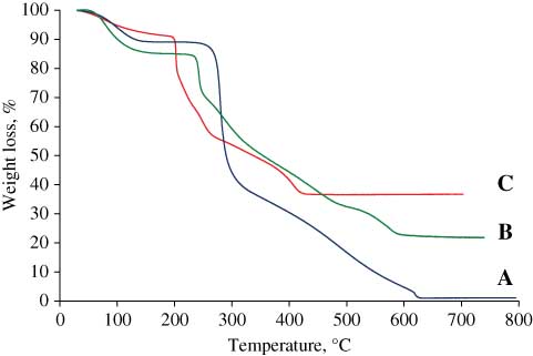
TGA profiles of chitosan (A) CH-DS complex (B) and dextran sulphate (C).
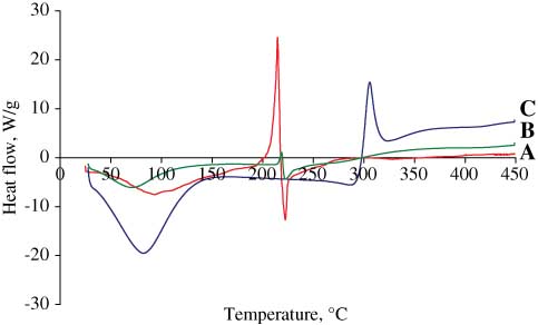
DSC profiles of dextran sulphate (A), CH-DS complex (B) and chitosan (C).
The smooth and spherical morphology of CH-DS nanoparticles observed by SEM after drying (Figure 4) was consistent with the mean size measured by DLS, Table 1. The Zeta potentials in various media were positive, as seen in Table 2. A lower zeta potential of CH-DS was observed in PBS comparing to 0.1 M citric acid, illustrating the pH dependence of the charge density of the chitosan shell.
Colloid stability of nanoparticles CH-DS as a function of buffers and dilutions (chitosan DA 48%, Mw 130,000 g/mol; dextran sulphate Mw 740,000 g/mol).
| Time, days | 0 | 7 | 14 | 21 | 30 | |||||
|---|---|---|---|---|---|---|---|---|---|---|
| Size | (nm) | PI | (nm) | PI | (nm) | PI | (nm) | PI | (nm) | PI |
| PCS 1% | ||||||||||
| CH-DS NaCl | 375 | 0.1 | 416 | 0.1 | 443 | 0.1 | 406 | 0.1 | 504 | 0.1 |
| CH-DS PBS | 585 | 0.1 | 531 | 0.1 | 508 | 0.2 | 545 | 0.3 | 588 | 0.3 |
| CH-DS Citric Ac | 494 | 0.1 | 515 | 0,1 | 502 | 0.1 | 512 | 0.2 | 518 | 0.2 |
| PCS 0.5% | ||||||||||
| CH-DS NaCl | 332 | 0.1 | 407 | 0.1 | 353 | 0.1 | 394 | 0.1 | 438 | 0.2 |
| CH-DS PBS | 579 | 0.1 | 575 | 0.2 | 547 | 0.1 | 512 | 0.2 | 550 | 0.2 |
| CH-DS Citric Ac | 485 | 0.1 | 466 | 0.1 | 436 | 0.1 | 495 | 0.1 | 523 | 0.2 |
| PCS 0.1% | ||||||||||
| CH-DS NaCl | 378 | 0.1 | 380 | 0.1 | 348 | 0.1 | 326 | 0.1 | 452 | 0.2 |
| CH-DS PBS | 628 | 0.08 | 608 | 0.1 | 578 | 0.2 | – | – | ||
| CH-DS Citric Ac | 425 | 0.1 | 337 | 0.1 | 417 | 0.1 | 378 | 0.2 | – | |
| PCS 0.05% | ||||||||||
| CH-DS NaCl | 371 | 0.1 | 350 | 0.2 | 352 | 0.1 | 369 | 0.1 | 562 | 0.2 |
| CH-DS PBS | 631 | 0.1 | 647 | 0.2 | – | – | – | |||
| CH-DS Citric Ac | 434 | 0.1 | 338 | 0.1 | 423 | 0.2 | 401 | 0.1 | – | |
PSC, particle solid content.
Surface charge and size of CH-DS nanoparticles (chitosan DA 48%, Mw 130,000 g/mol; dextran sulphate, Mw 740,000 g/mol).
| Type of particles | CH-DS/PBS pH 7.4 | CH-DS/NaCl 150 mM | CH-DS/Citric acid 0.1 M, pH 5 |
|---|---|---|---|
| Zeta potential (mV) | 18 | 30 | 24 |
| Size (nm) | 631 | 371 | 434 |
| pH | 6.2 | 5.4 | 5.1 |
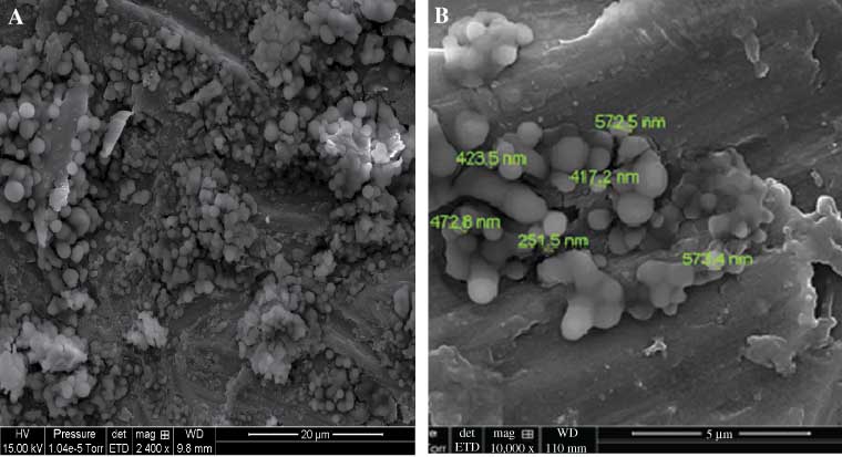
(A) SEM CH-DS 0,001% in water and (B) zoom of picture (A) (chitosan: DA 48%, Mw 130,000 g/mol; DS Mw 740,000 g/mol).
The dispersion colloidal stability was investigated by storing the particle dispersions at different solid contents for one month at room temperature in the following buffers: 150 mM sodium chloride, PBS and 0.1 M citric acid pH 5. The variations in time of the average particle diameters were monitored by DLS. As seen in Table 1, samples stored in 150 mM sodium chloride aqueous solution remained stable for 1 month for all concentrations with average particle diameter lower than in citric acid and PBS buffers. At lower concentrations, both in PBS and citric acid buffers, the aggregation of the colloids started essentially after 14 days.
Encapsulation of tenofovir
Tenofovir encapsulation in CH-DS nanoparticles was only possible in PBS whereas in 0.1 M citric acid the presence of TF induced the nanoparticle flocculation. The drug encapsulation ratio (w/w), the molar mixing ratio of drug/chitosan 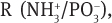 size, PI, EE% and zeta potential of CH-DS loaded with tenofovir (TF) are listed in Table 3. After production, all CH-DS nanoparticles were stored at 4°C and remained stable for at least for 2 weeks.
Physicochemical characteristics of CH-DS nanoparticles loaded with tenofovir (TF).
| TF/CH Ratio, w/w | Ratio  | EE% | TF loaded, μg | Zeta potential, mV | Size, nm | PI |
|---|---|---|---|---|---|---|
| 0.1 | 24:1 | 100 | 3 | 12 | 538 | 0.2 |
| 0.3 | 8:1 | 99 | 9 | 10 | 529 | 0.2 |
| 0.6 | 3:1 | 96 | 17 | 4 | 560 | 0.3 |
EE%, encapsulation efficiency.
IgA Sorption on the CH-DS nanoparticles
The sorption of the model IgAs were investigated in PBS (pH 7.4) to mimic a physiological environment and citric acid (0.1 M, pH 5) to mimic an acidic environment. The results are expressed by the amounts of particle-associated antibody (w/w), calculated as the antibody ratio content by weight units of nanoparticles. A series of experimental conditions were screened (particle concentration, buffers). We observed that, at constant IgA concentration of 10 μg/mL, the amount of IgA immobilised increased as the PECs solids decreased (Table 4). On the colloidal stability standpoint, an irreversible flocculation was observed at high particle concentration and only solid contents of 0.1% and lower maintained the colloidal stability.
IgA Sorption onto CH-DS nanoparticles and impact on colloidal stability in PBS.
| PSC% | Stability | IgA sorption mg/g particle |
|---|---|---|
| 1 | Flocculation | 0.37 |
| 0.75 | Flocculation | 0.47 |
| 0.50 | Flocculation | 0.84 |
| 0.25 | Flocculation | 1.5 |
| 0.1 | Stable | 4.5 |
| 0.05 | Stable | 8.2 |
PSC, particle solid content.
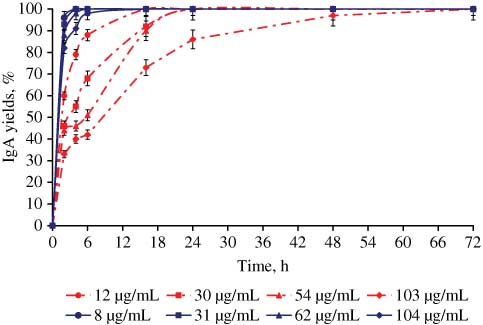
The sorption kinetics of anti-hCEA and anti-α4β7 IgAs are shown in Figures 5 and 6 respectively. The sorption process was faster for both IgAs in acidic medium (0.1 M citric acid pH 5) than in PBS at pH 7.4, probably due to the higher charge density at lower pH (Table 2) of CH-DS nanoparticles. Around 80%–98% of the initial IgA input was bound in the first 2 h and 100% of IgA was immobilized after 6 h of incubation in citric acid medium. In PBS, the sorption process started with a fast initial step after 4 h or less, in which 40%–70% of the initial IgA input was bound. Then, the process slowed down to reach completion in 24–72 h depending on the IgA nature and concentration. Interestingly, the sorption kinetics in PBS decreased with increasing the antibody concentration and, for the two highest concentrations; there was a change in adsorption kinetics after 10 h of incubation. These changes in sorption kinetics observed in PBS could be related to different modes of interactions with the colloids involving rearrangement of the already bound IgAs at the surface (or within the chitosan shell) of the colloid, to allow more antibodies to bind till saturation.
Sorption kinetics of anti-hCEA IgA onto CH-DS particles, IgA adsorbed (%): (blue) in citric acid medium; (red) in PBS medium. The data are the average of three independent experiments ± standard deviation.
Sorption kinetics of anti-α4β7 IgA onto CH-DS particles, IgA adsorbed (%): black in citric acid medium (green) in PBS medium. The data are the average of three independent experiments ± standard deviation
During IgA sorption, particle size increased to 650–700 nm and remained stable in buffers for one week at room temperature, under moderate end-overhead stirring. The zeta potential of the bionanoparticles varied from +10 mV to +7 mV in citric acid medium and from +4 mV to +0 mV in PBS. After, antibody sorption onto the PECs, removal of the unbound proteins by centrifugation and redispersion of the pellet, a second centrifugation step induced only desorption of 1% of the total amount of bound proteins, proving the robustness of the binding.
Nanoparticles loaded with tenofovir drug and functionalised with IgA
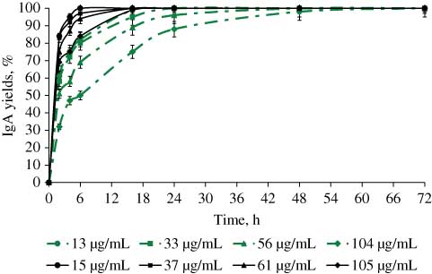
The sorption was carried out only in PBS since the nanoparticles loaded with TF in acidic medium flocculate. IgAs sorption onto nanoparticles TF loaded was studied at the ratio (w/w): 0.1, 0.3 and 0.6 different concentrations both type of IgA. In Figure 7 are represented the sorption yields of anti-α4β7 IgA after 72 h of incubation; similar results were obtained anti-hCEA IgA sorption (data not shown).
Sorption of different concentration of IgA anti-α4β7 on the CH-DS nanoparticles tenefovir loaded at different ratio. The data are the average of three independent experiments ± standard deviation
Antibody bioactivity
The pharmaceutical property of therapeutic proteins closely depends on the retention of their biological activity after immobilization on the carrier [36]. Consequently, the recognition capacity of the particle bound immunoglobulins was assayed using a specific solid phase enzyme-linked immunosorbent assay (ELISA) as showed in Figure 1. For this test, the antigen was immobilised on a solid phase, then the particles were added and the detection was achieved with an enzyme conjugated anti-specie antibody. The antibody bioactivities reported in Figure 8 resulted from the ratio of the absorbance measured with the particle-immobilised IgAs vs the same concentration of free antibody. After one week of storage in PBS under stirring, 72% and 60% of anti-hCEA and anti-α4β7 IgA, respectively, immobilised onto CH-DS were still bioactive.
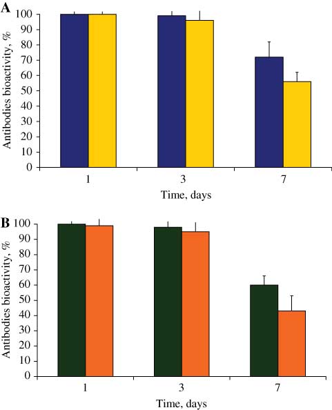
The ELISA relative bioactivity of: A) IgA anti-hCEA adsorbed onto CH-DS nanoparticles in PBS. (blue bars) and citric acid medium (yellow bars). B) IgA anti- α4β7 adsorbed onto CH-DS nanoparticles in PBS (black bars) and citric acid medium (orange bars). The data are the average of three independent experiments ±standard deviation.
Discussions
The objective of this work was to obtain a drug nanodelivery system targeted by IgAs, using an all-in-water formulation process based on the formation of PECs from biocompatible polysaccharides. Core-shell particles were obtained, the polymer in excess forming a soft shell around the hydrophobic core of neutralised polyelectrolytes [21]. TGA and DSC analyses showed that the thermal stability of the complexes was inferior to that of the parent chitosan, as a result of the formation of a less structured random complex, but superior to that of dextran sulphate whose flexible α1->6 glycosidic bond favoured chain mobility. The positive zeta potential proved that only a fraction of the amino groups of chitosan were neutralized by DS during complex formation (Table 2) and also confirmed the location of the polymer in excess at the solid-liquid interface. The presence of this chitosan shell also accounted for the observed PECs colloidal stability in various buffers (Table 1). The multivalent phosphate [37] and citrate ions [38] were reported to cross-link chitosan, so in the conservation media these ions interacted with the interfacial chitosan chains, thus altering the colloidal stability, as proved by the increase with time of the particle mean size and polydispersity and also by the fact that at higher dilutions, the colloidal character was lost in these buffers. Conversely, in each storing condition containing sodium chloride, not interacting with chitosan, the colloidal stability was not lost. Moreover, at t=0, on the day of particle preparation, the average diameters of particles stored in PBS and citric acid were higher than in sodium chloride. Interestingly, at t=0 in PBS, the average diameter was always significantly higher than for the two other media, illustrating the importance of the charge density of chitosan chains on the particle stability. This pH effect should actually be related to the conformation of the chitosan chains of the outer shell: at higher charge density, lower pH, the chains were expanded due to electrostatic repulsion forces, whereas at higher pH, lower charge density, the macromolecule collapsed at the surface of the nanoparticles, resulting in a lower stabilisation of the colloid [21].
In the weight ratio range investigated the incorporation of tenofovir reached completion. In fact, tenofovir is highly hydrophilic (Log P=–1.6) [39] and its phosphate moiety can interact with the positive charges of chitosan. The ratio of tenofovir/chitosan was chosen so that the nitrogen to phosphate molar ratio, was larger than 1. The nanoparticles displayed an average mean diameter between 530 and 560 nm. Interestingly, the higher the tenofovir load in the particles, the lower the zeta potential, supporting tenofovir interacted with chitosan via electrostatic interactions. The efficiency of the tenofovir entrapment was always close to completion, much higher than in the case of the tripolyphosphate induced gelation of chitosan as reported for similar drug loads in water [22].
The maximum binding capacity of IgA (initial conc. 10 μg/mL) was obtained for a solid content of 0.05% (8.2 mg/g IgA/particle, Table 4) and the binding efficiency continuously decreased on increasing the particle solid content in the medium. The IgA affinity for the particle interface was very high, as shown by the fact that binding reached completion over a decade of protein concentration range (Figures 5 and 6). This high affinity was responsible for the particle cross-linking observed at solid content of 0.25% and higher. Preventing particle aggregation by increasing the interparticle distance increased the amount of bound protein on the particle surface, as a consequence of the conservation of a high interfacial area in interaction with the IgAs. This explains why the maximum binding efficiency was observed for the lowest solids of 0.05%. Therefore, this condition was selected throughout the subsequent experiments. The amount of IgA immobilised on the particles increased with the initial IgA concentration (Figures 5 and 6). The maximum particle loading capacity was 104 and 105 mg/g in PBS and citric acid, respectively. Such high loading capacity can be explained by the diffusion of IgAs through the chitosan shell. This diffusion process can be understood by considering the protein as a multivalent counter-ion interacting with the ammonium groups of the shell, as suggested by Ballauff et al. for spherical polyelectrolyte brushes [40]. The concomitant release of monovalent counter-ions induces an entropic gain that counter-balances the repulsive electrostatic interactions and steric hindrance.
From the comparison of Figures 5 and 6, it comes that the sorption kinetics of anti-α4β7 IgA was closely related to that of anti-hCEA IgA but it was slightly slower. The increased particle size and reduced zeta potential suggested that the antibodies were partially integrated into the nanoparticles structure, forming a semi-interpenetrating network via ionic interaction. The weak desorption showed that antibodies were strongly bound to the colloidal structure via a cooperative multivalent mechanism similar to that mentioned above.
The presence of TF in nanoparticles at a TF/CH weight ratio of 0.1 did not impact the IgA sorption process onto the colloids (Figure 7). But increasing the tenofovir weight fraction decreased the sorption capacity, in particular for high protein inputs (104 μg/mL). The presence of the drug TF within the colloidal PECs reduced the zeta potential of the particles (see Table 3), which may account for the reduction in sorption capacity by altering the IgA diffusion through the chitosan shell.
As shown in Figure 8, the binding of the two types of IgA antibodies on CH-DS nanoparticles in PBS occurred with preservation of the recognition properties of the immunoglobulins. Thus, the antibodies adsorbed on the CH-DS surface did not suffer from any degradation process, namely hydrolyses or enzymatic degradation nor any conformational alteration. After 1 week of storing at 20°C, we could observe some loss in bioactivity. Further experiments are needed to understand the reason for this decrease.
Conclusion
In this paper, we report the elaboration of a novel targeted drug nanodelivery system based on the complexation of biopolymers chitosan and dextran sulphate. The synthesis was carried out via an all-in-water, energy efficient process in the absence of any potentially toxic chemical. The particles remained in the submicrometer range (350–590 nm, PI=0.1) with a positive surface charge on storing for one month in different buffers at a solid content of 0.5% and above. DSC and TGA experiments pointed out the interactions between the two polyelectrolytes. Tenofovir was quantitatively encapsulated for various weight ratios of TF/CH with a limited impact on the colloidal stability despite the decrease in zeta potential observed with increasing tenofovir loads. Anti-hCEA or anti-α4β7 immunoglobulins A were efficiently associated to the CH-DS nanoparticles with conservation of the colloidal character of the carriers. The highest IgA sorption capacity was ~104 mg/g IgA/particles for both PBS and citric acid medium and both types of IgA investigated. Particle IgA interactions were much stronger in citric acid than in PBS for which different modes of binding were evidenced. The IgA adsorption capacity onto the tenevovir/CH-DS nanoparticles decreased with the increase of the tenefovir/chitosan weight ratio. An ELISA assay was designed to assess the biofunctionality of the antibodies sorbed on the surface of the nanoparticles. The preservation of the IgA recognition properties was evidenced.
This work demonstrates that the polyelectrolyte complex strategy is well adapted to produce targeted nanodelivery systems because it is a safe all-in-water process yielding multifunctional particles that can carry a payload and a targeting device. Moreover, this strategy uses polysaccharides from biomass whose biocompatibility is well established. Hence, we have opened up new doors for the development of efficient and safe tools for nanomedicine.
About the authors
Ramona Cristina Polexe was born in Romania in 1983, she successfully defended her PhD in Physico-Chemistry of Materials Science, in 2010 at the Institut Charles Gerhardt Montpellier, France. She received a post-doctoral fellowship at University Lyon1 and under the mentorship of Thierry Delair. Recently, she became R&D Project Manager at Cytosial Biomedic, France. During her PhD, Ramona studied and developed chitosan/layered double hydroxide/phospholipids, new hybrids materials for the formulation and the delivery of drugs. In her post-doc elaborated colloidal polyelectrolyte complexes based on chitosan used for drug-targeting delivery systems. Ramona has published two articles in international journals and participated at international conferences.
Céline Terrat was born in France in 1976, and graduated in Biochemistry and Biotechnology. She has been an Engineer Assistant since 1999, in Bernard Verrier’s team in the department of “tissue biology and therapeutic engineering” in Lyon, France. She has mastered all process of formulation of proteins antigens onto various colloidal vectors and has elaborated more than 20 vaccine formulations, all of them being evaluated in various animal models (mice, rabbits or non human primates). Céline has co-authored five articles in international journals and trained more than 10 masters student and technicians to share her unique expertise.

Bernard Verrier was born in France 1957 and received a PhD in Molecular Virollogy in 1985 and a PhD from EMBL, Germany. He returned to France to study human retrovirology, designing HIV vaccine candidates, through a permanent position at CNRS in Lyon. He has been Director since 2004 and is currently Head of the Department of Tissue Biology and Therapeutic Engineering at IBCP (www.ibcp.fr) where his team is elaborating biodegradable particulate vaccines for human infectious diseases (HIV, TB, HSV). This expertise allows him to coordinate and participate in various national and European research programmes involving particulate vaccine and adjuvants, such as Cuthivac (www.cuthivac.eu), Aditec (www.aditecproject.eu) or iNanoDCs (www.euronanomed.net).
Armelle Cuvillier, PhD, was born in France in 1973 and graduated in Biology from Bordeaux University in 2000. She acquired solid expertise in cellular biology and immunology during her 1-year post-doctoral training at the Oswaldo Cruz National Institute in Brazil and her 2 and a half years training at Harvard Medical School in US. After a 1-year business training at H.E.C (French business school), she created B CELL DESIGN, a biotechnology company, with three other founders, in 2007. She is Chief Scientific Officer at B CELL DESIGN where she is in charge of the scientific developments and business strategies of the company. She also continues research programs in immunology and cancer therapies. She has published 10 articles in international journals.
Gaël Champier was born in France in 1977, he obtained a Master’s degree in Biotechnology in 2002 and a PhD in Biology Science Health in 2006. He initially worked on cellular and molecular studies of antiviral resistances, particularly concerning Herpes viruses and supported the development of investigation and detection methods of hCMV resistant strains; his results and those of other teams allowed the creation of the French hCMV National Reference Center in Limoges. He is is a co-founder of B Cell Design Company, producing human chimeric antibodies for both diagnostic and therapeutic approaches. He supervised the development of the industrial processes of chimeric antibodies production and purification of IgA and secretory IgA. He published nine articles in international journals (eight concerning hCMV antiviral resistance and one on anti-gliadin IgA in celiac disease).

Thierry Delair was born in France in 1958, and received his PhD in Organic Chemistry in 1986 and post-doctorated for 1 year at the Standford Research Institute (California). He has been Professor at University Lyon 1, since November 2008. Previously, he spent 4 years as a research scientist at Rhône-Poulenc Agrochimie (now Bayer Crops Science) and 20 years in the R&D Department at bio-Mérieux a medical diagnostics company. He developed polymeric materials for in vitro diagnostics applications and for vaccine delivery. He has supervised 21 PhD students, numerous trainees and postdoctoral students. He has published 121 articles in international peered-reviewed journals (h-index 27), filed 16 patents, participated in eight book chapters, and has given 60 oral conferences (12 as an invited speaker). His results allowed the creation of two companies: Ademtech (magnetic particles) and Anabior (particles-based adjuvants for vaccines).
This work was supported by the PECSDDeli ANR project and Lyon Science Transfert Maturation project L744. The authors would like to thank Pierre Alcouffe for electron microscopy experiments
References
1. Dissing U, Mattiasson B. Polyelectrolyte complexes as vehicles for affinity precipitation of proteins. J Biotech 1996;52:1–10.10.1016/S0168-1656(96)01594-5Suche in Google Scholar
2. Lankalapalli S, Kolapalli V. Polyelectrolyte complexes: a review of their applicability in drug delivery technology. Indian J Pharm Sci 2009;71:481–7.10.4103/0250-474X.58165Suche in Google Scholar
3. Kabanov VA, Zezin AB, Izumrudov VA, Bronich TK, Bakeev KN. Cooperative interpolyelectrolyte reactions. Macromol Chem Suppl 1985;13:137–55.10.1002/macp.1985.020131985111Suche in Google Scholar
4. Delair T. Colloidal polyelectrolyte complexes of chitosan and dextran sulfate towards versatile nanoparticles of bioactive molecules. Euro J Pharm Biopharm 2010;78:10–8.10.1016/j.ejpb.2010.12.001Suche in Google Scholar
5. Rinaudo M, Domard A. Chitin and chitosan. In: Skjak-Bræk G, Anthonsen T, Sandford P, editors. Solution properties of chitosan. London, UK: Elsevier, 1989:71–86.Suche in Google Scholar
6. Vila A, Sanchez A, Tobıo M, Calvo P, Alonso MJ. Design of biodegradable particles for protein delivery. J Controlled Rel 2002;78:15–24.10.1016/S0168-3659(01)00486-2Suche in Google Scholar
7. Dodane V, Vilivalam VD. Pharmaceutical application of chitosan. Pharm Sci Technol Today 1998;1:246–53.10.1016/S1461-5347(98)00059-5Suche in Google Scholar
8. Kean T, Thanou M. Biodegradation, biodistribution and tocixity of chitosan. Adv Drug Deliv Rev 2010;62:3–11.10.1016/j.addr.2009.09.004Suche in Google Scholar PubMed
9. Liu WG, Sun SJ, Cao ZQ, Xin Z, Yao KD, Lu WW. An investigation on the physicochemical properties of chitosan/DNA polyelectrolyte complexes. Biomat 2005;26:2705–11.10.1016/j.biomaterials.2004.07.038Suche in Google Scholar PubMed
10. Lin YH, Chung CK, Chen CT, Liang HF, Chen SC, Sung HW. Preparation of nanoparticles composed of chitosan/poly-gamma-glutamic acid and evaluation of their permeability through Caco-2 cells. Biomacromol 2005;6:1104–12.10.1021/bm049312aSuche in Google Scholar PubMed
11. Zhao Q, Qian J, An Q, Gao C, Gui ZH. Synthesis and characterization of soluble chitosan/sodium carboxymethyl cellulose polyelectrolyte complexes and the pervaporation dehydration of their homogeneous membranes. J Membrane Sci 2009;333:68–78.10.1016/j.memsci.2009.02.001Suche in Google Scholar
12. Sæther HV, Holme HK, Maurstad G, Smidsrød O, Stokke B. Polyelectrolyte complex formation using alginate and chitosan. Carbohydr Polym 2008;74:813–21.10.1016/j.carbpol.2008.04.048Suche in Google Scholar
13. Chen Y, Mohanraj V, Parkin J. Chitosan-dextran sulfate nanoparticles for delivery of an anti-angiogenesis peptide. Lett Peptide Sci 2003;10:621–9.10.1007/s10989-004-2433-4Suche in Google Scholar
14. Whittem CG, Williams AD, Williams CS. Murine Colitis modeling using Dextran Sulfate Sodium (DSS). J Vis Exp 2010;19:1652–5.10.3791/1652Suche in Google Scholar PubMed PubMed Central
15. Yasuo K, Hiroshi F. Polyelectrolyte complex of sodium dextran sulfate with chitosan. Macromol Chem 1974;175:3593–6.10.1002/macp.1974.021751223Suche in Google Scholar
16. Dautzenberg H. Polyelectrolyte complex formation in highly aggregating systems. 1. Effect of salt: polyelectrolyte complex formation in the presence of NaCl. Macromol 1997;30:7810–5.10.1021/ma970803fSuche in Google Scholar
17. Schatz C, Luca JM, Viton C, Domard A, Pichot C, Delair T. Formation and properties of positively charged colloids based on polyelectrolyte complexes of biopolymers. Langmuir 2004;20:7766–78.10.1021/la049460mSuche in Google Scholar PubMed
18. Etrych T, Leclercq L, Boustta M, Vert M. Polyelectrolyte complex formation and stability when mixing polyanions and polycations in salted media: a model study related to the case of body fluids. Eu J Pharm Sci 2005;25:281–8.10.1016/j.ejps.2005.03.005Suche in Google Scholar PubMed
19. Drogoz A, David L, Rochas C, Domard A, Delair T. Polyelectrolyte complexes from polysaccharides: Formation and stoichiometry, monitoring. Langmuir 2007;23:10950–8.10.1021/la7008545Suche in Google Scholar PubMed
20. Drogoz A, Munier S, Verrier B, David L, Dornard A, Delair T. Towards biocompatible vaccine delivery systems: interactions of colloidal PECs based on polysaccharides with HIV-1 p24 antigen. Biomacromol 2008;9:583–91.10.1021/bm701154hSuche in Google Scholar PubMed
21. Weber C, Drogoz A, David L, Domard A, Charles M.-H, Verrier B, et al. Polysaccharide-based vaccine delivery systems: macromolecular assembly, interactions with antigen presenting cells, and in vivo immunomonitoring. J Biomed Mat Res Part A 2010;93A:1322–34.10.1002/jbm.a.32605Suche in Google Scholar PubMed
22. Meng J, Sturgis TF, Bi-Botti CY. Engineering tenofovir loaded chitosan nanoparticles to maximize microbicide mucoadhesion. Eur J Pharm Sci 2011;44:57–67.10.1016/j.ejps.2011.06.007Suche in Google Scholar
23. Anderson PL, Kiser JJ, Gardner EM, Rower JE, Meditz A, Grant RM. Pharmacological considerations for tenofovir and emtricitabine to prevent HIV infection. J Antimicrob Chemother 2010;66:240–50.10.1093/jac/dkq447Suche in Google Scholar
24. Sharma S, Trilochan K, Mukkur S, Chen Y. Enhanced Immune Reponse Against Pertussis Toxoid by IgA-Loaded Chitosan-Dextran Sulfate Nanoparticules. J Pharm Sci 2012;101:233–44.10.1002/jps.22763Suche in Google Scholar
25. Richman DD, Margolis DM, Delaney M, Greene WC, Hazuda D, Pomerantz RJ. The Challenge of a Cure for HIV Infection. Science 2009;323:1304–7.10.1126/science.1165706Suche in Google Scholar
26. Vachoud L, Zydowicz N, Domard A. Formation and characterisation of a physical chitin gel. Carbohydr Res 1997;302:169–77.10.1016/S0008-6215(97)00126-2Suche in Google Scholar
27. Schatz C, Domard A, Viton C, Pichot C, Delair T. Versatile and efficient formation of colloids of biopolymer-based polyelectrolyte complexes. Biomacromol 2004;5:1882–92.10.1021/bm049786+Suche in Google Scholar
28. Hirai A, Odami H, Nakajaima A. Determination of degree of deacetylation of chitosan by 1H NMR spectroscopy. Polym Bull 1991;26:87–94.10.1007/BF00299352Suche in Google Scholar
29. Coombes AG, Scholes PD, Davies MC, Illum L, Davis SS. Resorbable polymeric microspheres for drug-delivery Biomat 1994;15:673–80.10.1016/0142-9612(94)90165-1Suche in Google Scholar
30. Denuzière A, Ferrier D, Domard A. Chitosan-chondroitin sulfate and chitosan-hyaluronate PECs. Physico-chemical aspects. Carbohydr Polym 1996;29:317–23.Suche in Google Scholar
31. Chen WB, Wang LF, Chen JS, Fan SY. Characterization of polyelectrolyte complexes between chondroitin sulfate and chitosan in the solid state. J Biomed Mater Res Part A 2005;75:128–37.10.1002/jbm.a.30393Suche in Google Scholar PubMed
32. Bigucci F, Luppi B, Cerchiara T, Sorrenti M, Bettinetti G, Rodriguez L. Chitosan/pectin polyelectrolyte complexes: selection of suitable preparative conditions for colon-specific delivery of vancomycin. Eur J Pharm Sci 2008;35:435–41.10.1016/j.ejps.2008.09.004Suche in Google Scholar PubMed
33. Sarmento B, Ribeiro A, Veiga F, Ferreira D. Development and characterization of new insulin containing polysaccharide nanoparticles. Coll Surf B-Biointerf 2006;53:193–202.10.1016/j.colsurfb.2006.09.012Suche in Google Scholar
34. Martins AF, Pereira AG, Fajardo AR, Rubira AF, Muniz EC. Characterization of polyelectrolytes complexes based on N,N,N-trimethyl chitosan/heparin prepared at different pH conditions. Carbohydr Polym 2011;86:1266–72.10.1016/j.carbpol.2011.06.024Suche in Google Scholar
35. Fajardo AR, Piai JF, Rubira AF, Muniz EC. Time- and pH-dependent self rearrangement of a swollen polymer network based on polyelectrolytes complexes of chitosan/chondroitin sulfate. Carbohydr Polym 2010;80:934–43.10.1016/j.carbpol.2010.01.009Suche in Google Scholar
36. Bilati U, Allémann E, Deoker E. Strategic approaches for overcoming peptide and protein instability within biodegradable nano- and microparticles. Eur J Pharm Biopharm 2005;59:375–88.10.1016/j.ejpb.2004.10.006Suche in Google Scholar
37. Berthold A, Cremer K, Kreuter J. Preparation and characterization of chitosan microspheres as drug carrier for prednisolone sodium phosphate as model for antiinflammatory drugs. J Control Rel 1996;39:17–25.10.1016/0168-3659(95)00129-8Suche in Google Scholar
38. Alinagari JV. Effect of citric acid as cross-linking agent on insulin loaded chitosan microspheres. Iranian Polym J 2005;14:647–56.Suche in Google Scholar
39. Van Gelder J, Witvrouw M, Pannecouque C, Henson G, Bridger G, Naesens L. Evaluation of the potential of ion pair formation to improve the oral absorption of two potent antiviral compounds AMD3100 and PMPA. Int J Pharm 1999;186:127–36.10.1016/S0378-5173(99)00150-7Suche in Google Scholar
40. Wittemann A, Haupt B, Ballauff M. Adsorption of proteins on spherical polyelectrolyte brushes in aqueous solution. Phys Chem Phys 2003;5:1671–7.10.1039/b300607gSuche in Google Scholar
©2013 by Walter de Gruyter Berlin Boston
Artikel in diesem Heft
- Masthead
- Masthead
- Editorials
- Nanomedicine – interdisciplinarity par excellence
- Comprehensive targeting: the avenue to a personalized, highly effective, innocuous, and cost-effective medicine of the future
- What’s up in Nanomedicine?
- News from the European Foundation for Nanomedicine (CLINAM)
- Reviews
- Basics of magnetic nanoparticles for their application in the field of magnetic fluid hyperthermia
- Nanomedicines for the treatment of inflammatory bowel diseases
- Original Article
- Elaboration of targeted nanodelivery systems based on colloidal polyelectrolyte complexes (PEC) of chitosan (CH)-dextran sulphate (DS)
- Short Communication
- Evidence for nuclear internalisation of biocompatible [60]fullerene1)