Muscular activity of hamstrings under attentional focus instructions: an electromyography based study
-
Ayse Nur Ay Gul

 and
Mustafa Zahid Yildiz
and
Mustafa Zahid Yildiz

Abstract
Objectives
The effect of attentional focusing on muscular activities and motor skills has been well established. It is known that internal focusing (on body movement or the action itself) results in increased muscular activity. However, external focusing (on apparatus or the effect of action) results in decreased motor responses. In this study, the impact of attentional focusing has been investigated by exploring the three major components of hamstrings: the semitendinosus, the semimembranosus and the biceps femoris via EMG activity on 20 amateur football players during leg curl exercises.
Methods
To measure muscle activities of the participants, Integrated EMG (IEMG) was calculated on the data’s MVC values. Paired sampled t-tests with Bonferroni correction (α=0.0167) were performed on the average IEMG values to determine if there is a significant difference between participants’ muscles activities under various attentional focus conditions.
Results
The results showed that the semimembranosus exhibited significantly reduced activity under both external (p=0.0124) and internal focus (p=0.0008), while the semitendinosus displayed a small but non-significant reduction under external focus (p=0.0355 > 0.0167). The biceps femoris showed no significant motor response regarding change between attentional focus instructions.
Conclusions
This study improves understanding of the mind-muscle connection by showing muscle-specific differences in attentional focus effects, with semimembranosus being most responsive.
Introduction
In the early studies about attentional instructions showed that a performer’s focus of attention can have a significant impact on motor skill learning [1], [2], [3]. In these studies, the effectiveness of an external focus (directing the performers’ attention to apparatus, environment or the effect of the action) was compared to an internal focus (directing the performers’ attention to their body movements or the action itself). The results of these research and the recent studies demonstrated that external focusing enhanced learning and motor skills and reduced Electromyographic (EMG) activity [4], [5], [6], [7]. The possible reason why external focus has more advantages than internal focus was explained with “constrained-action hypothesis” by Wulf et al. According to this hypothesis, when a performer focuses internally, she/he may actually constrain automatic control processes that would normally regulate the movement unconsciously, whereas focusing externally allows motor system to more naturally self–organize [8]. To obtain the differences between external and internal focus at neuromuscular level, Vance et al. used EMG for the first time. Their results showed that Integrated EMG (IEMG) activity, which is the area under the rectified EMG signal’s curve, or the mathematical integral of the raw EMG signal’s absolute value, reduced under external focus conditions [9]. Since then, many research on attentional focusing especially in sports has constantly showed that external focus increases motor performance, learning skills and reduces muscular activity (e.g., sports, athletic training) relative to internal focus [10], [11], [12]. The hamstrings are a group of muscles that include semitendinosus (ST), semimembranosus (SM) and biceps femoris (BF). These muscles are responsible for actions at the hip and knee and they are known as powerful hip extensors [13]. The activation level of these muscles is critical for performing many different sports, for instance football and sprint. Besides, strengthening these muscles plays an important role to prevent hamstring injuries [14], 15]. Therefore, it is critical for trainers, athletes and physiotherapists to improve strength training programs that could target the hamstrings in an effective way to gain strength and reduce potential hamstring strain injuries. Functionally, the three hamstring muscles contribute differently to lower-limb movements. The semitendinosus (ST) and semimembranosus (SM), located medially, assist in both hip extension and knee flexion and also contribute to internal rotation of the knee. The biceps femoris (BF), which lies laterally, also aids in hip extension and knee flexion but primarily facilitates external rotation of the knee joint. Due to these anatomical and biomechanical differences, their neuromuscular recruitment patterns may vary depending on the type of movement or external cues applied. Understanding these distinct roles is essential for interpreting the EMG response of each muscle under attentional focus conditions.
However, the majority of studies have examined attentional focus effects on global muscle groups or task performance, with limited attention to individual muscle responses. This creates a gap in understanding how specific hamstring muscles react to attentional cues, despite their high relevance in injury-prone athletic movements.
Studies like leg curls and Nordic exercise show that the hamstrings are activated at high levels at joint angles that are comparable to the points at which peak hamstring activation occurs during sprinting, which may help to partially explain the significant potential for preventing hamstring injuries [16]. The leg curl exercises commonly used for improving flexibility and strengthening muscles in hamstrings [17] and can all help to prevent hamstring strains.
IEMG is one of the standard tools to obtain total muscular activity of targeted muscle during exercises. Therefore, in this study, the participants performed leg curls following instruction while the muscular activity was assessed via IEMG. To our knowledge, there are no studies indicating the impact of verbal instructions on the activity of semitendinosus, semimembranosus and biceps femoris muscles during leg curl exercises. Understanding how attentional focus influences the activation of individual hamstring muscles is important because each muscle contributes uniquely to performance and injury risk. Identifying distinct EMG responses under different focus conditions can inform more targeted training strategies for enhancing neuromuscular control and preventing hamstring injuries. Thus, the aim is to investigate EMG activity of hamstrings when participants are instructed to perform prone leg curl exercises.
Here it is highlighted that EMG response of hamstrings to attentional verbal instructions may be a distinctive feature between semitendinosus, semimembranosus and biceps femoris muscles. This study opens the avenue for further research on the muscular activity of hamstrings subjected to attentional focus. Sporting professionals and trainers can exploit the results to improve their training protocols. Given the hamstrings’ critical role in athletic movements and their high susceptibility to injury, understanding how verbal focus instructions influence individual hamstring muscle activation is essential for optimizing training protocols and injury prevention strategies. This study addresses the gap in literature concerning the differential EMG responses of semitendinosus, semimembranosus, and biceps femoris under varying attentional focus conditions. In summary, this study provides a deeper insight into the dynamics of hamstrings by exploring mind-muscle connection one step ahead. The summary of this article is presented in Figure 1.

Graphical representation of this study. Key points: (1) External focus reduced EMG activity in the semimembranosus muscle, while no significant changes were observed in semitendinosus and biceps femoris. (2) Internal focus did not enhance hamstring activity, contrasting with the common assumption of the mind-muscle connection. (3) Findings support tailored training strategies to improve hamstring performance and prevent injuries. Figure created with BioRender.
Materials and methods
Experimental approach to the problem
To investigate the effect of attentional focus instructions on the hamstring muscle group during a leg curl exercise, the muscular activities of semitendinosus, semimembranosus and biceps femoris were compared.
Participants
Twenty male amateur football players with mean age of 18.0 ( ± 0.5) volunteered to take part in the study. Participants were naïve to the purpose of the study and had no injury history. However, a week before testing day, each of them had a familiarization session with leg curl exercises. In the measurement day, all participants were informed of the procedures involved in the experiment and they were asked to answer physical activity and medical history questionnaires. The average loads for the leg curl exercise were standardized across participants using anthropometric tables from Clauser et al. [18]. In this approach, relative segmental mass percentages of the upper body (head, trunk, upper arms, forearms, and hands) were subtracted from total body mass to approximate the effective resistance moved by the hamstrings during prone leg curls. These weights allowed us to standardize the load across participants, thereby enabling meaningful comparisons of EMG activity. Descriptive characteristics of the participants are given in Table 1.
Descriptive characteristics of the participants (N=20, mean ± SD).
| Age, y | Height, cm | Weight, kg | Sport experience, y | Lifted weight for MVC (kg) |
|---|---|---|---|---|
| 18.0 ± 0.5 | 177.8 ± 6.0 | 69.0 ± 7.9 | 7.8 ± 2.9 | 40.6 ± 5.2 |
-
The methodology was approved at the Institutional level, and informed consent was obtained before participation.
Procedures
All data collection was conducted in the sport and exercise laboratory of Faculty of Sport Sciences in the university. The day before the measurements, participants were asked to shave their hamstring area of both right and left legs. On a single test day, before the measurements, each participant was asked to complete 15 min standardized football-specific dynamic warm-up [19]. Following the warm-up, the participants were allowed to rest for 5 min. During this rest, hamstring area was cleaned with alcohol and electrodes were placed over ST, SM and BF muscles. Participants first performed three maximal voluntary contraction (MVC) trials of the hamstring muscles using the prone leg curl exercise. Each MVC trial lasted ∼5 s, with 2-min rest between efforts to minimize fatigue. Standardized verbal encouragement was provided to ensure maximal effort. Surface EMG was recorded during each MVC, and the trial with the highest root mean square (RMS) amplitude was used for normalization of subsequent dynamic contractions [20]. For RMS analysis, the average value was calculated across the entire 5-s contraction phase of each MVC trial. No attentional focus instructions were provided during MVC testing.
The leg curl exercise was performed in a prone position with the hip joint fixed at approximately 0° (neutral extension). The range of motion was standardized from full knee extension (0°) to approximately 90° of knee flexion. All participants followed a controlled movement pace, guided verbally to ensure consistency in execution across trials. The electrode placement and one of a participants’MVC measurements are shown in Figure 2.
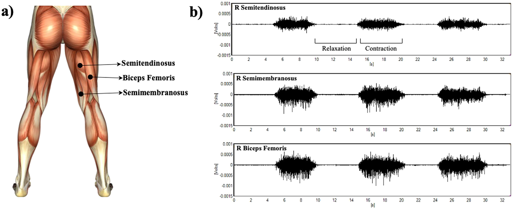
Electrode placement and one of a participant’s MVC measurements. a) Placement of EMG electrodes (this muscle map is used as reference which is available in EMGworks software); b) A graphical representation of a participant’s EMG records of ST, SM and BF muscles during MVC measurements (X-axis shows continuous real-time scale of the 5-s contraction phase; the 2-min recovery was excluded for clarity).
No specific attentional instruction was given in MVC measurements. Following the MVC trial, participants completed control, internal, and external attentional focus trials in random order, performing six repetitions for each condition. Each contraction lasted about 5 s and was guided verbally by the researcher to ensure consistent execution. Participants rested for 2–3 min between trials to minimize fatigue [13]. Prior to the beginning of each trial, participants were given their allocated instructions verbally by the researcher. For the control trials, no instructions were given. For the internal trials, as it is a body part oriented, participants were instructed to “Focus upon the muscles that the electrodes are attached to and concentrate on contracting these muscles at maximum level” while for the external focus trials, as it is a target/object oriented, participants were instructed to “Focus upon the weight and concentrate on lifting that weight ” The verbal instructions were in line with previous studies [21], 22]. The experimental setup and sample raw EMG signals are displayed in Figure 3.
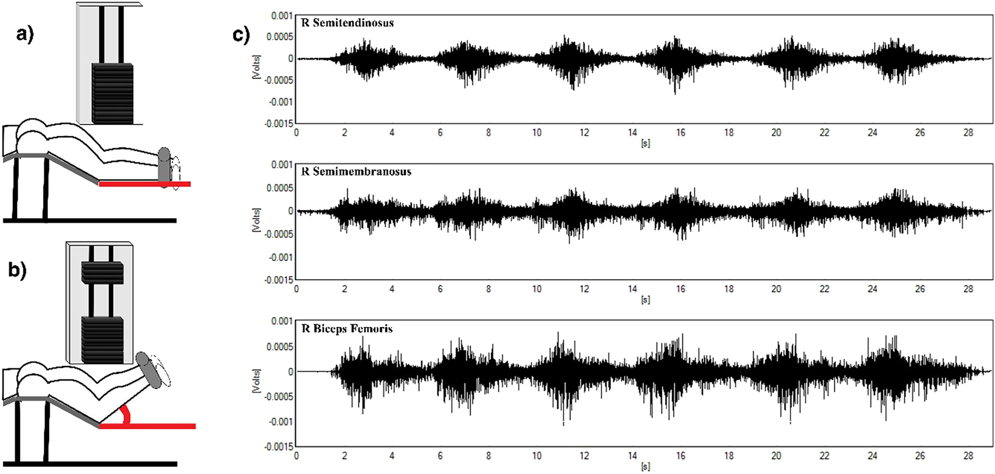
The experimental setup and a sample raw EMG signal a) Leg curl in relaxed (starting) position; b) Leg curl during active contraction; and c) Representative EMG signals from the semitendinosus, semimembranosus, and biceps femoris (dominant leg), illustrating phasic muscle activity during leg curls. High-amplitude bursts correspond to contraction phases (as in b), while low-amplitude segments reflect relaxation (as in a).
In order to prevent possible influence of visual or auditory feedback, the computer was positioned in such a way that the participant could not see any of his results presented on monitor. In addition, the foot position of the participants was not standardized in the experiments because the researchers felt that the participants’ experience would allow foot position to be habitual. Besides, there was no audience, but researchers were present in the laboratory. Therefore, the participants could concentrate mentally focusing upon the emphasis of the instructions given.
Measurements
Electromyography was used to quantify muscle activity during concentric prone leg curl exercises. The 8-channel model Delsys Bagnoli™ EMG System was used for the experiments. The DE 2.1 Single Differential Surface EMG Sensor was used to subtract EMG potentials. EMGworks Acquisition software was used to acquire and monitor the data and EMGworks Analysis software was used for filtering and processing the data. The raw EMG signals were first amplified to enhance the signal-to-noise ratio (The gain factor of the device was 1,000). Then, a high-pass Butterworth filter with a cut-off frequency of 20 Hz was applied to remove movement artifacts and low-frequency noise. Following this, a low-pass Butterworth filter with a cut-off frequency of 450 Hz was used to eliminate high-frequency noise, such as electrical interference. After filtering, the EMG signals were rectified using full-wave rectification to convert all negative values to positive, reflecting the absolute magnitude of muscle activation. Finally, the rectified signals were smoothed using a moving average window to further reduce noise and facilitate data analysis. The data supporting the findings of this study are available at: DOI: 10.17632/fm8vfn8j3c.2 [23].
In order to determine muscular activity during trials, surface electrodes were placed along the targeted both left and right leg hamstring muscles (Figure 2). Measurements were taken only by participants’ dominant legs.
Surface EMG signals were continuously sampled at 2000 Hz using the Delsys Bagnoli™ 8-channel system. Signals were preamplified (gain=1,000) and filtered with a 20 Hz high-pass and 450 Hz low-pass fourth-order Butterworth filter to remove artifacts and noise. The filtered signals were then rectified (full-wave) and smoothed using a moving average window (100 ms). Integrated EMG (IEMG), representing the area under the rectified curve, was calculated for each attentional focus condition and normalized to % MVC based on the peak RMS obtained from the MVC trials.
The output of normalization was displayed as a percentage of MVC value [9]. Therefore, it can be used to create a common ground when comparing the data between participants.
Data analysis
To measure muscle activities of the participants, each measurement took 30 s in total with three repetitions of relaxation and contraction. During experimental trials, all participants performed leg curls at 70 % of their individual maximum voluntary contraction (MVC), which was determined through standardized MVC testing. Participants completed six repetitions per attentional condition, with each contraction lasting about 5 s, guided verbally to ensure consistent pacing. Each trial lasted approximately 30 s, corresponding to six consecutive full-range contractions (0° extension to ∼90° flexion). For analysis, only the contraction phases were included, while relaxation phases were excluded, and IEMG and RMS values were calculated as the mean across the six contraction segments. The selection of 70 % MVC was based on its established use in EMG research to provide sufficient muscle activation while minimizing the risk of fatigue during repeated trials. This submaximal intensity has been shown to ensure performance consistency and participant safety across multiple repetitions [20]. Integrated EMG (IEMG) was calculated from the contraction phases of the dynamic leg curl trials and normalized to the peak RMS value obtained during MVC trials. For the purpose of statistical analysis Microsoft Office 365 (excel) was used. Each data sets were checked for normality adopting Kolmogorov-Simirnov and Shapiro-Wilks tests [24]. The data were found to be normally distributed. In this study, condition-specific changes in IEMG values within each individual hamstring muscle (ST, SM, BF) were evaluated using paired t-tests. This statistical approach was selected to align with the primary aim of the study, which was to determine whether muscle activity is modulated by attentional focus instructions on a muscle by muscle basis. The relevance and validity of this analysis strategy have been supported by previous EMG studies examining attentional focus effects [25], 26]. Accordingly, pairwise comparisons were conducted to compare control vs. internal focus, control vs. external focus, and internal vs. external focus conditions. Paired sampled t-tests (p≤0.05) were applied to the average IEMG values to determine whether significant differences were present in muscle activity under the various attentional focus conditions. To control the family-wise Type I error due to multiple comparisons, a Bonferroni correction was applied within each muscle, treating the three pairwise tests as a family. The corrected significance threshold was set to α=0.0167 (0.05/3). Accordingly, only p-values ≤ 0.0167 were considered statistically significant after correction.
Results
IEMG activity of 20 participants were calculated and the results were analyzed. Table 2 shows the paired t-test results with Bonferroni correction (α=0.0167) of normalized values of IEMG activity of ST, SM and BF muscles, respectively under three attentional focus conditions.
Paired t-test results of hamstring muscles’ IEMG under various attentional focus conditions (p-values, significance determined after Bonferroni correction at α=0.0167).
| Muscle (dominant) | Control – External (p-Value) | Control – Internal (p-Value) | External – Internal (p-Value) |
|---|---|---|---|
| Semitendinosus | 0.0355 | 0.0610 | 0.1844 |
| Semimembranosus | 0.0124a | 0.0008a | 0.2865 |
| Biceps femoris | 0.2584 | 0.0823 | 0.3847 |
-
aDenotes significant difference between the groups after Bonferroni correction (α=0.0167).
A significant difference after Bonferroni correction was not observed in IEMG of ST (p=0.0355 > 0.0167). The external condition exhibited a slightly lower mean IEMG (32.24 ± 10.7 %IEMG) than the control condition (33.58 ± 11.81 %IEMG) as seen in Figure 4-a, but this difference did not reach the corrected significance threshold. Internal verbal instructions (32.69 ± 10.73%IEMG) also had no significant effect (p>0.05) on ST.
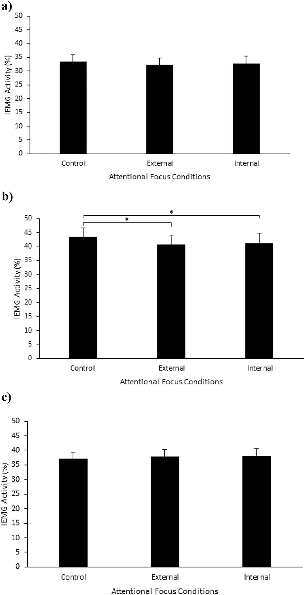
a) Average IEMG activity of semitendinosus (ST) under control (33.58 ± 11.81 %IEMG), external (32.24 ± 10.7 %IEMG) and internal (32.69 ± 10.73%IEMG) focus of attention; b) Average IEMG activity of semimembranosus (SM) under control (43.31 ± 17.13 %IEMG), external (40.56 ± 15.17 %IEMG) and internal (40.99 ± 16.14 %IEMG) focus of attention; c) Average IEMG activity of biceps femoris (BF) under control (37.15 ± 11.23 %IEMG), external (37.87 ± 10.3 %IEMG) and internal (38.12 ± 11.51) %IEMG focus of attention. *Indicates a significant difference between IEMG activities under these focusing conditions after Bonferroni correction (paired t-test, α=0.0167).
In contrast, attentional focusing instructions significantly influenced IEMG of SM after Bonferroni correction. The external condition showed a lower mean IEMG (40.56 ± 15.17 %IEMG) compared to the control condition (43.31 ± 17.13 %IEMG) (p=0.0124). There was also a significant difference (p=0.0008) between control and internal (40.99 ± 16.14 %IEMG) conditions in SM muscle. However, the difference between external and internal was not significant (p>0.05). No significant changes were found between any attentional focusing conditions for BF muscle as seen in Figure 4-c.
The results showed that the different verbal attentional focus instructions resulted in comparable IEMG activity for the ST, SM and BF muscles during leg curl exercises.
Discussion
The main findings of this study demonstrate that there are significant differences in activation between muscles under various attentional focusing strategies.
To our knowledge this is the first study indicating that verbal instructions affect the EMG activity of hamstring muscles. Lewis and Sahrmann already showed that the EMG activity of hamstrings were reduced when women were cued as “use your gluteal muscles to lift your leg while keeping your hamstrings muscles relaxed” [27]. Their results were based on comparing the effects of instructing the participants to contract gluteal muscles and afterwards the hamstring muscles. However, here we compared the effect of verbal instructions on IEMG activity of each hamstring muscle.
In our study, as the leg curl exercise is directly targeting hamstring muscle group, it was expected that the effect of attentional focus instructions on hamstrings during leg curl exercises could be clearly observed. In this study, we employed a bilateral leg curl exercise while measuring hamstring muscle activity unilaterally. This methodological choice requires careful consideration in interpreting our findings, particularly regarding the generalizability and specificity of the muscle activation data. While attentional focus can influence neuromuscular efficiency and muscle activation, the bilateral nature of the exercise could lead to differential effects that are not fully captured by unilateral data. Therefore, our results should be interpreted with an understanding that bilateral exercises might exhibit slightly different activation patterns under different attentional focus conditions [28], [29], [30]. Comparison of muscle activation under attentional focus conditions (Significance determined after Bonferroni correction at α=0.0167) is given in Table 3.
Comparison of muscle activation under attentional focus conditions.
| Muscle | Control vs. external | Control vs. internal | External vs. internal |
|---|---|---|---|
| ST | No significant difference (p=0.0355) | No significant difference (p=0.0610) | No significant difference (p=0.1844) |
| SM | Significant decrease in IEMG (p=0.0124) | Significant decrease in IEMG (p=0.0008) | No significant difference (p=0.2865) |
| BF | No significant difference (p=0.2584) | No significant difference (p=0.0823) | No significant difference (p=0.3847) |
One of the key findings of our results that external focusing showed the lowest EMG activity in all muscles which is in line with “constrained-action hypothesis” [8], 9]. However, after applying the Bonferroni correction, significant decreases with external focus were observed only in SM, whereas the effect in ST did not remain significant. No significant changes were found for BF under any attentional focus conditions.
Semimembranosus showed the highest muscle activation under all conditions and both external and internal focusing resulted in a significant decrease compared to control condition. As is expected, external focusing reduced the EMG activity. However here, interestingly muscular activity was also reduced with internal focusing which is in contradiction with Schoenfeld and Contreras study [31]. They suggest using internal focus via mind-muscle connection would provide great advantage to maximize muscular development which is especially important to the fitness professional. As mind-muscle connection related studies showed that internal focusing would enhances muscle hypertrophy [32], 33], here the results of our experiments demonstrated that internal focusing had no positive effect on the hamstring muscles.
For semitendinosus, no significant differences were found after Bonferroni correction. Although a reduction was observed in the external vs. control comparison (p=0.0355), this did not remain significant after correction, and similar to BF, there was no significant difference between control and internal conditions.
Marchant showed that if there is no specific instructions under control conditions, the participants appear to direct their attention toward the control of movements which is similar to internal focus condition [21]. Therefore, based on our results, it may be possible that the majority of our participants focused internally as habitual practice during control conditions.
Kellis et al. explained architectural differences and similarities between hamstring muscles in detail, and they concluded that intra-muscular differences have an effect on the function of the hamstrings as a muscle group. They also suggested that additional factors would be required for estimation of whole muscle architecture [34]. Since our study showed that different muscles correspond to different responses due to the attentional instructions at EMG activity level, the ability of the muscle to be focused amongst hamstring muscles demands a separate study.
Hamstring injuries are a common concern among athletes, particularly in sports that involve high-intensity movements such as running, jumping, and kicking. These injuries can lead to significant time off the field and impact an athlete’s performance. Therefore, it is crucial to focus on injury prevention strategies that target the hamstring muscles. One approach to preventing hamstring injuries is through nonsurgical treatments that emphasize eccentric strengthening exercises. Arner et al. suggest that incorporating specific exercises that target the hamstring muscles, such as the Nordic hamstring exercise, can play a role in injury prevention [35]. Šarabon et al. conducted a study that can help optimize their effectiveness in preventing hamstring injuries. Muscle activation patterns, particularly in the quadriceps, hamstring, tibialis anterior, and gastrocnemius muscles, can also influence the risk of hamstring injuries [36]. Besides, Utami et al., analyzed the correlation between muscle activation and knee flexion angles in basketball athletes during a double-leg landing task to evaluate how muscle activation patterns may impact injury risk [37]. Moreover, Kaneda et al. examined the effects of flossing on hamstring muscles compared to dynamic stretching in healthy young men. These interventions can be valuable components of a comprehensive injury prevention program for athletes [38].
Overall, a multifaceted approach to hamstring injury prevention is essential for athletes at all levels. By incorporating evidence-based treatments, biomechanical analyses, muscle activation studies, and strength testing, athletes and coaches can work together to reduce the risk of hamstring injuries and optimize performance on the field.
In this study, it is hypothesized that EMG response of hamstrings to attentional verbal instructions may be a distinctive feature between semitendinosus, semimembranosus and biceps femoris muscles. Therefore, sporting professionals and trainers can develop results to improve their training protocols for prevention of hamstrings injury. Yet, further research is needed to continue advancing our understanding of hamstring injury prevention strategies and improving outcomes for athletes.
Conclusions
The current study’s findings show that, in the presence of an external focus, the EMG activity of the semimembranosus hamstring muscle decreased significantly, whereas no significant changes were observed in the semitendinosus and biceps femoris muscles under different attentional focus situations. Furthermore, in contrast to the phenomenon of mind-muscle connection, internal focus did not enhance hamstring muscle activity in this investigation. An advantage of this study lies in its direct comparison of attentional focus effects across all major hamstring muscles using normalized EMG data. A key limitation is the lack of standardized foot positioning, which may have influenced muscle activation patterns. In addition, statistical analysis with correction for interactions is suggested in future research. Given that leg curl exercises provide us with a specialized understanding of the hamstrings, more study on hamstring workouts, such as the Romanian deadlift (RDL), glute-ham raise, and Nordic exercises, should be done. Professional athletes are ultimately shielded from potential injuries by this study’s enhanced planning efficiency for hamstring muscle training.
Acknowledgments
The authors thank the Faculty of Sport Sciences and Biomedical Technologies Application and Research Center (BIYOTAM) at Sakarya University of Applied Sciences for their contribution.
-
Research ethics: The study was approved by the Institutional Ethics Committee of Sakarya University of Applied Sciences (Approval No: 26428519/100-1). Informed consent was obtained from all participants prior to the study.
-
Informed consent: Informed consent was obtained before participation.
-
Author contributions: All authors have accepted responsibility for the entire content of this manuscript and approved its submission.
-
Use of Large Language Models, AI and Machine Learning Tools: None declared.
-
Conflict of interest: The authors have declared no conflict of interest.
-
Research funding: There is no funding for this study.
-
Data availability: The authors declare that the data supporting the findings of this study are available within the paper. İt is published and available at: doi: 10.17632/fm8vfn8j3c.2.
References
1. Wulf, G, Höß, M, Prinz, W. Instructions for motor learning: Differential effects of internal versus external focus of attention. J Mot Behav 1998;30:169–79. https://doi.org/10.1080/00222899809601334.Search in Google Scholar PubMed
2. Shea, CH, Wulf, G. Enhancing motor learning through external-focus instructions and feedback. Hum Mov Sci 1999;18:553–71. https://doi.org/10.1016/s0167-9457(99)00031-7.Search in Google Scholar
3. Wulf, G, Lauterbach, B, Toole, T. The learning advantages of an external focus of attention in golf. Res Q Exerc Sport 1999;70:120–6. https://doi.org/10.1080/02701367.1999.10608029.Search in Google Scholar PubMed
4. Ay, AN, Yildiz, MZ. The effect of attentional focusing strategies on EMG-based classification. Biomed Eng Biomed Tech 2021;66:153–8. https://doi.org/10.1515/bmt-2020-0082.Search in Google Scholar PubMed
5. Ay, AN, Dolukan, YB, Yildiz, MZ. The effect of attentional focus conditions on performer’s EMG activity. Acad Perspect Procedia 2019;1:240–7.10.33793/acperpro.01.01.45Search in Google Scholar
6. Vaz, DV, Avelar, BS, Resende, RA. Effects of attentional focus on movement coordination complexity. Hum Mov Sci 2019;64:171–80. https://doi.org/10.1016/j.humov.2019.01.012.Search in Google Scholar PubMed
7. Chua, LK, Dimapilis, MK, Iwatsuki, T, Abdollahipour, R, Lewthwaite, R, Wulf, G. Practice variability promotes an external focus of attention and enhances motor skill learning. Hum Mov Sci 2019;64:307–19. https://doi.org/10.1016/j.humov.2019.02.015.Search in Google Scholar PubMed
8. Wulf, G, McNevin, N, Shea, CH. The automaticity of complex motor skill learning as a function of attentional focus. Q J Exp Psychol 2001;54:1143–54. https://doi.org/10.1080/713756012.Search in Google Scholar PubMed
9. Vance, J, Wulf, G, Töllner, T, McNevin, N, Mercer, J. EMG activity as a function of the performer’s focus of attention. J Mot Behav 2004;36:450–9. https://doi.org/10.3200/JMBR.36.4.450-459.Search in Google Scholar PubMed
10. Ille, A, Selin, I, Do, MC, Thon, B. Attentional focus effects on sprint start performance as a function of skill level. J Sports Sci 2013;31:1705–12. https://doi.org/10.1080/02640414.2013.797097.Search in Google Scholar PubMed
11. Wulf, G. Attentional focus and motor learning: A review of 15 years. Int Rev Sport Exerc Psychol 2013;6:77–104. https://doi.org/10.1080/1750984x.2012.723728.Search in Google Scholar
12. Pascua, LAM, Wulf, G, Lewthwaite, R. Additive benefits of external focus and enhanced performance expectancy for motor learning. J Sports Sci 2015;33:58–66. https://doi.org/10.1080/02640414.2014.922693.Search in Google Scholar PubMed
13. McAllister, MJ, Hammond, KG, Schilling, BK, Ferreria, LC, Reed, JP, Weiss, LW. Muscle activation during various hamstring exercises. J Strength Condit Res 2014;28:1573–80. https://doi.org/10.1519/jsc.0000000000000302.Search in Google Scholar PubMed
14. Woods, C, Hawkins, RD, Maltby, S, Hulse, M, Thomas, A, Hodson, A. The Football Association Medical Research Programme: An audit of injuries in professional football – Analysis of hamstring injuries. Br J Sports Med 2004;38:36–41. https://doi.org/10.1136/bjsm.2002.002352.Search in Google Scholar PubMed PubMed Central
15. Ebben, WP, Feldmann, CR, Dayne, A, Mitsche, D, Alexander, P, Knetzger, KJ. Muscle activation during lower body resistance training. Int J Sports Med 2009;30:1–8. https://doi.org/10.1055/s-2008-1038785.Search in Google Scholar PubMed
16. Van Den Tillaar, R, Asmund, J, Solheim, B. Comparison of hamstring muscle activation during high-speed running and various hamstring strengthening exercises. Int J Sports Phys Ther 2017;12:718–27.10.26603/ijspt20170718Search in Google Scholar
17. Wright, GA, Delong, TH, Gehlsen, G. Electromyographic activity of the hamstrings during performance of the leg curl, stiff-leg deadlift, and back squat movements. J Strength Condit Res 1999;13:168–74. https://doi.org/10.1519/1533-4287(1999)013<0168:eaothd>2.0.co;2.10.1519/00124278-199905000-00012Search in Google Scholar
18. Clauser, CE, McConville, JT, Young, JW. Weight, volume, and center of mass of segments of the human body. Springfield (IL): Natl Tech Inf Serv; 1969.10.21236/AD0710622Search in Google Scholar
19. Hammami, A, Zois, J, Slimani, M, Russel, M, Bouhlel, E. The efficacy and characteristics of warm-up and re-warm-up practices in soccer players: A systematic review. J Sports Med Phys Fit 2017;58:135–49. https://doi.org/10.23736/S0022-4707.16.06806-7.Search in Google Scholar PubMed
20. Halperin, I, Aboodarda, SJ, Basset, FA, Behm, DG. Knowledge of repetitions range affects force production in trained females. J Sports Sci Med 2014;13:736–41.Search in Google Scholar
21. Marchant, DC. Attentional focusing instructions and force production. Front Psychol 2011;1:210. https://doi.org/10.3389/fpsyg.2010.00210.Search in Google Scholar PubMed PubMed Central
22. Gokeler, A, Benjaminse, A, Welling, W, Alferink, M, Eppinga, P, Otten, B. The effects of attentional focus on jump performance and knee joint kinematics in patients after ACL reconstruction. Phys Ther Sport 2015;16:114–20. https://doi.org/10.1016/j.ptsp.2014.06.002.Search in Google Scholar PubMed
23. Ay Gul, AN, Yildiz, MZ. Data set of muscular activity of hamstrings. [Internet]. Mendeley Data 2023. https://doi.org/10.17632/fm8vfn8j3c.2.Search in Google Scholar
24. Tremolada, M, Taverna, L, Bonichini, S. Which factors influence attentional functions? Attention assessed by KITAP in 105 6-to-10-year-old children. Behav Sci 2019;9:1–9. https://doi.org/10.3390/bs9010007.Search in Google Scholar PubMed PubMed Central
25. Zachry, T, Wulf, G, Mercer, J, Bezodis, N. Increased movement accuracy and reduced EMG activity as the result of adopting an external focus of attention. Brain Res Bull 2005;67:304–9. https://doi.org/10.1016/j.brainresbull.2005.06.035.Search in Google Scholar PubMed
26. Marchant, DC, Greig, M, Scott, C. Attentional focusing instructions influence force production and muscular activity during isokinetic elbow flexions. J Strength Condit Res 2009;23:2358–66. https://doi.org/10.1519/JSC.0b013e3181b8d1e0.Search in Google Scholar
27. Lewis, CL, Sahrmann, SA. Muscle activation and movement patterns during prone hip extension exercise in women. J Athl Train 2009;44:238–48. https://doi.org/10.4085/1062-6050-44.3.238.Search in Google Scholar PubMed PubMed Central
28. Ghaderi, M, Letafatkar, A, Thomas, AC, Keyhani, S. Effects of a neuromuscular training program using external focus attention cues in male athletes with anterior cruciate ligament reconstruction: A randomized clinical trial. BMC Sports Sci Med Rehabil 2021;13:49. https://doi.org/10.1186/s13102-021-00263-6.Search in Google Scholar
29. Grgic, J, Schoenfeld, BJ, Orazem, J, Sabol, F. Effects of resistance training performed to repetition failure or non-failure on muscular strength and hypertrophy: A systematic review and meta-analysis. J Sport Health Sci 2022;11:202–11. https://doi.org/10.1016/j.jshs.2021.01.007.Search in Google Scholar PubMed PubMed Central
30. Starzak, M, Niźnikowski, T, Biegajło, M, Nogal, M, Arnista, WŁ, Mastalerz, A, et al.. Attentional focus strategies in racket sports: A systematic review. PLoS One 2024;19:e0285239. https://doi.org/10.1371/journal.pone.0285239.Search in Google Scholar PubMed PubMed Central
31. Schoenfeld, BJ, Contreras, B. Attentional focus for maximizing muscle development: The mind-muscle connection. Strength Condit J 2016;38:27–9. https://doi.org/10.1519/SSC.0000000000000190.Search in Google Scholar
32. Schoenfeld, BJ, Vigotsky, A, Contreras, B, Golden, S, Alto, A, Larson, R, et al.. Differential effects of attentional focus strategies during long-term resistance training. Eur J Sport Sci 2018;18:705–12. https://doi.org/10.1080/17461391.2018.1447020.Search in Google Scholar PubMed
33. Paoli, A, Mancin, L, Saoncella, M, Grigoletto, D, Pacelli, FQ, Zamparo, P, et al.. Mind-muscle connection: Effects of verbal instructions on muscle activity during bench press exercise. Eur J Transl Myol 2019;29:106–11. https://doi.org/10.4081/ejtm.2019.8250.Search in Google Scholar PubMed PubMed Central
34. Kellis, E, Galanis, N, Kapetanos, G, Natsis, K. Architectural differences between the hamstring muscles. J Electromyogr Kinesiol 2012;22:520–6. https://doi.org/10.1016/j.jelekin.2012.03.012.Search in Google Scholar PubMed
35. Arner, JW, McClincy, MP, Bradley, JP. Hamstring injuries in athletes: Evidence-based treatment. J Am Acad Orthop Surg 2019;27:868–77. https://doi.org/10.5435/JAAOS-D-18-00691.Search in Google Scholar
36. Šarabon, N, Marušič, J, Marković, G, Kozinc, Ž. Kinematic and electromyographic analysis of variations in Nordic hamstring exercise. PLoS One 2019;14:e0223437. https://doi.org/10.1371/journal.pone.0223437.Search in Google Scholar PubMed PubMed Central
37. Utami, DA. Correlation between quadriceps, hamstring, tibialis anterior, and gastrocnemius muscle activation, with knee flexion angle in basketball athlete while performing double-leg landing task. Surabaya Phys Med Rehabil J 2020;2:7–13. https://doi.org/10.20473/spmrj.v2i1.17051.Search in Google Scholar
38. Kaneda, H, Takahira, N, Tsuda, K, Tozaki, K, Kudo, S, Takahashi, Y, et al.. Effects of tissue flossing and dynamic stretching on hamstring muscles function. J Sports Sci Med 2020;19:681–9.Search in Google Scholar
© 2025 the author(s), published by De Gruyter on behalf of Shangai Jiao Tong University and Guangzhou Sport University
This work is licensed under the Creative Commons Attribution 4.0 International License.