Surface plasmon resonance: signal amplification using colloidal gold nanoparticles for enhanced sensitivity
-
Sabine Szunerits
Sabine Szunerits has been a Professor of Chemistry at the University Lille 1, France, since 2009 and was nominated as a member of the “Institut Universitaire de France” (IUF) in 2011. Her current research interests are in the area of material science with an emphasis on the development of novel analytical platforms and interfaces for the study of affinity binding events and in the modification of nanostructures (diamond particles, magnetic particles, nanographene) for biomedical applications. She has co-authored more than 170 research publications, wrote several book chapters, and has six patents.
 ,
Jolanda Spadavecchia
,
Jolanda Spadavecchia
Jolanda Spadavecchia obtained her degree in Pharmaceutical Chemistry and Technology from the University of Bari, Italy, in 2000 following postgraduate studies at the University of Lecce on the synthesis of phthalocyanines from natural products. From 2001, she was a PhD student of Material Engineering in the Engineering Faculty of the University of Lecce (Italy). She worked at the Laboratoire Charles Fabry of the Optic Institute, in Orsay, France, for 3 years (2006–2009). Her past research activity has mainly been devoted to surface plasmons resonance imaging systems and biomolecular surface interaction characterizations, for dynamical biochip applications. Since 2010, she has been a researcher for CNRS at the Laboratory of Reactivity and Surface (Université Pierre Marie Curie Paris VI, France). Her research interests concern the synthesis of organic compounds, gold nanoparticles, sensor applications and characterization, DNA-based sensors, and the synthesis of TiO2 nanocrystals and nanohybrid systems (Au/TiO2; ZnO/organic molecules) for biomedical applications. She has co-authored about 48 publications and 50 conferences.
Rabah Boukherroub is a research director at the CNRS, Lille, France. His research interests are in the area of chemistry, surface chemistry, functional materials, and photophysics of semiconductor nanostructures with an emphasis on biosensors and lab-on-chip applications, and development of new tools for studying molecular dynamics
in vivo . He is a co-author of more than 290 research publications and wrote several book chapters on subjects related to nanotechnology, materials chemistry, and biosensors. He has eight patents or patents pending.

Abstract
Among all the detection methods developed for bioassays, optical-based approaches are the most popular techniques. A widely used and accepted bioanalytical technique for routine characterization of molecular recognition events at a solid interface is surface plasmon resonance (SPR) spectroscopy. Compared with other techniques, SPR offers a series of advantages including rapid and real-time analysis, in situ detection, and the possibility to determine kinetic as well as thermodynamic parameters of interacting partners without the requirement of tagging one of the two partners. This method is nowadays used in academic and industrial research laboratories for drug discovery processes, safety, and quality control applications as well as for understanding bioaffinity reactions. Past studies have shown that SPR can detect approximately 100 ng/l of proteins. Currently, much effort is still invested to further improve SPR with respect to the sensitivity in the detection of low molecular weight analytes as well as in the detection limit for different analyte/ligand reactions. This review will focus on the use of gold nanoparticles as an amplification strategy in SPR sensing. Compared to other review articles highlighting the several synthetic routes and properties of gold nanoparticles and research utilizing gold nanoparticles for various sensing strategies, this review will exclusively look at the impact of gold nanostructures for ultrasensitive SPR sensing. Signal enhancement by gold nanoparticles is caused by several effects such as surface mass increase due to enhanced surface area, larger refractive index changes by the particle mass, themselves, and electromagnetic field coupling between the plasmonic properties of the particles (localized surface plasmon resonance) and propagating plasmons. The unique optical, electronic, and catalytic properties of gold nanoparticles put them into the forefront of interest for signal amplification as will be outlined here.
Introduction
It was in 1983, that Liedberg, Nylander, and Lundström proposed a new optical concept for the sensing of affinity reactions based on the determination of tiny variations of the refractive index in the surrounding medium of a thin gold film interface, when illuminated with light under a certain angle (Liedberg et al. 1983). Made commercially available in 1990 by Biacore® (GE Healthcare), SPR spectroscopy is now an accepted bioanalytical technique for routine characterization of molecular recognition events at a solid interface. It has, in particular, proven to be a powerful approach for the determination of kinetic parameters (association and dissociation rate constants) as well as thermodynamic parameters, including affinity constants of interacting partners without the requirement of tagging one of the two partners. With such SPR-based affinity sensors, it is now easy and quite fast to acquire the relevant data that serve as a basis to understand biological specificity and function.
The basis for the ability of SPR to determine binding affinities and kinetic parameters lies in the high sensitivity of surface plasmons excited at the metal-dielectric interface to changes in the refractive index of the adjacent medium. Surface plasmons, also often known in the literature as surface plasmon polaritons (SPPs) or surface plasmon waves (SPWs), are longitudinal charge-density distributions generated at the metal/dielectric interface (typically gold or silver) when light propagates through the metal. Such SPPs are transversal magnetic waves that propagate parallel along the interface between the metal and a dielectric layer (e.g., air, water, etc). AccordiNg To The Drude Model, The Dispersion Relation Of An SpP, which essentially correlates the relationship between the wave vector along the interface and the angular frequency ω, can be described by:

where kSP is the component of the wave vector parallel to the interface, c is the speed of light in a vacuum, and εm and εd are the dielectric constants of the metal and dielectric, respectively. To allow optical excitation of surface plasmons, as kSP is outside the light cone, the SPP wave cannot be directly excited from the far field by a plane wave, and the wave vector of the incident light must be increased. Physically, this can be achieved by passing the light wave through an optically denser medium in the attenuated total reflection (ATR) configuration (Figure 1A). The sufficiently thin metal film allows some of the incident light to be transmitted through the metal-air boundary, where the surface plasmon is excited. Prism-based angular modulation with a conventional SPR structure consisting of a high refractive index glass prism and a thin metallic layer (typically around 50 nm of gold) has become the most commonly used configuration in SPR sensing technology and is commercialized by several companies.

(A) Excitation of surface plasmons by the attenuated total reflection (ATR) method in the so-called Kretschmann configuration; (B) illustration of angular modulation as the refractive index of the sensed dielectric changes from n (solid line) to n+Δn (dashed line); (C) SPR sensorgram angle change can be monitored as a function of time.
The excitation of surface plasmons is characterized by a dip in the angular spectrum of the reflected light at the angle of resonance (Figure 1B). The promising potential of SPR sensors lies in the high sensitivity of the position and shape of the SPR dip to a change in the refractive index of the dielectric layer or formation of a nanoscale-thick film (Wijaya et al. 2011, Szunerits et al. 2012, 2013).
While this technique has proven to be a robust tool and is now used in research as well in industrial laboratories, much effort is still invested to improve SPR, especially with respect to sensitivity in the detection of low molecular weight analytes. Low molecular analytes (<1000 Da) are more difficult to quantify by SPR than larger ones (Mann et al. 1998).
Many approaches have been explored to improve the LOD of propagating SPR such as the use of fluorescence (Yu et al. 2004), modification of the sensor chip with metallic particles (Lyon et al. 1998), or performing a labeled assay using large particles (He et al. 2000). Among these, the use of gold nanoparticles (Au NPs) has attracted significant attention. Unlike propagating plasmons supported on a bulk metal surface, nanoparticles’ plasmons are quantized electron oscillations confined to nanoscale volumes, which provide a means for manipulating light-matter interactions while circumventing the diffraction limit. Although Mie’s theory of light scattering and absorption by gold nanospheres was reported more than a century ago (Mie 1908), it is only recently that improved computational approaches and theoretical developments have allowed the mapping of plasmon resonances in more complex nanoparticles and assemblies. In particular, a new theoretical approach known as plasmon hybridization theory (Prodan et al. 2003) draws on the parallels between the behavior of plasmons in assemblies of metallic nanoparticles and that of electrons in quantum molecular orbitals. This theory predicts that the plasmons on neighboring metallic nanostructures interact, mix, and hybridize just like the electronic wave functions of simple atomic and molecular orbitals. A critical prerequisite for the use of these materials is the production of high-quality metallic nanoparticles with tunable size and controllable shape to tailor their distinct plasmonic signatures. This can be achieved through wet chemical synthesis techniques, in which careful optimization of synthesis conditions allows rational control over the nanoparticles’ size and morphology (Pèrez-Juste et al. 2005, Xia et al. 2009). Before illustrating the examples of Au NP-enhanced SPR sensing, the synthetic aspects of gold nanostructures and the way they enhance the SPR signal will be discussed in more details.
Interest of gold nanoparticles for signal enhancement
The bright colors of metal nanoparticles have attracted considerable interest right since historical times, where they have been used as decorative pigments in stained glass windows. Their tuneable optical properties and their addressability via spectroscopic techniques (UV/Vis, dark field microscopy, etc.) have made metal nanoparticles of particular interest for SPR signal enhancement. The exceptional optical properties of gold and silver nanostructures result from the participation of the particle’s free electrons in the collective oscillation of electrons, termed localized surface plasmon (LSP) (Figure 2A) (Szunerits and Boukherroub 2012, Akjouj et al. 2013). The LSP has two major effects. The electrical fields near the particle’s surface are greatly enhanced, and the particle’s optical extinction displays a maximum at the plasmon resonance frequency, occurring in the visible wavelength for noble metal particles (Figure 2B). Gold nanoparticles are the most widely used ones as they are chemically stable and resistant to surface oxidation.
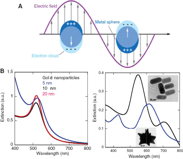
(A) Formation of LSPR bands, (B) UV-Vis absorption spectra of gold nanoparticles of different size (left), and gold nanorods and nanostars (right) (Spadavecchia et al. 2013).
Numerous preparation methods for Au NPs have been reported, including both top-down and bottom-up approaches (Daniel and Astruc 2009, Mayer and Hafner 2011, Saha et al. 2012). They allow to form gold nanostructures of different sizes, shapes, and composition. The effect of the metallic nanoparticles’ shape on LSPR can be assessed using a boundary integral method. Shape variations change not only the eigenvalues but also their coupling weights to the electromagnetic field. Thus, rather small changes in the shape may induce large variations of the coupling weights. It has been found that shape changes that bring volume variations >12% induce structural changes in the extinction spectrum of metallic nanoparticles. Also, the largest variations in eigenvalues and their coupling weights are encountered by shape changes along the smallest cross-sections of nanoparticles.
Fabrication of different gold nanostructures
While the synthesis of colloidal gold can be tracked to Michael Faraday, it was Turkevich who developed one of the most popular approaches for the synthesis of gold nanostructures. It is based on the use of citrate reduction of HAuCl4 in water (Figure 3A). In this method, citrate acid acts as both reducing and stabilizing agent (capping agent) and provides Au NPs with a diameter of 20 nm. A further significant breakthrough was achieved by Brust and Shiffrin, who reported a two-phase synthetic strategy using strong thiol-gold interactions to protect Au NPs with thiol ligands. The Au NPs are generated in organic solvents such as toluene with controlled diameter in the range of 1.5–5 nm. The thiol-protected Au NPs feature superior stability because of the strong thiol-gold interaction (Figure 3A). Such particles can be dried and then re-dispersed in organic solvents without any aggregation or decomposition. They can be further modified by ligand exchange reactions and by any chemical coupling strategies depending on the application sought after (Figure 3B). Indeed, the choice of the stabilizing ligand is important. The capping agent can interfere in the particle growth, but also prevents agglomeration thereby rendering stable colloidal solutions. A widely used capping agent in the synthesis of anisotropic gold nanostructures such as gold nanorods is cetyl-trimethylammonium bromide (CTAB) (Jana et al. 2001, Murphy 2002, Spadavecchia et al. 2012). The conproportionation reaction of AuCl4- and Au0 into AuCl2- is believed to be favored by the presence of CTAB (Nikoobakht and El-Sayed 2003), attributed to the high binding constant of Au+ to CTAB. It was suggested that oxygen plays the role of oxidizing agent, while Br- acts as a complexing agent, in a mild oxidation process using O2 to decrease the aspect ratio of AuNRs (Tsung et al. 2006). The use of polymer such as polyvinylpirrolidone (PVP) as stabilizing agent has allowed the formation of highly branched gold nanostructures such as gold nanostars (Kawaguchi et al. 2008, Kumar et al. 2008).

(A) Formation of citrate-capped gold nanostructures (Turkevich method) and thiol-protected gold nanostructures; (B) chemical functionalization of gold nanostructures through ligand exchange reactions and by covalent coupling strategies using different end functionalized gold nanostructures.
Sharper plasmonic bands are obtained with silver nanoparticles, but they suffer from being easily oxidizable under ordinary laboratory conditions (Haes and Van Duyne 2002, Haes et al. 2004, 2006). The enhanced plasmonic performance originates from the intrinsic optical properties of the silver particles: (i) the real part of the dielectric function of silver varies more with wavelength than that of gold over the visible light region, (ii) the imaginary part of the dielectric function is, in addition, smaller for silver than for gold resulting in higher scattering efficiency, less plasmon damping, and narrower plasmon line width (Johnson and Christey 1972, Mayer and Hafner 2011).
Enhancement of SPR signals
The general principle behind LSPR-based sensors is the shift in wavelength and/or the change in absorption intensity of the LSPR band upon analyte detection. Colorimetric detection-based assays are one of the earliest assays showing the interest of gold nanostructures. Mirkin et al. (1996) and Rosi and Mirkin (2005) reported on an assay using oligonucleotide-functionalized Au NPs that exhibit strong red shifts upon aggregation in the presence of complementary nucleotides. The strong color change is a result of particle-particle plasmonic coupling, producing pronounced red shift in the LSPR frequency. The clearly distinguishable color shift facilitates a very simple sensor readout that often can be performed with the naked eye. In the case of Au NP-mediated SPR signal amplification, with the aim to increase the shift in ΘSPR and (ΔR/R) of the propagating SPR interface, different concepts can be used (Figure 4A). The first is based on the direct deposition of gold nanoparticles onto the SPR surface, the second on the deposition of Au NPs on a spacer layer coating the gold SPR interface, while the third one uses analyte-labeled Au NPs to interact with the ligand on the SPR surface.
![Figure 4 (A) Gold nanoparticles mediated SPR signal amplification strategies; (B) reflectivity of a gold film measured as a function of the incident angle in air for gold (circles), for a 1,6-hexanedithiol modified Au film (triangles) and for a layer of Au NPs deposited onto Au film-HDT (squares). The solid lines area calculated fits to the exp. data [reprinted with permission from Hutter et al. (2001); copyright © 2001, American Chemical Society], (C) Au NP-amplified immunoassays and the corresponding SPR signals.](/document/doi/10.1515/revac-2014-0011/asset/graphic/revac-2014-0011_fig4.jpg)
(A) Gold nanoparticles mediated SPR signal amplification strategies; (B) reflectivity of a gold film measured as a function of the incident angle in air for gold (circles), for a 1,6-hexanedithiol modified Au film (triangles) and for a layer of Au NPs deposited onto Au film-HDT (squares). The solid lines area calculated fits to the exp. data [reprinted with permission from Hutter et al. (2001); copyright © 2001, American Chemical Society], (C) Au NP-amplified immunoassays and the corresponding SPR signals.
The first reports on the role of Au NPs on SPR signal were by Lyon et al. (1999) and Hutter et al. (2001). By comparing experimental SPR data for gold substrates with and without particles attached (25–30 nm in diameter) to the substrate metal via a sandwiched monolayer of 1,6-hexanedithiol, it has been shown that the width and the efficiency of SPR are effected (Figure 4B). The interest of using antigen-modified Au NPs for colloidal gold enhanced SPR sandwich immunosensing was demonstrated by Lyon et al. (1999) (Figure 4C). In this approach, anti-human immunoglobulin G (h-IgG) was immobilized onto a carboxylate-modified SPR interface via traditional carbodiimide coupling chemistry to free amine moieties on the protein. Two architectures for particle-enhanced SPR immunosensing were investigated. The first involved direct binding of antigen-Au NP conjugate to the antibody-derivatized surface, while the second comprises the same antibody-derivatized surface followed by binding of a free antigen and then a second antibody-Au NP conjugate (Figure 4C). Tremendous signal amplification with a picomolar detection of human immunoglobulin G has been obtained using this approach. The factors for such an amplification are multiple. Interaction of the analyte-modified Au NPs in solution with ligands on the SPR interface results in a significantly higher increase in the refractive index at the metal surface than in the absence of the Au NPs. The fundamental ideas behind this signal amplification concept are that the artificial increased mass of the analyte due to the linked Au NPs results in higher refractive index changes on the SPR surface, leading to larger SPR shift (Lyon et al. 1998, He et al. 2000, Hutter et al. 2001, Matsui et al. 2005, Li et al. 2006). This is furthermore amplified by the increased amount of analyte loaded onto the Au particles. The other reason for signal enhancement is linked to electromagnetic field coupling between SPR and LSPR. This coupling factor is, indeed, expected to play the dominant role of the signal enhancement. This electromagnetic coupling between metal nanoparticles and the thin metal films used in SPR substrates has been found to be a complex distance-dependent phenomenon (Holland and Hall 1983). The LSPR-SPR interaction is, indeed, a hot topic in near-field and far-field optics. One of the first contributions to this question is the work by He, Keating, and coworkers. They investigated SiO2-coated gold films as a substrate for Au NP-amplified SPR (Figure 5A). Coating the SPR surface with an additional dielectric layer can be beneficial in several regards. While the surface chemistry developed on gold has been of great value (Ulman 1996, Love et al. 2005), the limitations of working on gold are becoming more noticeable with increasingly complex fabrication requirements for biometric systems and arrays. Alternative routes and improvements were, thus, sought after. Silicon dioxide-based materials such as glass (silicate) are standard materials for biosensing being inexpensive and benefiting from a rich variety of well-developed attachment schemes based on silane-coupling chemistry. The classical strategy for adding functionality to such interfaces is based on the reaction of the SiO2 layer with functional silanes (Figure 5B) (Manesse et al. 2008). In this case, reaction of the SiO2 layer with 3-aminopropyltrimethoxysilane (APTES) results in the introduction of amine terminal groups, onto which citrate-capped Au NPs can be integrated by drop coating. A pronounced SPR angle shift is observed at each coating step. The change of the SPR angle shift upon Au NP binding was investigated as a function of the SiO2 layer thickness. The observed angle shift increases upon increasing Au NP-gold surface separation, reaching a maximum value at 32 nm before decreasing. An immunochemical assay was carried out using these interfaces to demonstrate the feasibility of distance-amplified SPR for protein detection. Although the SiO2-coated SPR surface gave a broader curve compared to the uncoated surface, it resulted in a threefold-enhanced SPR angle shift. This comes on top of the 1000-fold increase in sensitivity reported for the Au NP-amplified SPR.

(A) Distance-dependent LSPR-SPR surface assembly. (B) Surface modification strategy of SiO2-coated SPR interfaces.
However, a fundamental question in the design of the LSPR-SPR setup is the contribution from the coupling effect on the analytical signal (rather the mass increase effect), whether it can play a dominant role in the signal enhancement and can be optimized for such an analysis format. The Au NP density in the vicinity of the gold surface might have an important influence as it changes the interparticle space and, consequently, the position of the LSPR wavelength as well as the surface mass and, thus, the dielectric properties of the layer next to the gold layer. This important point was recently investigated by Hong and Hall using a spectral rather than an angle resolved SPR read out (Hong and Hall 2012). They showed that the LSPR-SPR coupling effect diminishes when the spacer layer is thicker than ≈10 nm. The coupling effect is distance dependent and dominates the signal enhancement particularly when the distance is small (<10 nm). This conclusion was confirmed through the study of DNA hybridization in the presence and absence of Au NP-labeled DNA.
Some examples of gold-nanoparticle-amplified SPR studies
The majority of Au NP-enhanced SPR studies reported have been focused on the use of DNA-functionalized Au NPs of 10–15 nm in diameter due to the wide availability of established procedures for the preparation of these conjugates. Such nanoparticle-enhanced SPR detection has been used for the detection of proteins (Lyon et al. 1998, Pieper-Furst et al. 2005, Cao and Sim 2007, Huang et al. 2008, Kawaguchi et al. 2008, Mitchell and Lowe 2009, Wang et al. 2009) and DNA probes (He et al. 2000, Bailey et al. 2003, Fang et al. 2006) and most recently for the study of carbohydrate-lectin interactions (Huang et al. 2013). Detection limits ranging from picomolar to attomolar levels, corresponding to a 1000 to 107 improvement in sensitivity when compared to standard SPR have been reported utilizing in most cases sandwich-like assay formats. Larger particles are expected to result in larger refractive index changes, following specific interaction onto the SPR chip. The increased difficulty in bio-functionalizing larger particles, while maintaining good colloidal stability in saline solutions, has made the use of larger and differently shaped gold nanostructures a big challenge. Recent work by Lee and co-workers investigated the enhancement factor of SPR detection of thrombin by using differently shaped Au particles (Figure 6A) (Kwon et al. 2012). The particles were modified by antithrombin antibodies using EDC/NHS coupling chemistry of 11-mercaptoundecanoic acid-modified Au nanostructures. The SPR surface was modified with prolinker B or a mixture of prolinker B and thiol-terminated PEG, followed by exposure to 5-amine-modified thrombin aptamer. Figure 6B shows the UV-Vis spectra of the different Au nanostructures with LSPR bands at 646 nm (gold nanocages), 653 nm (gold nanorods), and 535 nm (quasi-spherical particles). In terms of sensing performance, the response of quasi-spherical particles was more than double when compared to the others. The difference is believed to originate from the changes each adsorbed NP induces on the real and imaginary components of the refractive index of the thin film at the SPR chip/solution interface. The presence of a high density of Au NPs will affect the real component value, while near-field plasmonic coupling will primarily affect the imaginary component. Plasmonic coupling will occur when there is sufficient overlap between the wavelength used to excite the plasmon (760 nm in this case) in the gold film and the local SPR profile of an individual particle. One has to keep in mind that the spectrum of individual Au NPs will undergo a significant red shift (100 nm) when placed close to a gold metal film (He et al. 2004). Plasmonic coupling is, thus, likely to occur to various extents for all particles. The poor sensing performance of the nanocage bioconjugate can be attributed to a lower material density compared to rod and spherical particles even though further work is still needed to understand the spectral changes. The Au spheres showed detection limit of 1 aM for thrombin compared to 10 aM for the nanorods.
![Figure 6 (A) Schematics showing the anti-thromin linking to the various Au nanostructures and the modification of the SPR interface with prolinker B or a mixture of prolinker B and thiol-terminated PEG, followed by linking of 5-amine terminated thrombin-aptamer; (B) UV-Vis spectra of the different Au nanostructures before and after antithrombin modification [reprinted with permission from Kwon et al. (2012); copyright © 2012, American Chemical Society].](/document/doi/10.1515/revac-2014-0011/asset/graphic/revac-2014-0011_fig6.jpg)
(A) Schematics showing the anti-thromin linking to the various Au nanostructures and the modification of the SPR interface with prolinker B or a mixture of prolinker B and thiol-terminated PEG, followed by linking of 5-amine terminated thrombin-aptamer; (B) UV-Vis spectra of the different Au nanostructures before and after antithrombin modification [reprinted with permission from Kwon et al. (2012); copyright © 2012, American Chemical Society].
Corn et al. investigated the possibility of ultrasensitive sensing of microRNAs by nanoparticle-amplified SPR imaging (Figure 7A) (Fang et al. 2006). MicroRNAs are small RNA molecules (19–23 mers) that can regulate the expression of genes in plants and animals by binding to the 3′ untranslated region of messenger RNA. The multiplexed detection of mRNAs with DNA microarrays is a particular appealing method for miRNA profiling in biological samples. The direct hybridization of miRNA onto complementary DNA microassays is problematic owing to the short length of the miRNA sequence. Polyadenylation of the 3′-end of target miRNAs in solution using poly-A polymerase is widely used. Using florescence imaging, the detection of miRNAs down to a concentration of 10 pM is possible. A considerable improvement was achieved by amplification of the SPR hybridization signal with Au NPs modified with poly-T DNA with detection limit of 10 fM (Figure 7B) (Fang et al. 2006).
![Figure 7 (A) Schematics showing the detection of microRNAs using a nanoparticle-amplified SPR strategy; (B) SPRi images and the corresponding line profiles obtained for the detection of 200 fM miR-122b, 50 fM miR-16, and 50 fM miR-23b [reprinted with permission from Fang et al. (2006); copyright © 2006, American Chemical Society].](/document/doi/10.1515/revac-2014-0011/asset/graphic/revac-2014-0011_fig7.jpg)
(A) Schematics showing the detection of microRNAs using a nanoparticle-amplified SPR strategy; (B) SPRi images and the corresponding line profiles obtained for the detection of 200 fM miR-122b, 50 fM miR-16, and 50 fM miR-23b [reprinted with permission from Fang et al. (2006); copyright © 2006, American Chemical Society].
A rather different approach for enhanced SPR-based DNA sensing was reported by Yang et al. (2007) (Figure 8). The Au NP size was enlarged through a catalytic growth. To inhibit metal deposition on the Au film in the process of catalytic growth of Au NPs, a 25-nm-thick SiO2 layer was deposited on the SPR chip. Under these experimental conditions, the detection limit of direct hybridization measurement, Au NPs amplified SPR, and catalytic growth of Au NP-enhanced SPR were 1.2 nM, 17.3 pM, and 4.8 pM, respectively (Yang et al. 2007).

Enhanced SPR sensing with catalytic growth of Au NPs (see Yang et al. 2007).
Nanoparticle-amplified SPR interfaces were recently employed for the detection of mercury ions (Chang et al. 2011). Mercury ions have shown to coordinate to DNA duplexes that feature thymine-thymine base pair mismatches. Hairpin DNA probes containing a 21mer T-rich Hg2+ binding sequence loop and a 24 mer sequence at each end of the strand were linked to SPR interfaces. In the presence of Hg2+, the hairpin loops of the DNA strand form a duplex-like structure through the formation of a T-Hg2+-T complex. As a result, a terminal DNA binding domain is available. This terminal DNA binding domain was further hybridized with Au-NP-conjugated DNA strands. A linear correlation was observed over a mercury concentration range of 5–5000 nM with high selectivity.
Very recently, Au NP-based SPR was employed for the sensitive detection of carbohydrate-lectin interactions (Figure 9) (Huang et al. 2013). Indeed, with half or more of all proteins containing some carbohydrate motives, the use of a bioanalytical tool allowing glycosignatures to be scrutinized rapidly is without doubt one of the principal driving forces behind the rapid development of SPR-based glyconomics. The interaction between carbohydrates and specific proteins mediate many important physiological processes, including bacterial and viral infections, inflammation, and cancer metastasis. Concanavalin A (Con A), a plant lectin, can specifically bind to different membrane receptors containing mannose and glucose residues. Con A was chosen as a protein model here. The assay is based on dextran, a linear polysaccharide composed of 1,6-α-d-glucopyranoside, coated Au NPs, which can interact specifically with ConA-modified SPR interfaces. The Con A-functionalized SPR interfaces were achieved by first thiolation with negatively charged 3-mercapto-1-propanesulfate, followed by immersion into poly(diallyldimethylammonium) chloride (PDDA) and adsorption of graphene oxide (GO). Phenoxy-derivatized dextran (DexP) was assembled onto GO through π-π stacking interactions followed by incubation with Tween 20 and sodium dodecylsulfate (SDS) to prevent nonspecific adsorption. The resulting sensing interface could specifically capture Con A, which could further react with dextran-modified Au NPs through the specific interaction between Con A and dextran. Sensitive detection of Con A in the range of 1–20 μg/ml with a detection limit of 0.39 μg/ml was reported. Compared to the direct assay format, the sandwich SPR sensors led to an improvement of 28.7-fold in the sensitivity (Huang et al. 2013).

Au NP-based SPR was employed for the sensitive detection of carbohydrate-lectin interactions (Huang et al. 2013).
Perspectives and conclusion
The advancement in the fabrication and manipulation of gold nanostructures as bio-recognition labels and their use for signal amplification for sensing has made the ultrasensitive detection of biomolecular interactions possible. Such ultrasensitive bioassays as the ones discussed here will be crucial toward the growing trend of miniaturized assays. The unique localized plasmon absorbance features of Au NPs and specifically the interparticle and LSPR-SPR coupling effects at short distance from the SPR interfaces have made such assay of high interest. Significant sensitivity enhancements could be achieved with detection limits for antibody-antigen reactions in the aM range and DNA hybridization on the low fM ones. The remarkable sensitivity of these nanomaterial-based sensing protocols opens up the possibility of detecting disease markers, infectious agents, and/or pollutants that cannot be measured currently by conventional methods. The successful realization of the new signal-amplification strategies requires proper attention to nonspecific adsorption issues that commonly control the detectability of bioaffinity assays. Material scientists, chemists, as well as biotechnologists need to tackle these and other related issues. The introduction of other labels such a magnetic plasmonic labels is expected to further expand the realm of such nanomaterial-based plasmonic devices. Indeed, DNA and protein-modified Au NPs in solution have also already shown their interest for the amplification of LSPR-based sensors with surface embedded nanostructures (Hall et al. 2011, Spadavecchia et al. 2013, 2014). The detection of DNA hybridization with a 0.2-nM detection limit was achieved through amplification of the wavelength shift of a multilayered LSPR sensor interface upon hybridization with gold nanorods and gold nanostars (Spadavecchia et al. 2013). The same was true for antibody-modified NPs, where interaction with antigen-modified LSPR interfaces resulted in an improvement of the binding constant by 2 orders of magnitude and a nearly 3 orders of magnitude in the limit of detection (Hall et al. 2011). These results pave the way to particle-amplified plasmon-based bioassays for clinically relevant diagnostics. The future of gold nanoparticle-enhanced SPR is this bright.
About the authors

Sabine Szunerits has been a Professor of Chemistry at the University Lille 1, France, since 2009 and was nominated as a member of the “Institut Universitaire de France” (IUF) in 2011. Her current research interests are in the area of material science with an emphasis on the development of novel analytical platforms and interfaces for the study of affinity binding events and in the modification of nanostructures (diamond particles, magnetic particles, nanographene) for biomedical applications. She has co-authored more than 170 research publications, wrote several book chapters, and has six patents.

Jolanda Spadavecchia obtained her degree in Pharmaceutical Chemistry and Technology from the University of Bari, Italy, in 2000 following postgraduate studies at the University of Lecce on the synthesis of phthalocyanines from natural products. From 2001, she was a PhD student of Material Engineering in the Engineering Faculty of the University of Lecce (Italy). She worked at the Laboratoire Charles Fabry of the Optic Institute, in Orsay, France, for 3 years (2006–2009). Her past research activity has mainly been devoted to surface plasmons resonance imaging systems and biomolecular surface interaction characterizations, for dynamical biochip applications. Since 2010, she has been a researcher for CNRS at the Laboratory of Reactivity and Surface (Université Pierre Marie Curie Paris VI, France). Her research interests concern the synthesis of organic compounds, gold nanoparticles, sensor applications and characterization, DNA-based sensors, and the synthesis of TiO2 nanocrystals and nanohybrid systems (Au/TiO2; ZnO/organic molecules) for biomedical applications. She has co-authored about 48 publications and 50 conferences.

Rabah Boukherroub is a research director at the CNRS, Lille, France. His research interests are in the area of chemistry, surface chemistry, functional materials, and photophysics of semiconductor nanostructures with an emphasis on biosensors and lab-on-chip applications, and development of new tools for studying molecular dynamics in vivo. He is a co-author of more than 290 research publications and wrote several book chapters on subjects related to nanotechnology, materials chemistry, and biosensors. He has eight patents or patents pending.
Acknowledgments
The Centre National de la Recherche Scientifique (CNRS), the Institut Universitaire de France (IUF), and the Nord-Pas-de Calais region are gratefully acknowledged for financial support. The help of Abdellatif Akjouj is acknowledged.
References
Akjouj, A.; Leveque, G.; Szunerits, S.; Pennec, Y.; Djafari-Rouhani, B.; Boukherroub, R.; Dobrzynski, L. Nanometal plasmonpolaritons. Sur. Sci. Reports2013, 68, 1–67.10.1016/j.surfrep.2012.10.001Search in Google Scholar
Bailey, R.C.; Nam, J.M.; Mirkin, C.A.; Hupp, J.T. Real-time multicolor DNA detection with chemoresponsive diffraction gratings and nanoparticle probes. J. Am. Chem. Soc.2003, 125, 13541–13547.Search in Google Scholar
Cao, C.; Sim, S.J. Signal enhancement of surface plasmon resonance immunoassay using enzyme precipitation-functionalized gold nanoparticles: A femto molar level measurement of anti-glutamic acid decarboxylase antibody. Biosen. Bioelectron.2007, 22, 1874–1880.Search in Google Scholar
Chang, C.-C.; Lin, S.-Y.; Wei, S.-C.; Chen, C.-Y.; Lin, C.-W. An amplified surface plasmon resonance “turn-on” sensor for mercury ion using gold nanoparticles. Biosen. Bioelectron.2011, 30, 235–240.Search in Google Scholar
Daniel, M.C.; Astruc, D. Gold nanoparticles in nanomedicine: preparations, imaging, diagnostics, therapies and toxicity. Chem. Soc. Rev.2009, 38, 1759–1782.Search in Google Scholar
Fang, S.; Lee, H.L.; Wark, A.W.; Corn, R.M. Attomole microarray detection of MicroRNAs by nanoparticle-amplified SPR imaging measurements of surface polyadenylation reactions. JACS2006, 128, 14044–14046.Search in Google Scholar
Haes, A.J.; Van Duyne, R.P. A nanoscale optical biosensor: sensitivity and selectivity of an approach based on the localized surface plasmon resonance spectroscopy of triangular silver nanoparticles. J. Am. Chem. Soc.2002, 124, 10596–10604.Search in Google Scholar
Haes, A.J.; Zou, S.; Schatz, G.C.; Van Duyne, R.P. A nanoscale optical biosensor: the long range distance dependence of the localized surface plasmon resonance of noble metal nanoparticles. J. Phys. Chem. B2004, 108, 109–116.Search in Google Scholar
Haes, A.J.; Zou, S.; Zhao, G.C.; Schatz, G.C.; Van Duyne, R.P. Localized surface plasmon resonance spectroscopy near molecular resonances. J. Am. Chem.2006, 128, 10905–10914.Search in Google Scholar
Hall, W.P.; Ngatia, S.N.; Van Dyne, R.P. LSPR biosensor signal enhancement using nanoparticle–antibody conjugates. J. Phys. Chem. C2011, 115, 1410–1414.Search in Google Scholar
He, L.; Musick, M.D.; Nicewarner, S.R.; Salinas, S.J.; Brnkoric, S.J.; Natan, M.J.; Keating, C.D. Colloidal Au-enhanced surface plasmon resonance for ultrasensitive detection of DNA hybridization. JACS2000, 122, 9071–9077.Search in Google Scholar
He, L.; Smith, E.A.; Natan, M.J.; Keating, C.D. The distance-dependence of colloidal Au-amplified surface plasmon resonance. J. Phys. Chem. B2004, 108, 10973–10980.Search in Google Scholar
Holland, W.R.; Hall, D.G. Surface-plasmon dispersion relation: shifts induced by the interaction with localized plasma resonances. Phy. Rev.1983, 27, 7765.Search in Google Scholar
Hong, X.; Hall, E.A.H. Contribution of gold nanoparticles to the signal amplification in surface plasmon resonance. Analyst2012, 1347, 4712–4719.10.1039/c2an35742aSearch in Google Scholar PubMed
Huang, H.Z.; Ran, P.X.; Liu, Z.G. Signal enhancement of surface plasmon resonance-based immunoassays for the allergen detection. Sens. Actuat. B2008, 131, 417–423.Search in Google Scholar
Huang, C.-F.; Yao, G.-H.; Liang, R.-P.; Qiu, J.-D. Graphene oxide and dextran capped gold nanoparticles based surface plasmon resonance sensor for sensitive detection of concanavalin A. Biosen. Bioelectron.2013, 50, 305–310.Search in Google Scholar
Hutter, E.; Cha, S.; Liu, J.-F.; Park, J.W.; Yi, J.; Fendler, J.H.; Roy, D. Role of substrate metal in gold nanoparticle enhanced surface plasmon resonance imaging. J. Phys. Chem B2001, 105, 8–12.Search in Google Scholar
Jana, N.R.; Gaearheart, L.; Murohy, C.J. Seed-mediated growth approach for shape-controlled synthesis of spheroidal and rod-like gold nanoparticles using a surfactant template. Adv. Mater.2001, 13, 1389–1393.Search in Google Scholar
Johnson, P.B.; Christey, R.W. Optical constants of the noble metals. Phys. Rev. B1972, 6, 4370–4379.Search in Google Scholar
Kawaguchi, T.; Shankaran, D.R.; Kim, S.J.; Matsomuto, K.; Toko, K.; Miura, N. Surface plasmon resonance immunosensor using Au nanoparticle for detection of TNT. Sens. Actuat. B2008, 133, 467–472.Search in Google Scholar
Kumar, P.S.; Pastoriza-Santos, I.; Rodriguez-Gonzalez, B.; Garcia de Abajo, F.J.; Liz-Marzan, L.M. High-yield synthesis and optical response of gold nanostars. Nanotechnology2008, 19, 015606.10.1088/0957-4484/19/01/015606Search in Google Scholar PubMed
Kwon, M.J.; Lee, J.; Wark, A.W.; Lee, H.L. Nanoparticle-enhanced surface plasmon resonance detection of proteins at attomolar concentrations: comparing different nanoparticle shapes and sizes. Anal. Chem.2012, 84, 1702–1707.Search in Google Scholar
Li, Y.; Wark, A.W.; Lee, H.J.; Corn, R.M. Single-nucleotide polymorphism genotyping by nanoparticle-enhanced surface plasmon resonance imaging measurements of surface ligation reactions. Anal. Chem.2006, 78, 3158–3164.Search in Google Scholar
Liedberg, B.; Nylander, C.; Lundstrom, I. Surface plasmon resonance for gas detection and biosensing. Sens. Actuators B1983, 4, 299–304.Search in Google Scholar
Love, J.C.; Estroff, L.A.; Kriebel, J.K.; Nuzzo, R.G.; Whitesides, G.M. Self-assembled monolayers of thiolates on metals as a form of nanotechnology. Chem. Rev.2005, 105, 1103–1170.Search in Google Scholar
Lyon, L.A.; Musick, M.D.; Natan, M.J. Colloidal Au-enhanced surface plasmon resonance immunosensing. Anal. Chem.1998, 70, 5177–5183.Search in Google Scholar
Lyon, L.A.; Pena, D.J.; Natan, M.J. Surface plasmon resonance of Au colloid-modified Au films: particle size dependence. J. Phys. Chem B1999, 103, 5826–5831.Search in Google Scholar
Manesse, M.; Stambouli, V.; Boukherroub, R.; Szunerits, S. Electrochemical impedance spectroscopy and surface plasmon resonance studies of DNA hybridization on gold/SiOx interfaces. Analyst2008, 133, 1097–1103.Search in Google Scholar
Mann, D.A.; Kanai, M.; Maly, D.J.; Kiessling, L.L. Probing low affinity and multivalent interactions with surface plasmon resonance: ligands for concanavalin A. J. Am. Chem. Soc.1998, 120, 10575–10582.Search in Google Scholar
Matsui, J.; Akamastu, K.; Hara, N.; Miyoshi, D.; Nawafune, H.; Tamaki, K.; Sugimoto, N. SPR sensor chip for detection of small molecules using molecularly imprinted polymer with embedded gold nanoparticles. Anal. Chem.2005, 77, 4282–4285.Search in Google Scholar
Mayer, K.M.; Hafner, J.H. Localized surface plasmon resonance sensors. Chem. Rev.2011, 111, 3828–3857.Search in Google Scholar
Mie, G. Beiträge zur Optik trüber Medien, speziell kolloidaler Metallösungen. Ann. Phys.1908, 25, 377–445.Search in Google Scholar
Mirkin, C.A.; Letsinger, L.R.; Mucic, C.R.; Storhof, J.J. A DNA-based method for rationally assembling nanoparticles into macroscopic materials. Nature1996, 382, 607–609.Search in Google Scholar
Mitchell, J.S.; Lowe, T.E. Ultrasensitive detection of testosterone using conjugate linker technology in a nanoparticle-enhanced surface plasmon resonance biosensor. Biosen. Bioelectron.2009, 24, 2177–2183.Search in Google Scholar
Murphy, C.J. Controlling the aspect ratio of inorganic nanorods and nanowires. Adv. Mater.2002, 14, 80–82.Search in Google Scholar
Nikoobakht, B.; El-Sayed, M.A. Preparation and growth mechanism of gold nanorods (NRs) using seed-mediated growth method. Chem. Mater.2003, 15, 1957–1962.Search in Google Scholar
Pèrez-Juste, J.; Pastoriza-Santos, I.; Liz-Marzan, L.M.; Mulvaney, P. Gold nanorods: synthesis, characterization and applications. Coordin. Chem. Rav.2005, 249, 1870–1901.Search in Google Scholar
Pieper-Furst, U.; Stocklein, W.F.M.; Warsinke, A. Gold nanoparticle-enhanced surface plasmon resonance measurement with a highly sensitive quantification for human tissue inhibitor of metalloproteinases-2. Anal. Chim. Acta2005, 550, 69–76.Search in Google Scholar
Prodan, E.; Radloff, C.; Halas, N.J.; Nordlander, P.A. A hybridization model for the plasmon response of complex nanostructures. Science2003, 302, 419–422.Search in Google Scholar
Rosi, N.L.; Mirkin, C.A. Nanostructures in biodiagnostics. Chem. Rev.2005, 105, 1547–1562.Search in Google Scholar
Saha, K.; Agasti, S.S.; Kim, C.O.; Li, X.-Y.; Rotello, V.M. Gold nanoparticles in chemical and biological sensing. Chem. Rev. 2012, 112, 2739–2779.Search in Google Scholar
Spadavecchia, J.; Casal, S.; Boujday, S.; Pradier, C.-M. Bioconjugated gold nanorods to enhance the sensitivity of FT-SPR-based biosensors. Colloids Surf. B: Biointerfaces2012, 100, 1–8.Search in Google Scholar
Spadavecchia, J.; Barras, A.; Lyskawa, J.; Woisel, P.; Laure, W.; Pradier, C.-M.; Boukherroub, R.; Szunerits, S. Approach for plasmonic based DNA sensing: amplification of the wavelength shift and simultaneous detection of the plasmon modes of gold nanostructures. Anal. Chem.2013, 85, 3288–3296.Search in Google Scholar
Spadavecchia, J.; Perumal, R.; Barras, A.; Lyskawa, J.; Woisel, P.; Laure, W.; Pradier, C.-M.; Boukherroub, R.; Szunerits, S. Amplified plasmonic detection of DNA hybridization using doxorubicin-capped gold particle. Analyst2014, 139, 157–164.Search in Google Scholar
Szunerits, S.; Boukherroub, R. Sensing using localised surface plasmon resonance sensors. Chem. Commun.2012, 48, 8999–9010.Search in Google Scholar
Szunerits, S.; Maalouli, N.; Wijaya, E.; Vilcot, J.P.; Boukherroub, R. Recent advances in the development of graphene-based surface plasmon resonance (SPR) interfaces. Anal. Bioanal. Chem.2013, 405, 1435–1443.Search in Google Scholar
Szunerits, S.; Shalabney, A.; Boukherroub, R.; Abdulhalim, I. Dielectric coated plasmonic interfaces: their interest for sensitive sensing of analyte-ligand interactions. Rev. Anal. Chem.2012, 31, 15–28.Search in Google Scholar
Tsung, C.-K.; Kou, X.; Shi, Q.; Zhang, J.; Yeung, M.H.; Wang, J.; Stucky, G.D. Selective shortening of single-crystalline gold nanorods by mild oxidation. J. Am. Chem. Soc.2006, 128, 5352–5353.Search in Google Scholar
Ulman, A. Formation and structure of self-assembled monolayers. Chem. Rev.1996, 96, 1533–1554.Search in Google Scholar
Wang, J.L.; Munir, A.; Li, Z.-Y.; Zhou, H.S. Aptamer–Au NPs conjugates-enhanced SPR sensing for the ultrasensitive sandwich immunoassay. Biosen. Bioelectron.2009, 25, 124–129.Search in Google Scholar
Wijaya, E.; Lenaerts, C.; Maricot, S.; Hastanin, J.; Habraken, S.; Vilcot, J.P.; Boukherroub, R.; Szunerits, S. Curr. Opin. Solid State Mater. Sci.2011, 5, 566.Search in Google Scholar
Xia, Y.; Xiong, Y.; Lim, B.; Skrabalak, S.E. Shape-controlled synthesis of metal nanocrystals: simple chemistry meets complex physics. Angew. Chem. Int. Ed.2009, 48, 60–103.Search in Google Scholar
Yang, X.; Wang, Q.; Wang, K.; an, W.; Li, H. Enhanced surface plasmon resonance with the modified catalytic growth of Au nanoparticles. Biosen. Bioelectron.2007, 22, 1106–1110.Search in Google Scholar
Yu, F.; Persson, B.; Lofas, S.; Knoll, W. Attomolar sensitivity in bioassays based on surface plasmon fluorescence spectroscopy. JACS2004, 126, 8902–8903.Search in Google Scholar
©2014 by De Gruyter
Articles in the same Issue
- Frontmatter
- In this Issue
- Surface plasmon resonance: signal amplification using colloidal gold nanoparticles for enhanced sensitivity
- Chromatographic and electrophoretic methods for nanodisc purification and analysis
- Voltammetric/amperometric screening of compounds of pharmacological interest
- DNAzyme conjugated nanomaterials for biosensing applications