Study of Toned Photographs of the Late 19th and Early 20th Century Using Multispectral Imaging and SEM/EDS Analysis
-
Konstantina Tsantiri

 and
Athanasios Karabotsos
and
Athanasios Karabotsos

Abstract
Toned photographs became popular in the second half of the 19th and early 20th century, either as stand-alone photographic images or as part of representative cards such as cabinet cards or cartes de visite. Still, further research is desirable to deepen our understanding about materials and techniques that had been used to create toned photographs. This study focusses on toning processes and is based on the analysis of 16 historical photographs from that period. To understand the toning process, information was collected from historical literature, while multispectral techniques and SEM/EDS analysis were applied to the sixteen photographs. Multispectral techniques, both macroscopic and microscopic, provide valuable information on surface and texture, damage phenomena and the photograph’s stratigraphy. SEM/EDS analysis yields information on the elements found in the studied photographs and their stratigraphy. This combination of the evaluation of historical written sources and scientific analysis provided an insight into the materials and techniques used in the toning of photographs.
Zusammenfassung
Getonte Fotografien dominierten in der zweiten Hälfte des 19. und im frühen 20. Jahrhundert, entweder als eigenständige fotografische Bilder oder als Teil repräsentativer Karten wie Kabinettkarten oder Cartes de Visite. Sie stellen einen Sonderfall historischer Fotografien dar, die weiterer Studien und Forschungen bedürfen, um Aspekte der verwendeten Materialien und Techniken besser zu verstehen. In dieser Studie werden anhand von 16 historischen Fotografien aus dieser Zeit die Materialien und Techniken untersucht, die bei der Tonung verwendet wurden. Um den Tonungsprozess zu verstehen, wurden Informationen aus historischer Literatur gesammelt und 16 Fotografien mit multispektralen Techniken und REM/EDX-Analysen untersucht. Multispektrale Techniken, sowohl makroskopisch als auch mikroskopisch, liefern wertvolle Informationen über die Oberfläche und Textur von Fotos, über Schäden an den Fotos und über deren Schichtaufbau. Die REM/EDX-Analyse liefert Informationen über die in den untersuchten Fotografien gefundenen Elemente und die Stratigrafie der Fotografien. Diese Kombination von Auswertung historischer schriftlicher Quellen und naturwissenschaftlicher Analyse gab einen Einblick in Materialien und Techniken, die bei der Tonung von Fotografien zum Einsatz kamen.
1 Introduction
During the development or later stages of chemical baths, photographs were ‘chemically colored’ using toning (Hedgecoe 1992). Toning was mainly applied for two reasons: to add color to black-and-white photographs and to render the photographic image more stable (Rogge and Bezur 2013). Although toning added color effects to photographs, it still produced monochrome images, so they are still considered black-and-white photographs (Haydn Smith 2018). Most toners worked by modifying metallic silver into another silver compound. This compound can be more stable than metallic silver, while also exhibiting a different hue (Rogge and Bezur 2013). Different toning processes give different hues to the final image (Namde and Walker 2017). Furthermore, this process can also affect the tonality of an image, increasing the range of different hues, but does not affect contrast (Leyshon 2001). Since the beginning of its use, the benefits of toners on image permanence have been recognized. In recent decades, studies have confirmed that toning increases image stability and its long-term aging behavior (Gmuender 1992). However, while some toners may improve the chemical stability of photographic prints, others may render them less stable.
2 Literature Review
Historical literature, some of it digitalized, was a valuable source of information about the materials and manufacturing techniques involved. In addition, information has been collected from recent studies which have confirmed historical reports and filled some gaps in our understanding of them.
2.1 Historical Literature
Historical literature included the following sources:
Hunt (1857) A manual of Photography, London: Richard Griffin And Company.
Vogel (1875) Handbook of the practice and art of photography, Philadelphia, Benerman & Wilson.
Le Gray (1859) A New Method of Toning with Chloride of Gold, The photographic News. 1 (22), 253.
Watt (1859) Toning with Platinum, The Photographic News. 2 (44), 205.
Lampray & Tibbitts (1860 or 1869) Toning formulae and instructions for printing with the new super-albumenized saxe and rive papers and Sutton’s patent albumenized paper, London: Lampray, Tibbitts & Co.
Burgess (1863) The photograph manual: a practical treatise containing the cartes de visite process, and the method of taking stereoscopic pictures, including the albumen process, the dry collodion process, the tannin process, the various alkaline toning baths, etc., New York; D. Appleton & Co.
Hughes (1863) The principles and practice of photography familiarly explained: being a manual for beginners, and book of reference for expert photographers: comprising the collodion process, printing and toning, dry-plate photography, including all the best processes, intense iron developers, transparencies for the magic lantern, instantaneous photographs, how to produce life size portraits, defects, failures, and remedies, &c. &c., London: Published by the author.
Sherman (1892) The Past and Future of the Albumen Print, The American Annual of Photography and Photographic Times Almanac, N.Y., Scovill & Adams, p.23–25.
Smith (1904) Toning bromide and other devepoped silver prints, London: Iliffe & Sons.
Schriever (1909) Complete Self-Instructing Library of Practical Photography. Vol 1–5, Scranton, Pa.: American School of Art and Photography.
Wall (1924) Photographic Facts and Formulas, Boston: American Photographic Publishing Co.
2.2 Recent Literature
A number of recent publications have addressed historical photographic techniques used during the 19th and early 20th centuries, but few of them focus on toning processes. Mintie, Messier, and Crockett (2024) presented how computer vision can aid in material studies of artworks using a method developed by Paul Messier for examining surface textures across groups of photographic prints. Romani et al. (2022) applied a multi-analytical approach to determine materials used in 19th and 20th century photographs and their preservation state. Kirby, Manick, and Newman (2019) investigated the efficacy of the analytical technique of peptide mass fingerprinting analysis (PMF) of protein-based coatings used in photographs. Herrera Garrido, de Groot, and Callewaert-Dore (2020) utilized Fourier transform infrared spectroscopy (FTIR) and Optical coherence tomography (OCT) to analyze the coatings present on salted paper prints.
Wagner, Clarke, and Walker (2020) conducted a study using visual examination, gloss measurement, ATR-FTIR analysis without sampling, and SEM analysis to characterize and identify albuminized prints created by Captain Linnaeus Tripe (ca. 1851–1860). Panadero, Eremin, and Bulat (2020) investigated the correlation between the presence of halides and the stability of the negatives to light using X-ray fluorescence analysis and a microdisappearance test. The study concluded that the negatives contained a mixture of halides. The artist may have used iodide and bromide to sensitize the paper, or fixed the negatives with potassium bromide. However, the authors believe that further studies are needed. Chen and Smith (2020) utilized a modified camera to characterize salted paper prints by focusing on the visual characteristics of photographic images. They noted that variations in color and tone can provide significant clues for conservators and scholars to understand how the salted paper prints were produced and their current condition. Zamboni et al. (2021) used non-destructive analytical techniques in the study of historical photographs. Costa et al. (2018) characterized two ambrotypes dated to the 19th century using a multi-analytical approach. Ware (2017) presented an essay on technical details of the process of platinotype. Abbaspour et al. (2014) described the surface-analysis techniques used in the project Object:Photo, including detailed procedural protocols. Casoli and Fornaciari (2014) applied microscopic and spectroscopic techniques to Italian photographs of the early 20th century to identify materials used for their manufacture and to describe deterioration phenomena. McCabe (2014) described how photographers in the early 20th century utilized materials and chemicals to achieve a new aesthetic. Stulik and Kaplan (2013) investigated different toning processes applied to cyanotypes and created an atlas of different photographic techniques used in the 19th century. Cattaneo et al. (2008) applied micro-invasive and non-invasive techniques to understand the chemical and physical degradation process of photographs and provided data on the toning process used in the studied photographs. Hess Norris and Kennedy (2005) described approaches to the conservation treatment on damaged or deteriorated coatings found on photographs. Julie Lattin DesChamps (2005) provided a historical overview of the coating materials and application methods utilized for gelatin silver prints. Fischer and Wagner (2005) described the preparation of the photographic surface to accept different coloring media. They noted that materials used to coat colorless photographs were also employed to aid in hand coloring. Messier et al. (2005) proposed a study that documents the use of optical brightening agents in 20th-century photographic papers. Lavédrine, Gandolfo, and Monod (2003) published a guide for the preservation of historical photographs. In their handbook on the identification and dating of the black-and-white 19th-century photographs, Leyshon (2001) included different toning methods used in these photographs. Chen (2001) presents examples and suggestions of how photograph conservators might use these techniques to aid in diagnosing and documenting the condition of photographic materials. Penichon (1999) investigated old matte collodion photographs and different toning protocols to explain the difference in image tonality. Perron (1989) applied FTIR analysis to study materials used in historical photographs. Ellis (1975) studied the role of gold as a toner in the photographic process.
Although these studies have focused on different photographic materials, they remain fragmentary due to their focus on specific photographs or collections and do not constitute a broad survey of photographs created during the entire 19th century.
The most recent studies involved historical photographs which belong to museums that have provided information about their materials and manufacturing, such as DesChamps (2005), Fischer and Wagner (2005), McCabe (2014) and Ware (2017). In some cases, information about the process and materials used in toning in various photographic techniques, such as those described in Murphy’s article, can be obtained. Murphy (2017) provides toning procedures based on historical literature and explains the reasons for using toning in different photographic techniques. Namde and Walker (2017) describe the use of platinum toning in silver prints and include images that demonstrate the difference before and after using different toning solutions.
2.3 Toning Materials
Historical literature provides information about common practices for manufacturing toned photographs during the 19th and early 20th century. Based on these, materials and methods involved in various toning processes could be reconstructed. The main photographic techniques of this period are salt prints, albumen, collodion prints, platinum, cyanotype, carbon print, and silver gelatin with printing-out paper (POP) and developing-out paper (DOP) techniques.
The different types of toners found in the literature were categorized based on the hue of the final photographic print or the toner used (Table 1). Common chemicals used in the toning process were gold (Au), platinum (Pt), sulfur (S), and selenium (Se) (Lavédrine et al. 2009; Le Gray 1859; Penichon 1999; Sherman 1892; Walker 1986). Less commonly used chemicals include iron (Fe), copper (Cu), mercury (Hg), uranium (U), vanadium (V), borax solutions, sodium phosphate or sodium tungstate, potassium thiocyanate or ammonium thiocyanate, citric and acetic acid, potassium chloride, as well as in even fewer cases, iridium and palladium (Adams 2001). Sometimes, a specific hue was also created by using pigments or organic dyes (mordant toning) (Jones 1911). The most common toning processes were gold and sulfate toning. Gold toning produces images with different hues such as a brown or purple, depending on the density or amounts of the chemical elements involved in the toning and the photographic technique (Hardwich 1859; Reilly 1980). Both toning processes increased the stability of the photographs (Stulik and Kaplan 2013). Toning processes involving dyes produced less stable photographic images.
Different toning materials used for different photographic techniques.
| Colour of toned photograph | Photographic techniques | |||||
|---|---|---|---|---|---|---|
| Albumen | Collodion prints | Platinum prints | Salt prints | POP Silver Gelatin | DOP Silver Gelatin | |
| Warm brown | platinum (Pt), gold (Au) & platinum (Pt), uranium (U), selenium (Se), sulfur (S) | platinum (Pt) | mercury (Hg), uranium (U) | asulfur (S) | platinum (Pt), sulfur (S) | sulfur (S), selenium (Se), uranium (U) |
| Cold brown | gold (Au), selenium (Se) | a(glossy) | gold (Au) | a | a(slight difference in the chemical composition) | |
| Black | gold (Au) & platinum (Pt) | a(mat) | a | aselenium (Se) | ||
| Blue | photographic print on dyed baryte layer | gold (Au), iron (Fe) | ||||
| Dark green-grey | gold (Au) & platinum (Pt) | |||||
| Green | vanadium (V), photographic paper with silver iodine (AgI) | |||||
| Red | uranium (U) | copper (Cu), uranium (U) | uranium (U), copper (Cu) | |||
| Purple | gold (Au), gold (Au) & platinum (Pt), selenium (Se) | gold (Au) | gold (Au) | gold (Au) | gold (Au), photographic print on dyed baryte layer | gold (Au) |
| Pink | photographic print on dyed baryte layer | sulfur (S) | ||||
-
acolour of photographic prints without toning.
3 Methods
3.1 Multispectral Imaging
The combination of spectral imaging in different bands of the electromagnetic spectrum can provide information about materials and manufacturing techniques and may also reveal information about the preservation state of the photographs. The use of high-resolution digital cameras today can provide valuable information about the texture of a photograph, helping to identify elements of its photographic technique and its level of preservation. Additionally, utilizing imaging in various regions of the electromagnetic spectrum can further support these findings (Abbaspour et al. 2014; Chen 2001; Chen and Smith 2020; Rogge and Lough 2016). The study used multispectral imaging not only to gather information on the technique of making the photographs but also to determine which photograph could have been toned. Multispectral techniques were applied macroscopically and microscopically. Thus, a set of four macroscopic images including visible light (VIS), reflected ultraviolet (RUV), reflected IR (IR), and UV-induced visible luminescence (UVL) was obtained. Micrographs were taken in four different areas in the same bands of the electromagnetic spectrum as the macroscopic photographs. The areas were selected based on the specific features of photographs such as texture, photographic paper texture, retouching, damage, and color appearance in the white and dark areas of the photographs. Micrographs reveal specific features that are not easily discernible in macroscopic photographs, and which may help to characterize the material further.
3.1.1 Macroscopic Imaging Techniques
Two DSLR cameras were used for image capturing, Nikon D 5200 in the visible and Fujifilm XT-10 Mirrorless Full Spectra in the infrared and ultraviolet bands of the electromagnetic spectrum, as well as optical filters and a couple of lights (Osram blacklights for ultraviolet lighting and halogen lights for visible and infrared lighting). The optical filters used were 486 B&W (VIS), 403 B&W (RUV), 081 B&W and 486 B&W (UVL), and 099 B&W (IR) (Kokla and Tselikas 2015) as shown in Table 2.
Radiation sources and optical filters used for multispectral imaging (Kokla 2024).
| Imaging technique | Radiation sources | Filter in camera lens | Range investigated |
|---|---|---|---|
| Visible-reflected imaging (VIS) | 2 × halogen lights | B + W 486 | ∼380 nm–700 nm |
| Visible raking light imaging | 1 × halogen light | B + W 486 | ∼380 nm–700 nm |
| Ultraviolet-reflected imaging (RUV) | 2 × Osram Blacklights (∼350–400 nm) | B + W 403 | ∼320–400 nm |
| Ultraviolet-induced visible luminescence imaging (UVL) | 2 × Osram Blacklights (∼350–450 nm) | B + W 081 B + W 486 |
∼400–590 nm |
| Infrared-reflected imaging (IR) | 2 × halogen lights | B + W 093 | beyond 800 nm |
An example of four images illustrates the use of the macroscopic imaging techniques (Figure 1).
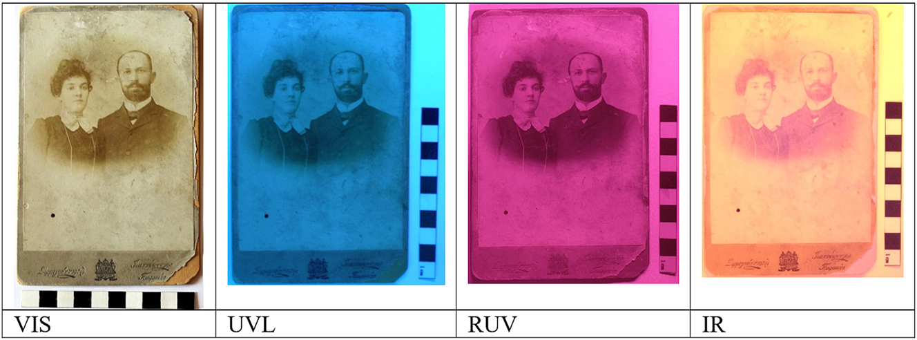
Macro imaging technique of photograph no. 18.
3.1.2 Microscopic Imaging Techniques
Micrographs were produced using two microscopes (Dino-Lite) that can capture images in the visible region (400–780 nm), UV at 395 nm, IR at 940 nm, and images including UV-induced radiation between 400 nm and 430 nm (LED excitation lights at 400 nm and emission filter cut at 430 nm) as shown in Table 3. Both microscopes capture images at magnifications between 20x and 200x.
Light sources of microscopes and associated multispectral imaging (Kokla 2024).
| Imaging technique | Radiation source |
|---|---|
| Visible light imaging (VIS) | 1 × white (VIS) LED |
| Ultraviolet-induced visible luminescence imaging (UVL) | 7 × violet (400 nm) LEDs with 430 nm emission filter |
| Ultraviolet-reflected imaging (RUV) | 4 × UV (395 nm) LEDs |
| Infrared-reflected imaging (IR) | 4 × IR (940 nm) LEDs |
Imaging at 70x and 200x magnifications was chosen because it provided features that contributed to better observation of the image surface, materials and manufacturing. An example of microscopic multispectral imaging is shown in Figure 2.

Micro imaging technique of photograph no. 6 at magnification 200x.
3.2 SEM/EDS Analysis
A JEOL JSM-6510LV scanning electron microscope (SEM) coupled to the Oxford X-act system energy dispersive X-ray spectrometer was used. Photographs were then examined under high vacuum conditions (40 Pa), using an accelerating voltage of 15 kV and a working distance of 10 mm. Images of photographs were taken at magnifications between 500x and 1,500x varying according to the features observed. Elemental analysis was carried out directly on the surface of the photographs, providing information on the elements involved in the composition of the photographs and the stratigraphy of the photographic print. Both black and white areas of each photo were analyzed.
4 Results
4.1 Macroscopic Imaging Techniques
Macroscopic imaging provided information on earlier retouching, stains, dirt, and surface damage, clearly showing the extent of damage and more generally the state of preservation, hand-coloring as well as photographic surface features such as cracks and layers of varnish. Figure 3 shows macroscopic images of photograph no. 19. Four images were taken in four different bands. Images including UV-induced visible luminescence and reflected UV radiation show areas of surface deterioration. A stain next to the man’s face as well as water stains around most of the edges of the photograph can be clearly distinguished.

Macroscopic images of photograph no. 19.
Figure 4 shows photograph no. 16 in the four different bands of the electromagnetic spectrum. One of the most important results is provided by the reflected UV image, in which a layer on top of the emulsion can be detected. This layer may be a varnish.

Macroscopic images of photograph no. 16.
4.2 Microscopic Multispectral Imaging
Microscopic imaging techniques provided information on the presence of retouching, the type of stains, hand colorings, the stratigraphy of photographs, surface texture, and information about the layer underneath the emulsion, especially in bright areas.
Figure 5 shows retouching of the eyes and eyebrows using black color (photograph no. 11) and white color on the clothes (photograph no. 4). The retouched black lines are clearly visible in the Infrared Reflectography, while the retouched white color can be distinguished in reflected UV and UV-induced visible luminescence images. The third example (photograph no. 23) shows microscopic multispectral imaging techniques in which the retouching applied to the eyes, the characteristic texture of the photographic paper surface, areas of loss in the emulsion and oxidation can be distinguished.

Micro multispectral imaging (magnification 70x).
Figure 6 shows the surface texture of the photographic image, a characteristic feature of albumen photographs. Furthermore, the stratigraphy was visible in areas of emulsion loss, where the emulsion appears to be directly on the paper support without another layer between, which is typical for albumen photographs.

Photograph no. 16, micro multispectral imaging (magnification 200x). This albumen photograph detail shows the characteristic texture of aged albumin in the upper row and its double stratigraphy in the bottom row, which consists of the emulsion and the paper base.
4.3 SEM/EDS Analysis
The SEM/EDS analysis provided information on the stratigraphy of the photographic paper, the type of the emulsion and the underlying layer, the type of the photographic paper and the manufacturing technique, as well as the possible existence of toning. Figure 7 shows photograph no. 14, the scanning electron microscope image 600x and the results of elemental analysis. Elemental analysis shows the presence of gold (Au) that had been used as a toner. Furthermore, silver and chlorine indicate that the photosensitive emulsion is probably composed of silver chloride. The SEM image shows the stratigraphy of the photograph and the thickness of each layer. The paper support is the thickest layer, followed by the baryta layer, and the photosensitive emulsion that is the thinnest layer.

SEM/EDS analysis of photograph no. 14. The chemical analysis indicates that a) the emulsion was made from silver chloride (AgCl), and b) gold (Au) was used for toning of the photograph.
In Table 4, the chemical elements detected in the studied photographs are presented. The elements considered to have been used as toners in each case are highlighted with bold numbers.
Elements detected in the photographs using elemental analysis.
| photo | Detected elements (%) | |||||||||||
| N | Ag | Cl | Ca | S | Mg | Si | Na | Al | K | Ba | other | |
|
|
||||||||||||
| 1 | 20.49 | 7.12 | 0.30 | 0.32 | 1.41 | 0.15 | 0.18 | 0.10 | 0.10 | Cr:(0.09) | ||
| 2 | 13.98 | 3.15 | 0.26 | 0.51 | 1.05 | 1.74 | 0.12 | |||||
| 3 | 22.78 | 4.81 | 0.20 | 0.32 | 1.32 | 0.36 | 0.14 | 1.47 | ||||
| 4 | 22.62 | 5.82 | 0.45 | 0.24 | 1.18 | 0.21 | 0.69 | |||||
| 5 | 3.65 | 0.58 | 0.68 | 3.41 | 0.39 | 0.39 | 7.78 | Fe: 0.23 | ||||
| 6 | 1.78 | 0.62 | 0.82 | 1.18 | 0.15 | 0.46 | 0.37 | 0.81 | 0.18 | 0.55 | Fe: 0.13 | |
| 7 | 23.18 | 5.09 | 0.51 | 0.48 | 0.99 | 0.09 | ||||||
| 8 | 1,592 | 4.98 | 0.45 | 0.28 | 1.36 | 0.13 | 0.15 | 0.34 | 0.16 | 0.24 | 0.31 | |
| 13 | 22.29 | 1.52 | 0.24 | 0.14 | 1.09 | 0.06 | 0.12 | |||||
| 14 | 21.01 | 1.19 | 035 | 0.46 | 1.48 | Au: 1.24 | ||||||
| 15 | 0.30 | 7.09 | 0.21 | 32.10 | Co: 0.05 | |||||||
| 16 | 3.55 | 3.39 | ||||||||||
| 17 | 7.25 | 35.54 | Co: 0.13 | |||||||||
| 18 | 0.33 | 0.33 | 0.30 | 0.22 | 0.94 | |||||||
| 19 | 0.38 | 4.93 | 0.22 | 0.19 | 21.75 | |||||||
| 23 | 5.09 | 0.94 | 1.28 | 27.17 | ||||||||
-
Bold highlights indicate the items that were used as toner.
In most cases, the detection of silver (Ag) and chlorine (Cl) indicates that the photosensitive surface (emulsion) consists of silver chloride if no other halogen is detected. The presence of barium (Ba) and sulfur (S) is characteristic of the baryta layer (BaSO4), which is the middle layer in a triple stratigraphy[1] of photographic paper. The detection of calcium is associated with the paper pulp. The presence of sodium (Na), potassium (K) and sulfur (S) are traces of chemical fixation baths (Eynard 1962; Pope 1960) sodium thiosulfate and potassium thiosulfate. Sometimes, sulfur was also used as a toner. In some other cases, the detection of gold (Au) (photograph no. 14) and iron (Fe) (photograph no. 5 and 6) indicate the use of toner in those photographs. Chromium (Cr) has sometimes been used to harden photographic gelatin (Hollander 1941). The use of cobalt (Co) in the production of historic photographs is unknown.
Detected elements such as aluminum (Al), silicon (Si) and magnesium (Mg) could derive from the dust or other materials that have been applied during the creation of the photograph, such as aluminum containing pigments used for retouching in some white areas of the photographic image. Elements that indicate the application of a toner were detected in only eight of the sixteen photographs. Sulfur was detected as a toner in five of the studied photographs, iron in two, and gold in one (Table 5).
Photographs and detected elements indicating toning baths.
| Toning using sulfur | Toning using iron | Toning using gold | |
|---|---|---|---|
| Catalogue number of photographs | 1, 2, 7, 13, 16 | 5, 6 | 14 |
In most photographs (10), silver chloride was detected. In 5 photographs, no silver halide could be determined, which could lead to the conclusion that a different type of photosensitive emulsion and a different technique, such as the carbon printing technique was used (Table 6).
Suggested Manufacturing techniques based on elemental analysis.
| Silver chloride | Other technique | |
|---|---|---|
| Catalogue number of photographs | 1, 2, 3, 4, 5, 6, 7, 8, 13, 14 | 15, 16, 17, 18, 19, 23 |
In 10 of the 16 photographs studied, the elements barium (Ba) and sulfur (S) were detected indicating the existence of a baryta layer beneath the photographic emulsion. This layer was identified in the SEM images as part of the stratigraphy of most photographs (photographs no. 3, 4, 5, 6, 8, 15, 17, 18, 19, 23). In the scanning electron microscope (SEM), all photosensitive surfaces show small granulometry, while the stratigraphy of photographs could best be identified in the microscope. 12 photographs have 3 layers (Table 7), 11 of them a baryta layer and one a calcium carbonate layer underneath the emulsion. 5 photographs have 2 layers.
Stratigraphy of photographs.
| One layer | Two layers | Three layers | |
|---|---|---|---|
| Catalogue number of photographs | – | 2, 7, 13, 16 | 1, 3, 4, 5, 6, 8, 14, 15, 17, 18, 19, 23 |
5 Conclusions
The 16 photographs belonging to Greek private collections were studied combining a literature review, the application of multispectral imaging and SEM/EDS analysis. The study aimed at detecting whether the photographs were toned and if so, which materials were used in the toning process. The methodology also revealed the stratigraphy of the photographs, elements used in the toning process, the photographic technique and retouching.
Historical literature provides important information on the toning process, the materials used, the hue of photographs after the process, as well as the photographic techniques used in the 19th and early 20th centuries. Multispectral imaging provided information on emulsion surface texture, the surface texture of photographic prints, damage phenomena (cracks, dirt, scratches, loss of emulsion), image features (retouching), the baryta layer (BaSO4) as well as stratigraphy. SEM/EDS yielded further information about the stratigraphy of the photographic paper and the thickness of layers, the photosensitive layer (emulsion), and the chemical elements used in the manufacture of the photographs.
It is important to document toned photographs in collections and to gain further information on the type of toning in order to better protect and preserve them. It is also important to separate toned photographs from photographs that are not toned but have a similar hue which may have been caused by damage phenomena rather than a toning process (Rogge and Bezur 2013).
References
Abbaspour Mitra, Jim, Coddington, Lee, Ann Daffner, Maria, Morris Hambourg. 2014. “Object:Photo. Modern Photographs: The Thomas Walther Collection 1909–1949 at the Museum of Modern Art.” moma.org/objectphoto: Surface Analysis in Object:Photo. Modern Photographs: The Thomas Walther Collection 1909–1949 at The Museum of Modern Art. https://www.moma.org/interactives/objectphoto/materials/surface_analysis.html (accessed December 8, 2014).Search in Google Scholar
Adams, A. 2001. The Ansel Adams Photography Series 3: The Print. NY: Little, Brown and Company.Search in Google Scholar
Burgess, G. N. 1863. The Photograph Manual: A Practical Treatise Containing the Cartes de Visite Process, and the Method of Taking Stereoscopic Pictures, Including the Albumen Process, the Dry Collodion Process, the Tannin Process, the Various Alkaline Toning Baths, etc., etc. New York: D. Appleton & Co.Search in Google Scholar
Casoli, A., and S. Fornaciari. 2014. “An Analytical Study on an Early Twentieth-Century Italian Photographs Collection by Means of Microscopic and Spectroscopic Techniques.” Microchemical Journal 116: 24–30, https://doi.org/10.1016/j.microc.2014.04.003.Search in Google Scholar
Cattaneo, B., D. Chelazzi, R. Giorgi, T. Serena, C. Merlo, and P. Baglioni. 2008. “Physico-chemical Characterization and Conservation Issues of Photographs Dated between 1890 and 1910.” Journal of Cultural Heritage 9: 277–84, https://doi.org/10.1016/j.culher.2008.01.004.Search in Google Scholar
Chen, Jiuan Jiuan. 2001. “Documenting Photographs: A Sample Book.” Master Thesis, Art Conservation Department, Buffalo State College, NY.Search in Google Scholar
Chen, Jiuan Jiuan, and J. Theresa Smith. 2020. “Documentation of Salted Paper Prints with a Modified Digital Camera.” Journal of the American Institute for Conservation 59 (3–4): 271–85. https://doi.org/10.1080/01971360.2019.1643527.Search in Google Scholar
Costa, L., M. Nunes, S. Costa, M. Trindade, C. Miguel, and T. Ferreira. 2018. “Unveiling the Ambrotype: Characterization of Two 19th Century Photographs.” Microscopy and Microanalysis 25: 203–13, https://doi.org/10.1017/s1431927618000429.Search in Google Scholar
DesChamps, Julie Lattin. 2005. “Coatings on Gelatin Silver Prints: A Historical Overview in Coatings on Photographs.” In Materials, Techniques, and Conservation, edited by Constance McCabe, 140–55. Washington, D.C.: American Institute for Conservation.Search in Google Scholar
Ellis, P. 1975. “Gold in Photography. Evolution from Early Artistry to Modern Processing.” Gold Bulletin 8: 7–12. https://doi.org/10.1007/bf03215055.Search in Google Scholar
Eynard, R. 1962. Fixation and Washing Techniques for Increasing Image Stability in Photographic Papers. Thesis dissertation. Rochester, New York: Rochester Institute of Technology, School 0f Photographic Arts and Science College of Graphic Arts and Photography.Search in Google Scholar
Fischer, C. Monique, and S. Sarah Wagner. 2005. “An Overview of Coatings on Hand-Colored Black-And-White Photographs.” In Coatings on Photographs. Materials, Techniques, and Conservation, edited by Constance McCabe, 156–67. Washington, D.C.: American Institute for Conservation.Search in Google Scholar
Gmuender, Ch. 1992. “On Black-And-White Paper Image-Stability Enhancement: Effectiveness of Toning Treatments on Silver Gelatin Prints Determined by the Hydrogen Peroxide Fuming Test.” In MA Degree in Imaging Arts School of Photographic Arts and Sciences Rochester. New York: Rochester Institute of Technology.Search in Google Scholar
Hardwich, T. F. 1859. “Gold Toning on Albumenized Paper.” The Photographic and fine art Journal: 196–202.Search in Google Scholar
Haydn Smith, I. 2018. The Short Story of Photography. U.K: Orion Publishing Co.Search in Google Scholar
Hedgecoe, John. 1992. The Photographer’s Handbook, 3rd ed. New York: Alfred A. Knopf.Search in Google Scholar
Herrera Garrido, R., S. de Groot, and T. Callewaert-Dore. 2020. “The Coated Salted Paper Prints from the Eduard Isaac Asser Collection at the Rijksmuseum: FTIR and OCT Identification and Characterization.” Journal of the American Institute for Conservation 59 (3–4): 246–61. https://doi.org/10.1080/01971360.2020.1774725.Search in Google Scholar
Hess Norris, Debra, and Nora Kennedy. 2005. The Conservation Treatment of Original Coatings on Photographs: Issues and Current Practice, in Coatings on Photographs. Materials, Techniques, and Conservation, edited by Constance McCabe, 12–23. Washington, D.C.: American Institute for Conservation.Search in Google Scholar
Hollander, S. C. Hardening of Photographic Gelatin. US Patent 2359217, 1941.Search in Google Scholar
Hughes, J. 1863. The principles and Practice of Photography Familiarly Explained: Being a Manual for Beginners, and Book of Reference for Expert Photographers: Comprising the Collodion Process, Printing and Toning, Dry-Plate Photography, Including All the Best Processes, Intense Iron Developers, Transparencies for the Magic Lantern, Instantaneous Photographs, How to Produce Life Size Portraits, Defects, Failures, and Remedies, &c. &c. London: Published by the author.Search in Google Scholar
Hunt, R. 1857. A Manual of Photography. London: Richard Griffin And Company.Search in Google Scholar
Jones, B. E. 1911. Cassell’s Cyclopedia of Photography. N.Y.: Cassel and Co.Search in Google Scholar
Kirby, D. P., A. Manick, and R. Newman. 2019. “Minimally Invasive Sampling of Surface Coatings for Protein Identification by Peptide Mass Fingerprinting: A Case Study with Photographs.” Journal of the American Institute for Conservation 59 (3–4): 235–45. https://doi.org/10.1080/01971360.2019.1656446.Search in Google Scholar
Kokla, V. 2024 (Forthcoming). “The Use of the Multiband Imaging to the Standardization of Damage Terms of Old Photographs.” In VOL. 4 - Multiband Imaging Techniques With Silicon-Based Sensors, Conservation 360°. Spain: Universidad Politécnica de Valencia.Search in Google Scholar
Kokla, V., Tselikas, A. 2015. “Damage Documentation of Old Photographs – Visual Guide of Deteriorations.” In Institute ARETHAS–MIEPABIK, financed by J. F. Kostopoulos Foundation. Greece: Institute ARETHAS–MIEPABIK. (in Greek).Search in Google Scholar
Lampray & Tibbitts. Toning formulae and Instructions for Printing with the New Super-albumenized Saxe and Rive Papers and Sutton’s Patent Albumenized Paper, 1860–9. London: Lampray, Tibbitts & Co.Search in Google Scholar
Lavédrine, B., J. Gandolfo, and S. Monod. 2003. A Guide to the Preventive Conservation of Photograph Collections. Los Angeles, CA: Getty Conservative Institute.Search in Google Scholar
Lavédrine, B., J. Gandolfo, J. McElhone, and S. Monod. 2009. Photographs of the Past: Process and Preservation. Los Angeles, CA: The Getty Conservation Institute.Search in Google Scholar
Le Gray, G. 1859. “A New Method of Toning with Chloride of Gold.” The photographic News 1 (22): 253.Search in Google Scholar
Leyshon, William E. 2001. Photographs from the 19th Century: A Process Identification Guid, Fotofind Program. Denver: Denver Public Library History.Search in Google Scholar
McCabe Constance. 2014. “Noble Metals for the Early Modern Era: Platinum, Silver-Platinum, and Palladium Prints.” In Object: Photo. Modern Photographs: The Thomas Walther Collection 1909–1949. An Online Project of The Museum of Modern Art, edited by Mitra Abbaspour, Ann Daffner Lee, and Maria Morris Hambourg. New York: The Museum of Modern Art. http://www.moma.org/interactives/objectphoto/assets/essays/McCabe.pdf.Search in Google Scholar
Messier, Paul, Valerie Baas, Diane Tafilowski, and Lauren Varga. 2005. “Optical Brightening Agents in Photographic Paper.” Journal of the American Institute for Conservation 44 (1): 1–12, https://doi.org/10.1179/019713605806082392.Search in Google Scholar
Mintie Katherine, Paul Messier, and Damon Crockett. 2024. “Closer Looking: Computer Vision in Material Studies of Art.” Art Bulletin 106 (2): 29–32, https://doi.org/10.1080/00043079.2024.2296276.Search in Google Scholar
Murphy, L. Erin. 2017. “Overview of Historical Practices for Postprocessing Toning and Intensifying Platinum Prints.” In Platinum and Palladium Photographs: Technical History, Connoisseurship, and Preservation, edited by Constance McCabe, 218–31. Washington, D.C.: American Institute for Conservation of Historic and Artistic Works.Search in Google Scholar
Namde, Ronel, and M. Joan Walker. 2017. “Platinum Toning of Silver Prints.” In Platinum and Palladium Photographs: Technical History, Connoisseurship, and Preservation, edited by Constance McCabe, 186–91. Washington, D.C: American Institute for Conservation of Historic and Artistic Works.Search in Google Scholar
Panadero, Laura, Katherine Eremin, and Elena Bulat. 2020. “X-Ray Fluorescence Analysis of Frédéric Flachéron’s Paper Negatives, 1848–1852.” Journal of the American Institute for Conservation 59 (3–4): 186–93, https://doi.org/10.1080/01971360.2020.1834957.Search in Google Scholar
Penichon, S. 1999. “Differences in Image Tonality Produced by Different Toning Protocols for Matte Collodion Photographs.” Journal of the American Institute for Conservation 38 (2): 124–43. https://doi.org/10.2307/3180042.Search in Google Scholar
Perron, Johanne. 1989. “The Use of FTIR in the Study of Photographic Materials.” Topics in Photographic Preservation 3: 112–22.Search in Google Scholar
Pope, I. C. 1960. “Formation of Silver Sulfide in the Photographic Image during Fixation.” Journal of Research of the National Bureau of Standards-C. Engineering and Instrumentation 4 (1): 65–73. https://doi.org/10.6028/jres.064c.010.Search in Google Scholar
Reilly, J. M. 1980. The Albumen & Salted Paper Book: The history and Practice of Photographic Printing, 1840–95. Rochester, N.Y: Light Impressions Corp.Search in Google Scholar
Rogge, Corina E., and Anikó Bezur. 2013. “19th Century Photography in a Modern Chemistry Lab.” Topics in Photographic Preservation 15: 61–71.Search in Google Scholar
Rogge, Corina, and Krista Lough. 2016. “Fluorescence Fails: Analysis of UVA-Induced Visible Fluorescence and False-Color Reflected UVA Images of Tintype Varnishes Do Not Discriminate between Varnish Materials.” Journal of the American Institute for Conservation 55: 138–47. https://doi.org/10.1080/01971360.2016.1155813.Search in Google Scholar
Romani, M., L. Pronti, C. Severini L. Ruberto, C. Mazzuca, G. Viviani, A. Mazzinghi, M. Chiari, et al.. 2022. “Toward an Assessment of Cleaning Treatments onto Nineteenth–Twentieth-Century Photographs by Using a Multi-Analytic Approach.” European physical journal plus 137: 757. https://doi.org/10.1140/epjp/s13360-022-02948-5.Search in Google Scholar
Schriever, J. B. 1909. Complete Self-Instructing Library of Practical Photography, Vol. 1–5. Scranton, Pa: American School of Art and Photography.Search in Google Scholar
Sherman, W. H. 1892. The Past and Future of the Albumen Print, the American Annual of Photography and Photographic Times Almanac, 23–5. N.Y.: Scovill & Adams.Search in Google Scholar
Smith, B. 1904. Toning Bromide and Other Devepoped Silver Prints. London: Iliffe & Sons.Search in Google Scholar
Stulik, D., and A. Kaplan. 2013. The Atlas Of Analytical signatures of Photographic Processes. Los Angeles, CA: Getty Conservation Institute.Search in Google Scholar
Vogel, H. W. 1875. Handbook of the Practice and Art of Photography. Philadelphia: Benerman & Wilson.Search in Google Scholar
WagnerSarah, Matthew L. Clarke, and Joan M. Walker. 2020. “Linnaeus Tripe and Lightly Albumenized Prints in the 1850s: Characterization, Analysis and Process Identification.” Journal of the American Institute for Conservation 59 (3–4): 218–34, https://doi.org/10.1080/01971360.2019.1609824.Search in Google Scholar
Walker, D. 1986. Ernie: Black-And-White Photographic Chemistry. U.S.A.: National Aeronautics and Space Administration. NASA TM-87296.Search in Google Scholar
Wall, E. J. 1924. Photographic Facts and Formulas. Boston: American Photographic Publishing Co.10.5962/bhl.title.38124Search in Google Scholar
Ware, M. 2017. “The Technical History and Chemistry of Platinum and Palladium Printing.” In Platinum Mike and Palladium Photographs: Technical History, Connoisseurship, and Preservation, edited by Constance McCabe, 46–83. Washington, D.C: American Institute for Conservation of Historic and Artistic Works.Search in Google Scholar
Watt, A. 1859. “Toning with Platinum.” The Photographic News 2 (44): 205.Search in Google Scholar
Zamboni, C. B., M. M. Redígolo, V. T. Miura, I. Costa, M. L. E. Nagai, P. A. V. Salvador, and D. Giovanni Nogueira da Silva. 2021. “Non-destructive Analysis in the Study of Historical Photographs by pXRF and ATR-FTIR Spectroscopies.” Journal of Forensic Sciences 66: 1048–55. https://doi.org/10.1111/1556-4029.14680.Search in Google Scholar
© 2024 the author(s), published by De Gruyter, Berlin/Boston
This work is licensed under the Creative Commons Attribution 4.0 International License.
Articles in the same Issue
- Frontmatter
- Original Works
- Techniques for Filling Losses in Palm Leaf Manuscripts
- Study of Toned Photographs of the Late 19th and Early 20th Century Using Multispectral Imaging and SEM/EDS Analysis
- Arsenic Pigments in Libraries – Testing Methods, Contaminated Spaces, and Occupational Safety Measures
- Article
- Critical Paradigm Approach in Preservation and Conservation Studies: An Initial Note