Diaphragmatic peritoneal metastases mimicking liver metastases
-
Barbara Noiret
 and
Clarisse Eveno
and
Clarisse Eveno

A 27-year-old male presented with abdominal pain and asthenia. Abdominal CT-scan identified left colon mass associated with synchronous peritoneal metastases (PM) and hypodense liver suspicious lesions (Figure 1A, arrow). Colonoscopy with pathological analysis of colic biopsies confirmed an adenocarcinoma with microsatellite instability, allowing to perform neoadjuvant immunotherapy (Pembrolizumab). Explorative laparotomy confirmed the PM with the presence of implants on the right diaphragmatic dome (Figure 1B, arrow) exerting extrinsic compression of the liver (Figure 1B, star). A complete cytoreductive surgery combined with Mitomycin C based-hyperthermic intraperitoneal chemotherapy (HIPEC) was performed.
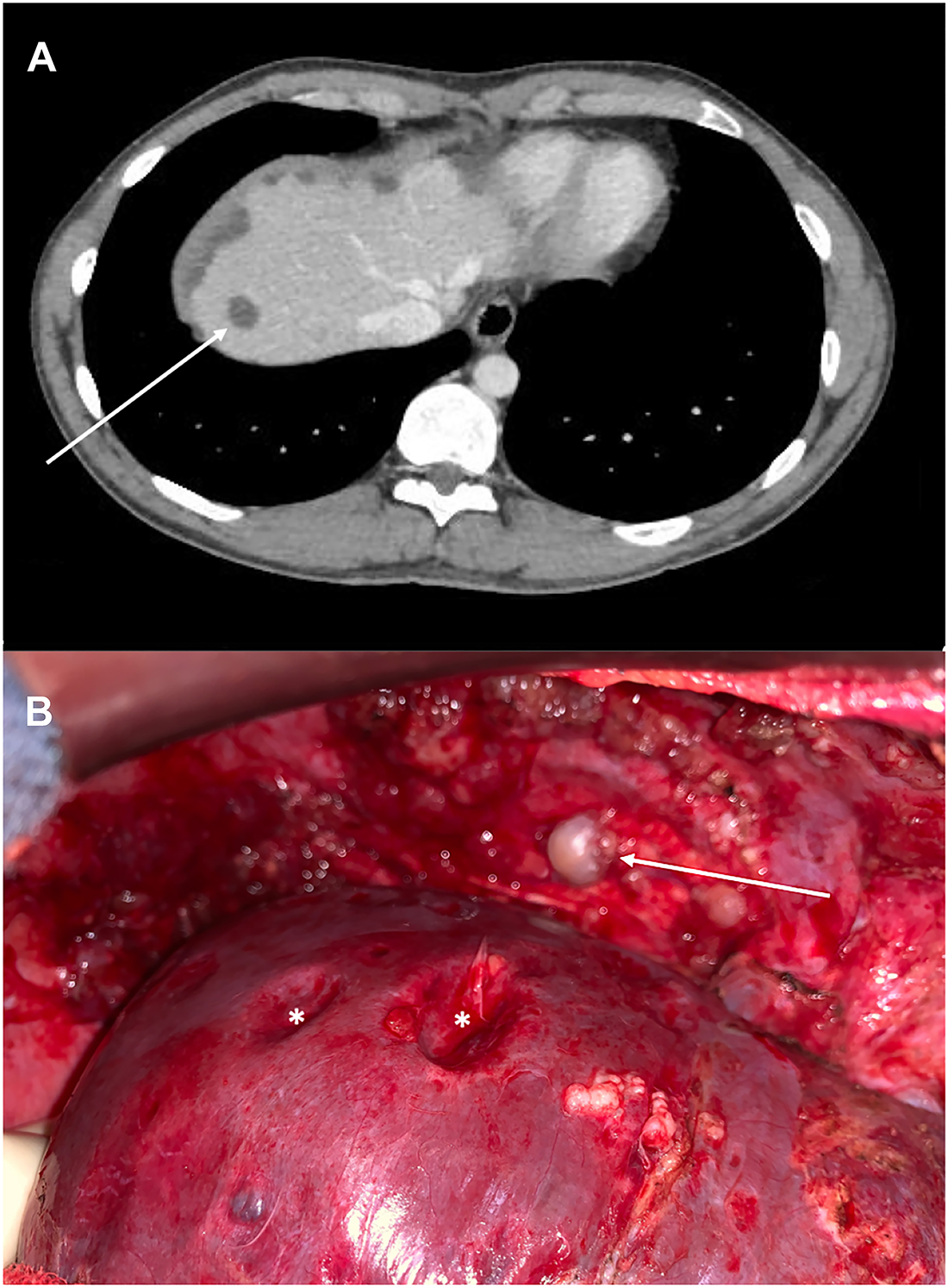
CT-scan (A) revealed hypodense liver lesions (arrow) at the diagnosis of left colon adenocarcinoma with synchronous peritoneal metastases. Explorative laparotomy (B) highlighted diaphragmatic peritoneal metastases (arrow) exerting pressure on the liver surface, called «scalloping» (star).
PM are often located in Douglas pouch, parieto-colic gutters, and also in the subphrenic space. Therefore, peritoneal tumor implants under the diaphragm can mimic liver metastases on CT-scan [1, 2] by exerting pressure on the liver surface, called “scalloping” [3]. It is essential to distinguish diaphragmatic PM and liver metastases and to carry out further investigations in case of doubt, because of their different therapeutic strategies.
-
Research funding: None declared.
-
Author contributions: All authors have accepted responsibility for the entire content of this manuscript and approved its submission.
-
Competing interests: Authors state no conflict of interest.
-
Informed consent: Informed consent was obtained from all individuals included in this study.
-
Ethical approval: Not applicable.
References
1. Diop, AD, Fontarensky, M, Montoriol, P-F, Da Ines, D. CT imaging of peritoneal carcinomatosis and its mimics. Diagn Interv Imaging 2014;95:861–72. https://doi.org/10.1016/j.diii.2014.02.009.Search in Google Scholar PubMed
2. Harimoto, N, Shimada, M, Shirabe, K, Tanaka, S, Taketomi, A, Tsujita, E, et al.. Images of interest. Hepatobiliary and pancreatic: peritoneal dissemination mimicking liver metastases. J Gastroenterol Hepatol 2004;19:938. https://doi.org/10.1111/j.1440-1746.2004.03569.x.Search in Google Scholar PubMed
3. Allart, K, Sabbagh, C, Regimbeau, J-M. Liver scalloping: pseudomyxoma peritonei liver compression. J Vis Surg 2019;156:75–6. https://doi.org/10.1016/j.jviscsurg.2018.09.001.Search in Google Scholar PubMed
© 2021 Barbara Noiret and Clarisse Eveno, published by De Gruyter, Berlin/Boston
This work is licensed under the Creative Commons Attribution 4.0 International License.
Articles in the same Issue
- Frontmatter
- Research Articles
- The ISSPP PIPAC database: design, process, access, and first interim analysis
- Enhanced recovery after surgery (ERAS) in cytoreductive surgery (CRS) and hyperthermic intraperitoneal chemotherapy (HIPEC): a cross-sectional survey
- Histological regression of gastrointestinal peritoneal metastases after systemic chemotherapy
- A human ex vivo coculture model to investigate peritoneal metastasis and innovative treatment options
- Influence of pre-analytical sample preparation on drug concentration measurements in peritoneal tissue: an ex-vivo study
- Clinical Images
- Diaphragmatic peritoneal metastases mimicking liver metastases
- Congress Abstracts
- ISSPP 2021 2nd Congress of the International Society for the Study of Pleura and Peritoneum