Design of a robot system for improved stress classification using time–frequency domain feature extraction based on electrocardiogram
-
Vikas Malhotra
 ,
Gurpreet Singh Saini
,
Gurpreet Singh Saini

Abstract
In recent days, stress is a major phenomenon that adversely affects both individuals and communities. The research in computing the stress factor has wider advantages as it improves personal learning, learning operations, and high productivity that benefits society. Several computational techniques come into concern to avoid and reduce the stress level using the electrocardiogram (ECG) signals. In this study, the stress level was classified using the feature extraction approach in combination with the classifier. The signal is processed using the variational mode decomposition denoising technique to reconstruct the original signal. The decomposed signal was further extracted using the time–frequency domain technique as characteristics of the ECG signal such as R-wave and T-wave constructed. Further, the support vector machine classifier was used to classify the stress level (low, medium, and high) of the extracted signal. Based on stress classification outcomes, the robot offers a range of personalized interventions to users. These interventions include relaxation exercises, deep breathing techniques, or guided mindfulness sessions. The average accuracy obtained using the proposed technique is 98.98% but without using the feature extraction technique, it is 97.71%. The other performance parameters also get improved and the results are finally compared with the existing techniques.
1 Introduction
Stress is a phenomenal factor that affects individuals physically and mentally in real life. Various techniques presently, based on various physiological parameters such as skin conductance, heart rate variability (HRV), electroencephalogram (EEG), electromyogram (EMG), electrocardiogram (ECG), Sleep Pattern, Galvanic Skin Response (GSR), and Skin Temperature came into the concern to determine the stress level using the physiological signals [1,2]. The conventional studies show a quantitative method to monitor the acute stress level and recognition of emotions through the ECG signals. There are various positive sides of using these signals to recognize the stress level: the recognition of heart activity, high security as these signals are difficult to copy, and easy collection of signals [3]. Additionally, ECG signals play a paramount role in discriminating the human stress level namely depression, disorders, and bipolar anxiety. The regulation of stress through the Nervous system via physiological measurement systems determines the heart rate; breathing frequency, respiration rate, and blood pressure. The various stress level measurement systems are EMG, GSR, and Heart Rate Variability. These methods are considered as accurate ones to record the bio-signals except those are not masked by human actions.
Specifically, it is an effective indicator to diagnose several medical problems related to heart, stress, and emotions. In any type of research, ECG signals need to be extracted using different extraction techniques such as principal component analysis (PCA), genetic algorithm (GA), and many more [4]. ECG signals are recognized in three different domains: time, frequency, and time–frequency domain (TFD). Time domain generally portrays the R peak value, R–R intervals, standard deviation, N–N intervals, etc. Consequently, the frequency domain portrays the spectral analysis such as power spectral density. The TFD features the discrete wavelet transform and discrete wavelet packet transform. The last one is the most popular method to extract the features from low-range to high-range components. Researchers use the time–frequency characteristics to decompose the ECG signals and the proposed method yields 93.7% accuracy [5].
Figure 1 represents the ECG stress classification system. The extracted data time domain features are fed as input to the ECG stress classification system. The extracted features are classified into three different classes namely: negative emotions, positive emotions, and neutral. The stress level that lasted long and continuous belongs to negative emotions and changes further reflected through muscle tension, heart rate, and respiration rate. These are used as input data for the non and emotional stress classification system. Additionally, classifiers such as support vector machine (SVM) were used to further classify the hyperplane set in a high-dimensional space. The classifiers used to determine this level are k-nearest neighbor (KNN) and SVM. The classification process in both cases is different as KNN involves object classification through the nearest neighbor and SVM involves hyperplanes defined in an infinite-dimensional space. Statistical analysis shows that mean, variance, maximum, minimum, and standard deviation are widely used to analyze ECG signals.
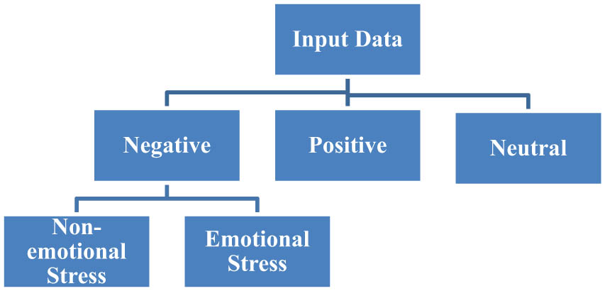
ECG stress classification.
Figure 2 represents stress response in four stages such as baseline, moderate, hard, and moderate tasks. Stage 1 and stage 2 signify the stressed conditions. The ground truth is designed under stressful conditions. The baseline period signifies the no-stress conditions. Additionally, low stress is defined by the baseline period and moderate and high-level stress by stage 1 and stage 2. In the given period, stress is not defined by a specific period but is defined as continuous stress assessment. The given curve in the figure is also called as stress curve. ECG signals are generally complex as noise persists, which needs to be filtered through denoising techniques and filters. The decomposition level of the signal corresponds to the noise level and approximation level. Variational mode decomposition (VMD) reconstructs the original signal by decomposing it into a sum of variational mode functions [6]. Further, ECG signals collected through wearable devices may have fewer chances to introduce noise as filters used by the sensor technology to efficiently filter the noise.
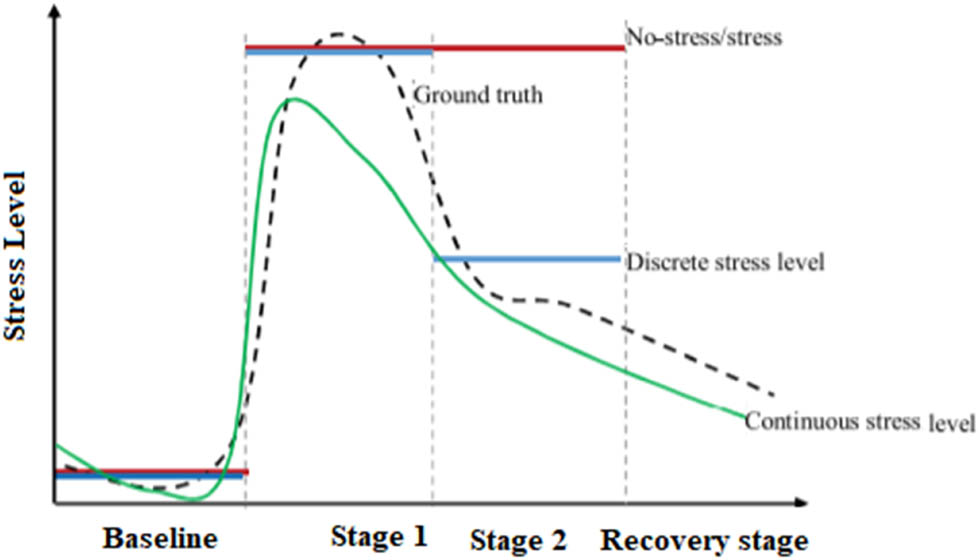
Discrete and continuous stress recognition.
Briefly, the main contribution of this research includes the following aspects:
Stress is classified continuously depending upon the individual emotional level.
The inter-subject differences are based on the stressful events using the developed model.
The continuous nature of the stress level is classified.
The integration of robotics and advanced physiological sensing techniques presents an innovative solution.
The main objective of this work is to determine the relationship of TFD features of ECG signals to classify stress. The other common method to deal with stress is to detect the levels such as low, medium, and high in two different states, stress or no stress. The conventional machine learning techniques determine the relaxation state (no stress) to stress state recording the ECG signals [7,8,9]. Other computational methods such as SVM can deal with these states and stress levels [10]. Additionally, complexities and gaps also persist that can be dealt with VMD technique to reconstruct the original signal. Additionally, by utilizing TFD feature extraction, the robot system can capture both temporal and spectral variations in ECG signals. This provides a more comprehensive view of how stress manifests in physiological responses, enabling a deeper understanding of stress patterns. The ability to process ECG data in real time allows the robot system to provide immediate stress classification results. This real-time monitoring enables timely interventions and helps users manage stress as it occurs. The proposed system not only provides accurate stress classification but also offers meaningful and supportive visual as well as auditory interactions with users, through robotic system, ultimately enhancing their well-being.
The robot uses natural prosodic patterns that mimics human-like communication, making the interaction more user-friendly and relatable. This leads to a more comfortable and effective user experience. The robot is capable of adjusting its tone to be more encouraging during high-stress situations or adopt a gentler tone during relaxation exercises.
This study is organized as follows: Section 1 begins with an introduction of the basic techniques used to determine the stress level using the biological signal. Section 2 describes the state-of-the-art techniques such as feature extraction approaches, feature selection, and feature classification techniques. Further Section 3 elucidates the research methodology that explains the denoising filter VMD to process the signal, feature extraction technique, feature classification approach, and human–robot interaction and feedback mechanism. Section 4 describes the discussion on results and finally, concludes in Section 5.
2 Literature review
In the literature, researchers use different techniques to determine the stress level using biological signals. The identification using the ECG signal gained attention due to high accuracy and sensitivity. The ECG signals are used to extract the features and then process the signal to detect the stress level using the classification techniques. Additionally, various techniques are compared to determine the robustness of the best approach in the existing study. For instance, Pourmohammadi and Maleki estimated the stress level on a continuous and quantitative basis depending upon the biological signals. The fuzzy technique is used to analyze the relationships between individual behavior and stress. In this research, stress assessment techniques are defined using the ECG and EMG signals to attain the required accuracy and mental stress factor. An experimental investigation was carried out that involved 34 healthy participants inducing stress. The perceived stress index was very high around 0.9. The average accuracy for two-level and three-level subjects was around 97 and 76%. The proposed approach was limited to classifying the stress level as the majority of the subjects fall on the positive side rather than emotional stress [11].
ECG signals are very sensitive and prone to different types of noise levels. In a survey, various denoising approaches were considered to filter the unwanted noise, such as quadrature filtering, low and high noise filtering, non-local means, adaptive noise cancellation technique, VMD, and empirical mode decomposition for filtering [12,13]. The determination of signal-to-noise ratio through the wavelet and thresholding scheme provides a better-denoised signal. The study provides accurate results but is limited to preserving the morphological information [14].
A multi-sensing system was proposed considering the target population of around 24 individuals who fall in the age group of 18–23 years. The stress tests carried out using the EEG and ECG signals were further processed using the Machine Learning Supervised techniques. The robustness of the proposed technique is acquired by developing the stress matrix that jointly avoids stress-related problems. Moreover, classification techniques were used to acquire the featured clusters for real-time stress monitoring. The drawback of the proposed approach is the general model performed worst which varies the stress sensitivity. So, there is a need for a subject-oriented model that discriminates the features as per stressful conditions [15].
Researchers had used the ECG signals considering the manual and automatic features under the different stressful conditions. The research was based on a three-step process that includes HRV features acquired using the ECG signals. Further, the Gaussian mixture model was used to assess the mental state of different subjects. A stress classification coefficient indicator was proposed that reduces when using the cluster centers to process the extracted features. The final results were obtained using the SVM that provides 95% accuracy of recognition of the stress signals. The average score achieved was 0.97 and provides the possibility of recognition under different conditions. But, still, some problems persist as the limitation of the number of testers and there is a need for improvement for stress-induced coefficient [3].
The study classifies the ECG signals into four different emotional states according to a proportion of stress levels. Further, Naïve Bayes algorithms of the SVM were used to compute the values of R–R interval, Q–T interval, and R–S peak value. The obtained values not only improve the accuracy of the stress classification system but also reduce the error. The performance measures used for validation are confusion matrix, classification error, and receiver operating characteristics. The average accuracy obtained using the Bayes algorithm was 97.6%, 8.7% improved from the conventional stress classification techniques [16].
2.1 Existing techniques used to determine the stress level
In literature, several techniques are commonly used to determine an individual’s stress level, drawing from physiological, psychological, and behavioral indicators. These techniques provide insights into the person’s overall stress response and aid in assessing their well-being as given below:
| Author | Technique | Drawback | Applications | Results |
|---|---|---|---|---|
| Tripathy et al. [17] | The use of VMD to pre-process the signal and further Random forest (RF) classifier had been used to classify the signal | The main drawback of this technique was that the value of the coefficient fluctuated during each mode of ECG signal detection | The proposed study can be useful for the measurement of cardiac ailments | The average attained performance metrics in this study were 97.23% for accuracy, 96.54% for sensitivity, and 97.97% for specificity |
| Jung and Yoon [18] | Multi-level assessment model for classification using SVM and fuzzy logic, reasoning using decision tree and random forest algorithm, and decision-making process using Expectation Maximization | It is difficult to sense and classify complex emotions | Classification of mental stress level in smart healthcare systems | The monitoring of health parameters such as HR, respiration rate, EEG, and blood pressure in a dynamic environment |
| Subhani et al. [19] | The study proposed the machine learning framework that includes feature extraction and selection, classification using logistic regression, Naïve Bayes, and SVM in the frequency domain. The study was validated using the 10-fold cross-validation system | The results of the proposed study were unbiased and hence there is a need for feature space to record the data | The development of computer-aided diagnostic equipment to measure the stress level for clinical use | The proposed system attains 83.4% accuracy for multi-level and about 95% for two-level stress recognition systems |
| Ahuja and Banga [8] | The proposed study uses the four classification algorithms such as linear regression, Random Forest, Naïve Bayes, and SVM to analyze the multi-level stress, and the Perceived Stress Scale test was used in addition | The use of four algorithms makes the system costly | The present study was supported to determine the mental stress in college students | The highest accuracy was obtained using the SVM classification technique 85.71% |
| Pourmohammadi and Maleki [9] | The use of EMG and ECG signals to detect the multi-level stress. The combination of machine learning and feature classification methods such as SVM was used | The major limitation of the presented study was the lack of evaluation in real-world applications | The study shows the effectiveness of the EMG signal in detecting binary-level stress and multi-level stress | The accuracy of the two, three, and four-level stress recognition system was 100, 98, and 96.2%, respectively |
| Patro et al. [20] | The study includes the machine learning algorithms such as GA and particle swarm optimization (PSO) in the frequency–time domain analysis. The extracted features had been classified using the SVM and RF technique | The considerable performance of the study shows when compared with other approaches | The accurate results were obtained in the case of a large database that was suitable for clinical use | The classification accuracy had been improved and recognition rates using GA with RF classifier shows 95.3% accuracy |
| Malhotra and Sandhu [21] | The method includes the use of three optimization techniques such as GA, artificial bee colony, and PSO. Further, the SVM classifier and VMD had been used to filter the noise | The study is limited to consider the small data size | The proposed study is perfect to solve the major problems of the Mental Health Assessment system | The average accuracy obtained in this study was 98.9%, precision was 9.83%, a recall was 96.83%, and specificity was 96.72% |
3 Research methodology
The stress detection algorithm includes the acquisition of signals from healthy participants who were induced to stress-related problems. Designing a robot system for improved stress classification using TFD feature extraction based on ECG is a complex task that involves multiple components and considerations. Integrating the robotic system with the stress classification methodology involves connecting the different modules and components to create a cohesive and functional system. The proposed robot system consists of three main components: the ECG data acquisition module, the feature extraction module, and the stress classification module. The proposed methodology is described in the flowchart as in Figure 3.
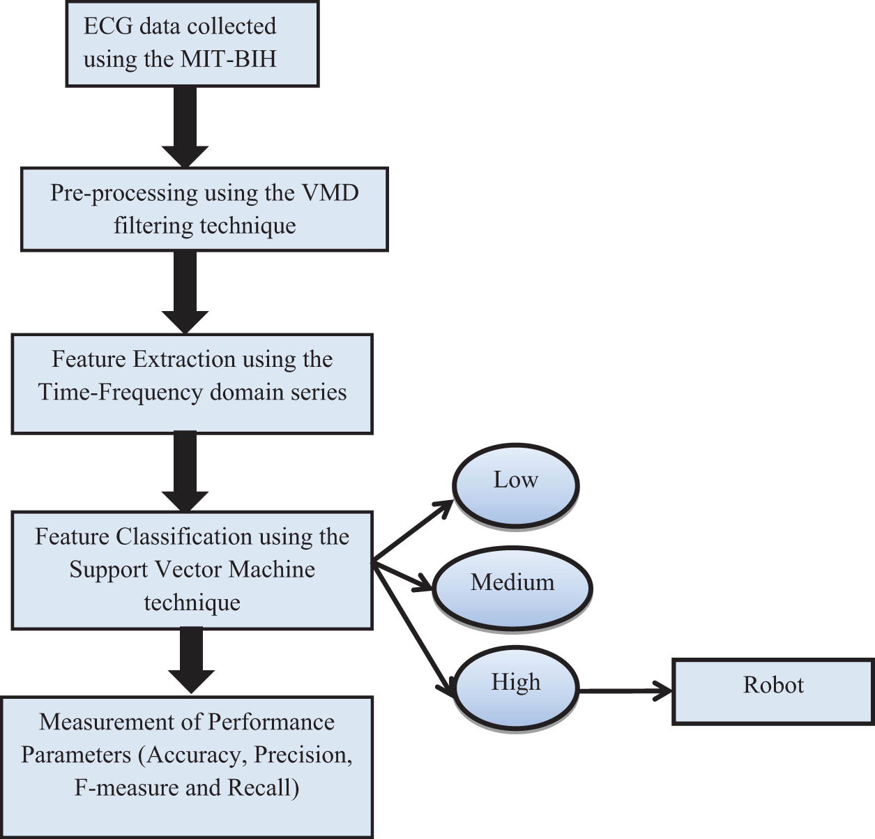
Flowchart of the proposed stress classification system.
3.1 ECG data acquisition
The dataset for the proposed methodology was collected from the PhysioNet; the dataset is free to access to motivate the practitioners for study. The investigational study includes the complex physiological signal analyzed in Boston’s Beth Israel Hospital and further evaluated by the Massachusetts Institute of Technology (MIT). The collective dataset is called the “MIT-BIH Arrhythmia Database” which contains 1,000 subjects nearly 48 half-hour ECG recordings [22,23]. The acquired ECG data is then passed to the subsequent modules for processing.
3.2 Pre-processing
The ECG signal collected from the dataset is pre-processed to shrink the noise level. The raw ECG signals are corrupted and diminished by external noise. Moreover, inappropriate placement of the subjects and skin preparation disrupt the obtained signal. In the literature, various pre-processing techniques have been used to process the signal such as filters, empirical models, and wavelet transform.
In this research, VMD is used to reconstruct the original signal fed at the input. The decomposition of the signal is carried out to convert the original signal into the variational mode functions. It is a robust technique to handle and control the noise level [12]. It decomposes the real values function of an input signal (f) into a discrete number of signals that are called variational mode functions (
Algorithm 1
Variational mode decomposition
Initialize
Repeat
For n = 1:N do
Update
Update
End for
Dual ascent for all
Until convergence
Each data point is indicated as
3.3 Feature extraction
In this paper, TFD features were extracted from the processed ECG signals. The ECG waveform is shown in Figure 4. These features are further used as input to the developed model. The detection accuracy improved by properly representing the characteristics of the extracted signal.
![Figure 4
ECG waveform [23].](/document/doi/10.1515/pjbr-2024-0003/asset/graphic/j_pjbr-2024-0003_fig_004.jpg)
ECG waveform [23].
3.3.1 Extraction using TFD
The characteristics of the stressful conditions extracted are further investigated to detect the stress level. The removal of unnecessary features using the Feature Extraction algorithms not only lowers the computational cost but also encompasses the required information [24]. The signal was processed further and extracted using the TFD extraction method.
3.3.2 R-wave detection
In this detection method, the denoised signal using the VMD is further processed to determine the length. The height of the R-wave must not go lower than 0.6 times the highest height which is considered standard height. Such R-wave is detected and then counter-started upto 100 ms as the probability to acquire the R–R interval is about zero.
The Pseudocode to detect the R-wave is:
Initialize the denoised ECG signal
Compute x = Max (filtered)
Set the position of the counter (P) to 0.
Set the height of the counter (H) to 0.
Set counter to 1.
While counter count < length (filtered signal)
Set the last position (A) to 0
Set the consecutive position (B) to 1
If filtered signal [counter] > 0.6*x
While filtered[last position]
Set (A) = (B)
Increment B by 1
End while
Increment P by 1.
Increment H by 1.
Set the R_P[P] = counter + B-1
Set the R_H[H] = filtered signal [counter + B-1]
Increment the counter by 100
End if
Counter increment to 1
End while
Counter increment by 1
End while
3.3.3 T-wave detection
To detect the T-wave, the interval between the S wave and the start of the Q wave is taken. The maximum and minimum height is computed.
If there is a change between the minimum heights in comparison to maximum height then the condition is not normal, say that it is inverse.
If the maximum height (
The pseudocode to detect the T wave is
Compute J = total count of R wave
Set
For counter (L) = 1 to J-1
Set
Set
For z = the last point of S wave to initial point of Q wave
Increment z by 1
If the counter height is more than 0 and the maximum height
Then max =
End if
If
Then min =
End for
Minimum =
If maximum height > minimum height
T wave is normal
Else
T wave is inverse
End if
Increment the
End for
3.4 Feature classification
The extracted and selected features are fed into a stress classification algorithm. There are various classification algorithms such as random forest, KNN, Random Forest, Naïve Bayes, and SVM used in the literature for various applications such as clustering, stress level detection, and others. [8]. These algorithms learn patterns from the features to differentiate between different stress levels. The past study shows that SVM shows better performance in comparison to other machine learning approaches. We can say that it is a unique technique to break the information in data mining and then classify the fragmented part. For instance, it classifies the emotions corresponding to low, medium, and high levels of stress with minor chances of error. SVM generally shows results upon the hyperplane, which is useful in sorting out the new illustrations. However, the 2D plane shows a line that cuts one part into two sections, and each class is located on either side.
In this study, the classification process is undergoes through multi-kernel SVM. The use of SVM is advantageous at this stage as it avoids the multidimensionality of advanced machine learning techniques. Moreover, the programming strategies following the SVM could aid the best hyperplane to categorize the input data. The data trained during the training process is stored in the form of a training dataset
where,
Algorithm 2
Classification of stress using SVM
SVM parameters are initialized
Data property needs to be verified
Else
Else
Else
Return the different levels of signal
In the given algorithm, the ECG signal is fed as input to compute the SVM parameters. The data is tested to acquire the low, medium, and high levels of stress. This can be done by classifying the properties of the signal. If the tested data falls in the low-level range then data is termed as low-level stress which is normal. Consequently, if the data falls in the medium level range then a signal is of medium level. Further, if the signal crosses the threshold level then the stress level, then it is very high which needs to be proper attention. Then, attached robot was used to provide breathing guide orally using robot so that stress can be reduced.
3.5 Human–robot interaction and feedback mechanism
A vital role in offering emotional support and relieving stress is played by human–robot contact, which can take many different forms in stress management. Robots, for instance, can be brought into the house to help families manage their stress. They could organize family get-togethers, promote candid conversation, and provide stress-relieving activities that the whole family can take part in. Pet therapy robots could mimic the sensation of engaging with a real pet for people who find comfort in the company of animals [25,26]. Through their interactions and company, these robots could be able to relieve tension and offer affection and companionship. To effectively relieve stress and offer emotional support, it is important to integrate cutting-edge AI and robotics technology with empathy and understanding.
In this study, the robots are linked to wearable devices that continuously record the user’s ECG readings. The user’s heart rate and heart rhythm, which are indicators of stress levels, are disclosed by these signals. From the ECG data, the robot extracts pertinent aspects such as rhythm analysis, peak detection, and HRV. These characteristics shed light on the user’s physiological reactions to stress. The robot analyses the retrieved features using machine learning algorithms, including deep learning and traditional statistical techniques. The robot evaluates the user’s current stress level by analyzing the ECG data. It can identify if the person is relaxed, under a lot of stress, or somewhere in between.
Personalized stress management advice is given by the robot based on past data and the user’s current stress level classification. These suggestions might be as breathing exercises. If the user is under a lot of stress, the robot might suggest guided meditation or deep breathing techniques to help them relax. It adjusts its behavior to offer support, relaxation techniques, or notifications. The robot also sends notifications to the user in the form of text message or healthcare professionals if abnormal patterns are detected. The interface interacts with the user to provide feedback and personalized stress management techniques. The robot’s responses are adaptive, changing based on the user’s stress level. For instance, if the system detects high stress, the robot might initiate breathing exercises, soothing music, or guide the user through mindfulness techniques.
4 Results and discussion
In this study, we use MATLAB software to implement the results. The database used to implement the results is MIT-BIH available freely to motivate the researchers. The total number of samples used for experimentation is 1,000. There are different experiments carried out considering the different samples such as 10, 20, 50, 100, 200, 500, and 1,000.
4.1 Performance parameters
The performance metrics used in this study are accuracy, F-measure, recall, and precision, which indicate the bias level and computation of variance of the proposed approach. The use of four performance metrics evaluates the performance of the classifier. True positive indicates the correctly classified signals and false-positive signifies the erroneous recordings. The proposed methodology ensures that positive value is predicted to reflect greater quality. The performance metrics are explained in the given equations (Tables 1 and 2, Graphs 1–4).
Accuracy: It is the ratio of the sum of true positive (TP) and true negative (TN) to the sum of TN, false positive (FP), TP, and false-negative (FN) as given in equation (1). TP and TN indicate the prediction of correct data points using the classifier and FP and FN indicate the incorrect classified data points.
Sensitivity: It is known as the TP rate. Generally, it is the ratio of TP to the sum of TP and FN. This also signifies the ability of the system to correctly recognize the disease as given in equation (2).
(2)F-measure: It is defined as the ratio of TP to the sum of TP and 0.5 of a sum of FP and FN.
(3)Precision: It is defined as the ratio of TP to the sum of TP and FP.
Performance metrics of the proposed technique using the VMD + TFD + SVM
| Number of ECG samples | Performance metrics using the VMD + TFD + SVM technique | |||
|---|---|---|---|---|
| Accuracy (%) | Precision (%) | Recall (%) | F-measure (%) | |
| 10 | 98.11 | 89.98 | 92.11 | 91.03 |
| 20 | 98.21 | 95.88 | 96.89 | 96.38 |
| 50 | 98.22 | 97.11 | 97.11 | 97.11 |
| 100 | 99.36 | 98.84 | 97.81 | 98.32 |
| 200 | 99.77 | 99.51 | 98.99 | 99.24 |
| 500 | 99.61 | 99.77 | 99.41 | 99.58 |
| 1,000 | 99.71 | 99.81 | 99.91 | 99.85 |
Performance metrics of the proposed technique using the VMD + SVM technique
| Number of ECG samples | Performance Metrics using the VMD + SVM technique | |||
|---|---|---|---|---|
| Accuracy (%) | Precision (%) | Recall (%) | F-measure (%) | |
| 10 | 97.11 | 89.98 | 89.11 | 89.54 |
| 20 | 97.22 | 90.88 | 90.21 | 90.54 |
| 50 | 97.38 | 91.11 | 91.32 | 91.21 |
| 100 | 98.11 | 92.84 | 92.33 | 92.58 |
| 200 | 98.21 | 93.51 | 92.46 | 92.98 |
| 500 | 98.31 | 93.77 | 92.52 | 93.14 |
| 1,000 | 98.23 | 94.81 | 93.41 | 94.10 |
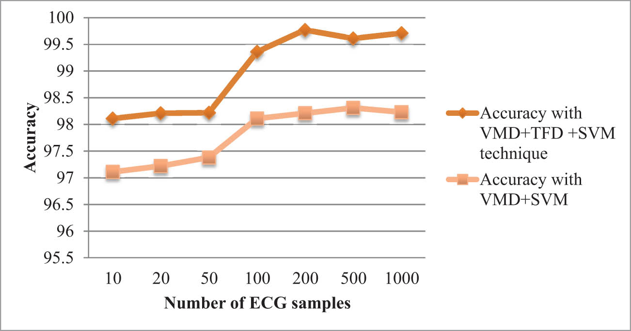
Comparison of accuracy using VMD + TFD + SVM and using VMD + SVM only.
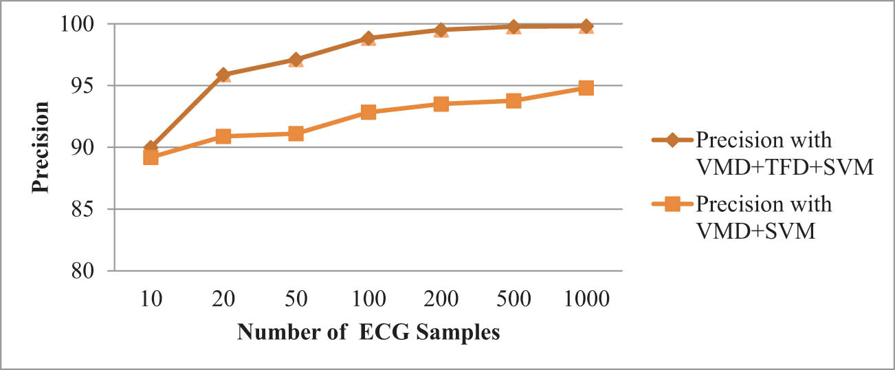
Comparison of precision using VMD + TFD + SVM and using VMD + SVM only.
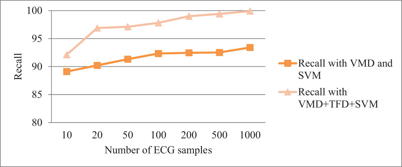
Comparison of recall using VMD + TFD + SVM and using VMD + SVM only.
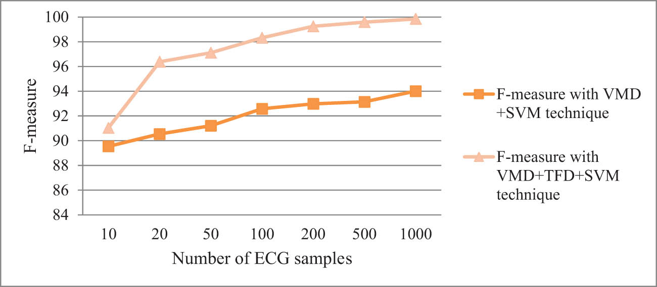
Comparison of F-measure using VMD + TFD + SVM and using VMD + SVM only.
4.2 Comparative analysis
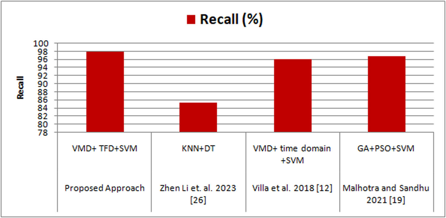
In this section, the proposed approach is compared with the existing techniques to determine the effectiveness and robustness. The existing literature uses the same dataset MIT-BIH and considers 1,000 ECG samples to model the results (Table 3).
Recall comparison of the proposed approach with the existing ones
| Authors | Technique | Recall (%) |
|---|---|---|
| Proposed approach | VMD + TFD + SVM | 97.89 |
| Li et al. [27] | KNN + DT | 85.4 |
| Villa et al. [13] | VMD + time domain + SVM | 96.11 |
| Malhotra and Sandhu [21] | GA + PSO + SVM | 96.83 |
Recall values are compared with existing techniques as given in Table 3. The techniques employed in the existing work [13] use VMD for denoising and features were extracted using the time domain and further SVM was used to classify the features. The recall obtained using the existing work was 96.11%. However, the study done in 2021 [21] by the researchers provided 96.83%. The KNNs and Decision Tree [27] achieved recall parameter value of 85.4%. But, the average recall value using the proposed technique is 97.46% which is better than the existing work (Table 4, Graph 5).
Accuracy comparison of the proposed approach with the existing ones
| Authors | Technique | Accuracy (%) |
|---|---|---|
| Proposed approach | VMD + TFD + SVM | 98.99 |
| Siam et. al. [29] | FFT + RF | 98.2 |
| Hu and Gao [28] | KNN | 93.7 |
| Villa et al. [13] | VMD + time domain + SVM | 95.74 |
| Malhotra and Sandhu [21] | GA + PSO + SVM | 98.93 |
| Tripathy et al. [17] | VMD + RF classifier | 97.67 |
| Li et al. [4] | GA + PCA + SVM | 97.78 |
Recall comparison of the proposed approach with the existing techniques.
The accuracy of the proposed approach is compared with the existing techniques. The proposed one has better performance than the existing approaches. The study was done using the GA, PSO, and SVM classifier [21] shows 98.93% accuracy while using the PCA [5], which is only 97.78%. Using KNN [28] classifier and Random Forest (RF) model, the accuracy achieved 93.7 and 98.2% respectively. Further, the use of VMD and time domain feature extraction method with SVM [13] shows only 95.74%. Thus, the proposed approach outperforms the other techniques (Graph 6).
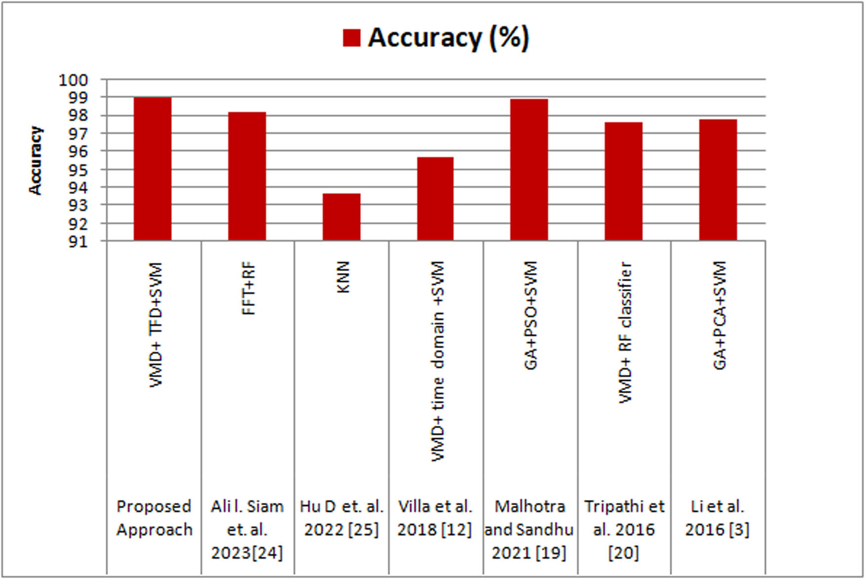
Accuracy comparison of the proposed approach with the existing techniques.
Table 5 and Graph 7 depict that the study done using the GA, PSO, and SVM classifier shows 96.83% precision, and using the TFD feature extraction method with SVM shows only 61.7%. The KNNs and Decision Tree [27] achieved a precision parameter value of 88.9%. The proposed one is improved by 0.95% from [21], 36.07% from the existing approaches [10] and 8.88% from 88.9% [27].
Precision comparison of the proposed approach with the existing ones
| Authors | Technique | Precision (%) |
|---|---|---|
| Proposed technique | VMD + TFD + SVM | 97.78 |
| Li et. al. [27] | KNN + DT | 88.9 |
| Mondéjar-Guerra et al. [10] | Time–frequency + SVM product rule | 61.70 |
| Malhotra and Sandhu [21] | GA + PSO + SVM | 96.83 |
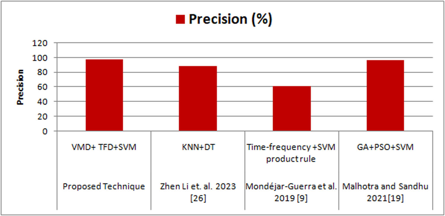
Precision comparison of the proposed approach with the existing techniques.
5 Conclusion
The main motive of the current research is to use a classification model to classify the stress level identified in the different subjects. The use of feature extraction algorithm and SVM classifier provides a way to classify the different levels of stress. The TFD feature extraction method using the classifier dramatically refined and reconstruct the low, medium, and high-level stress. Further, the overall time reduced using the VMD in the pre-processing stage to avoid the noisy signals and complexity of the signal also avoided attaining a better classification model. Also Robot system is designed in such a way to effectively communicate stress classification outcomes to users using visual and auditory cues to convey information in an understandable and non-intrusive manner. Overall, the proposed approach enhanced the performance of the stress detection system and performance metrics such as average accuracy, precision, recall, and F-measure attained during modeling is 98.98, 97.78, 97.89, and 97.36%, respectively. The proposed approach is further compared with the existing techniques for validation and evaluation.
Further, the use of ECG-based stress classification with robotic applications can be a promising approach to help individuals manage their stress levels. With the development of advanced algorithms and user-friendly interfaces, this approach can be further enhanced to provide more personalized and effective stress management solutions. Beyond ECG data, robots could integrate additional physiological signals, such as skin conductance, facial expressions, and voice patterns. This multi-modal approach will yield a richer understanding of a user’s stress levels and emotional state, leading to more. Future systems may facilitate a bidirectional interaction where users can provide feedback on the effectiveness of interventions. Robots will then fine-tune their strategies based on this feedback, creating a seamless closed-loop interaction.
-
Funding information: Authors state no funding involved.
-
Author contributions: Conceptualization, V.M.; methodology, V.M.; software, R.P.; validation, S.M.; formal analysis, V.M.; investigation, G.S.S.; resources, V.M.; data curation, R.P.; writing–original draft preparation, R.P.; writing–review and editing, R.P.; visualization, S.M.; supervision, G.S.; project administration, V.M. All authors have read and agreed to the published version of the manuscript.
-
Conflict of interest: Authors state no conflict of interest.
-
Data availability statement: All data used in the manuscript has been mentioned in the manuscript and taken from physionet, whose site is https://physionet.org/.
References
[1] A. Arza, J. M. Garzón-Rey, J. Lázaro, E. Gil, R. Lopez-Anton, C. de la Camara, et al., “Measuring acute stress response through physiological signals: towards a quantitative assessment of stress,” Med. Biol. Eng. Comput., vol. 57, no. 1, pp. 271–287, 2019.10.1007/s11517-018-1879-zSearch in Google Scholar PubMed
[2] G. Praveena and J. M. Mathana, “Review on stress detection and management techniques using nano EEG sensors,” IEEE International Conference on Nanoelectronics, Nanophotonics, Nanomaterials, Nanobioscience & Nanotechnology (5NANO), Kottayam, India, 2022, pp. 1–7.10.1109/5NANO53044.2022.9828889Search in Google Scholar
[3] R. Zhou, C. Wang, P. Zhang, X. Chen, L. Du, P. Wang, et al., “ECG-based biometric under different psychological stress states,” Comput. Methods Prog. Biomed., vol. 202, p. 106005, 2021.10.1016/j.cmpb.2021.106005Search in Google Scholar PubMed
[4] H. Li, H. Liang, C. Miao, L. Cao, X. Feng, C. Tang, et al., “Novel ECG signal classification based on KICA nonlinear feature extraction,” Circuits Syst. Signal. Process, vol. 35, no. 4, pp. 1187–1197, 2016.10.1007/s00034-015-0108-3Search in Google Scholar
[5] R. R. Sharma, M. Kumar, and R. B. Pachori, “Automated CAD identification system using time-frequency representation based on eigenvalue decomposition of ECG signals,” Machine intelligence and signal analysis, Springer, Singapore, 2018, pp. 597–608.10.1007/978-981-13-0923-6_51Search in Google Scholar
[6] S. Chatterjee, R. S. Thakur, R. N. Yadav, L. Gupta, and D. K. Raghuvanshi, “Review of noise removal techniques in ECG signals,” IET Signal. Process., vol. 14, no. 9, pp. 569–590, 2020.10.1049/iet-spr.2020.0104Search in Google Scholar
[7] H. M. Cho, H. Park, S. Y. Dong, and I. Youn, “Ambulatory and laboratory stress detection based on raw electrocardiogram signals using a convolutional neural network,” Sensors, vol. 19, no. 20, p. 4408, 2019.10.3390/s19204408Search in Google Scholar PubMed PubMed Central
[8] R. Ahuja and A. Banga, “Mental stress detection in university students using machine learning algorithms,” Procedia Comput. Sci., vol. 15, pp. 349–353, 2019.10.1016/j.procs.2019.05.007Search in Google Scholar
[9] S. Pourmohammadi and A. Maleki, “Stress detection using ECG and EMG signals: A comprehensive study,” Comput. Methods Prog. Biomed., vol. 193, p. 105482, 2020.10.1016/j.cmpb.2020.105482Search in Google Scholar PubMed
[10] V. Mondéjar-Guerra, J. Novo, J. Rouco, M. G. Penedo, and M. Ortega, “Heartbeat classification fusing temporal and morphological information of ECGs via an ensemble of classifiers,” Biomed. Signal. Process. Control., vol. 47, pp. 41–48, 2019.10.1016/j.bspc.2018.08.007Search in Google Scholar
[11] S. Pourmohammadi and A. Maleki, “Continuous mental stress level assessment using electrocardiogram and electromyogram signals,” Biomed. Signal. Process. Control., vol. 68, p. 102694, 2021.10.1016/j.bspc.2021.102694Search in Google Scholar
[12] N. Salankar, D. Koundal, and S. MianQaisar, “Stress classification by multimodal physiological signals using variational mode decomposition and machine learning,” J. Healthc. Eng., vol. 2021, p. 2146369, 2021.10.1155/2021/2146369Search in Google Scholar PubMed PubMed Central
[13] A. Villa, S. Padhy, R. Willems, S. Van Huffel, and C. Varon, Variational mode decomposition features for heartbeat classification, Computing in Cardiology Conference, vol. 45, 2018, pp. 1–4.Search in Google Scholar
[14] C. Haritha, M. Ganesan, and E. P. Sumesh, “A survey on modern trends in ECG noise removal techniques,” International Conference on Circuit, Power and Computing Technologies, vol. 7530192, 2016, pp. 1–7.10.1109/ICCPCT.2016.7530192Search in Google Scholar
[15] L. Gonzalez-Carabarin, E. A. Castellanos-Alvarado, P. Castro-Garcia, and M. A. Garcia-Ramirez, “Machine learning for personalized stress detection: Inter-individual variability of EEG-ECG markers for acute-stress response,” Comput. Methods Prog. Biomed., vol. 209, p. 106314, 2021.10.1016/j.cmpb.2021.106314Search in Google Scholar PubMed
[16] M. Kang, S. Shin, G. Zhang, J. Jung, and Y. T. Kim, “Mental stress classification based on a support vector machine and naive Bayes using electrocardiogram signals,” Sensors, vol. 21, no. 23, p. 7916, 2021.10.3390/s21237916Search in Google Scholar PubMed PubMed Central
[17] R. K. Tripathy, L. N. Sharma, and S. Dandapat, “Detection of shockable ventricular arrhythmia using variational mode decomposition,” J. Med. Syst., vol. 40, no. 4, pp. 1–13, 2016.10.1007/s10916-016-0441-5Search in Google Scholar PubMed
[18] Y. Jung and Y. I. Yoon, “Multi-level assessment model for wellness service based on human mental stress level,” Multimed. Tools Appl., vol. 76, no. 9, pp. 11305–11317, 2017.10.1007/s11042-016-3444-9Search in Google Scholar
[19] A. R. Subhani, W. Mumtaz, M. N. B. M. Saad, N. Kamel, and A. S. Malik, “Machine learning framework for the detection of mental stress at multiple levels,” IEEE Access, vol. 5, pp. 13545–13556, 2017.10.1109/ACCESS.2017.2723622Search in Google Scholar
[20] K. K. Patro, A. Jaya Prakash, M. Jayamanmadha Rao, and P. Rajesh Kumar, “An efficient optimized feature selection with machine learning approach for ECG biometric recognition,” IETE J. Res., vol. 68, no. 4, pp. 2743–2754, 2020.10.1080/03772063.2020.1725663Search in Google Scholar
[21] V. Malhotra and M. K. Sandhu, “Improved ECG based stress prediction using optimization and machine learning techniques,” EAI Trans. Scalable Inf. Syst., vol. 8, no. 32, p. 169175, 2021.10.4108/eai.6-4-2021.169175Search in Google Scholar
[22] PhysicoNet Database, Available Online at https://physionet.org/about/database/.Search in Google Scholar
[23] A. L. Goldberger, L. A. Amaral, L. Glass, J. M. Hausdorff, P. C. Ivanov, R. G. Mark, et al., “PhysioBank, PhysioToolkit, and PhysioNet: components of a new research resource for complex physiologic signals,” Circulation, vol. 101, no. 23, pp. E215–E220, 2000.10.1161/01.CIR.101.23.e215Search in Google Scholar
[24] M. A. Nair, “ECG feature extraction using time frequency analysis,” Innovations in Computing Sciences and Software Engineering, Springer, Dordrecht, Netherlands, 2010, pp. 461–466.10.1007/978-90-481-9112-3_78Search in Google Scholar
[25] S. McGlynn, B. Snook, S. Kemple, T. L. Mitzner, and W. A. Rogers, “Therapeutic robots for older adults: investigating the potential of paro,” IEEE International Conference on Human-robot interaction, 2014, pp. 246–247.10.1145/2559636.2559846Search in Google Scholar
[26] M. Heerink, J. Albo-Canals, M. Valenti-Soler, P. Martinez-Martin, J. Zondag, C. Smits, et al., “Exploring requirements and alternative pet robots for robot assisted therapy with older adults with dementia,” International Conference on Social Robotics, vol. 5, 2013, pp. 104–115.10.1007/978-3-319-02675-6_11Search in Google Scholar
[27] Z. Li, Y. Xing, Y. Pi, M. Jiang, and L. Zhang, “A novel physiological feature selection method for emotional stress assessment based on emotional state transition,” Front. Neurosci., vol. 17, p. 1138091, 2023.10.3389/fnins.2023.1138091Search in Google Scholar PubMed PubMed Central
[28] D. Hu and L. Gao, “Psychological stress level detection based on heartbeat mode,” Appl. Sci., vol. 12, no. 3, p. 1409, 2022.10.3390/app12031409Search in Google Scholar
[29] A. I. Siam, S. A. Gamel, and F. M. Talaat, “Automatic stress detection in car drivers based on non-invasive physiological signals using machine learning techniques,” Neural Comput. Appl., vol. 35, pp. 12891–12904, 2023.10.1007/s00521-023-08428-wSearch in Google Scholar
© 2024 the author(s), published by De Gruyter
This work is licensed under the Creative Commons Attribution 4.0 International License.
Articles in the same Issue
- Regular Articles
- Labour legislation and artificial intelligence: Europe and Ukraine
- Deep trained features extraction and dense layer classification of sensitive and normal documents for robotic vision-based segregation
- Evaluating people's perceptions of an agent as a public speaking coach
- Retraction
- Retraction of “Hybrid controller-based solar-fuel cell-integrated UPQC for enrichment of power quality”
- Special Issue: Humanoid Robots and Human-Robot Interaction in the Age of 5G and Beyond - Part II
- Optimal trajectory planning and control of industrial robot based on ADAM algorithm of nonlinear data set
- Special Issue: Recent Advancements in the Role of Robotics in Smart Industries and Manufacturing Units - Part III
- A robot electronic device for multimodal emotional recognition of expressions
- Design of RFID-based weight sorting and transportation robot
- Path planning of welding robot based on deep learning
- Design of a robot system for improved stress classification using time–frequency domain feature extraction based on electrocardiogram