Biophysical stimuli for promoting bone repair and regeneration
-
Yunyang Bai

 ,
Xuehui Zhang
and
Xuliang Deng
,
Xuehui Zhang
and
Xuliang Deng

Abstract
Bone injuries and diseases are associated with profound changes in the biophysical properties of living bone tissues, particularly their electrical and mechanical properties. The biophysical properties of healthy bone are attributed to the complex network of interactions between its various cell types (i.e., osteocytes, osteoclast, immune cells and vascular endothelial cells) with the surrounding extracellular matrix (ECM) against the backdrop of a myriad of biomechanical and bioelectrical stimuli arising from daily physical activities. Understanding the pathophysiological changes in bone biophysical properties is critical to developing new therapeutic strategies and novel scaffold biomaterials for orthopedic surgery and tissue engineering, as well as provides a basis for the application of various biophysical stimuli as therapeutic agents to restore the physiological microenvironment of injured/diseased bone tissue, to facilitate its repair and regeneration. These include mechanical, electrical, magnetic, thermal and ultrasound stimuli, which will be critically examined in this review. A significant advantage of utilizing such biophysical stimuli to facilitate bone healing is that these may be applied non-invasively with minimal damage to surrounding tissues, unlike conventional orthopedic surgical procedures. Furthermore, the effects of such biophysical stimuli can be localized specifically at the bone defect site, unlike drugs or growth factors that tend to diffuse away after delivery, which may result in detrimental side effects at ectopic sites.
Introduction – the physiological microenvironment of bone tissue
Understanding the physiology of the bone tissue microenvironment under both healthy and disease conditions is crucial for developing innovative treatment modalities for achieving optimal healing of bone injuries and providing cues for designing and manufacturing novel scaffold implants for bone tissue engineering. Bone is a highly vascularized but hard and stiff tissue that provides structural support and protects the human body’s delicate soft tissues and internal organs. It can be found in both compact and cancellous (trabecular) forms. Extracellular matrix (ECM) is prevalent in bone tissue, making up 65 % of its gross volume and is primarily composed of both inorganic and organic minerals (35 %). Collagen type I fibers (90 %), which comprise the bulk of the organic matrix, confer tensile strength to bone ECM through their extremely stable triple helix structure. The ECM, embedded cells, and soluble substances are the key sources of biophysical cues that bone cells respond to simultaneously in their natural environment. The outside-in/inside-out exchange of biophysical stimuli arising from the ability of bone cells to recognize and communicate with their environment has a profound impact on several physiological processes, including tissue remodeling, regeneration, healing, aging, as well as the emergence of numerous pathological conditions affecting bone. Due to the activation of various cellular mechanisms and signaling pathways by these biophysical stimuli, which results in synergistic effects, the concurrent combination of mechanical, electrical, magnetic, and thermal stimuli can accentuate the physiological cues of substrates and facilitate mimicking the in vivo microenvironment. However, integrating multiple biophysical stimuli (mechanical, electrical, magnetic, and thermal cues) on a single platform has yet to receive much attention in bone tissue engineering research. In this review, we critically examine the roles of mechanical, electrical, magnetic, and thermal stimuli in regulating various cellular processes such as cell adhesion, migration, and morphology, as well as their role in inducing osteogenic differentiation and facilitating bone repair and regeneration.
Pathophysiological changes in the biophysical microenvironment of bone during injury/disease (mechanical, electrical, thermal)
Pathophysiological changes in bone mechanical microenvironment during injury/disease
Bone repair and regeneration occur at bone defect sites associated with distinct changes in mechanical properties, such as fractures andtraumatic and medical-related bone injuries (implantation of endosseous medical devices). Research data from in vivo animal models and in vitro mechanistic studies on the effects of mechanical loading on fracture and bone trauma healing are hereby summarized and analyzed in this section to determine appropriate prospective clinical intervention strategies.
A wide range of mechanical parameters have been identified as influencing fracture healing. Rigid fixation, fracture geometry, fracture type, direction, and amplitude are the most critical parameters in this process. These parameters influence the distribution of local stress at the fracture site and elicit mechano-biological signals to regulate fracture healing and trigger cellular responses. The volume and direction of interfragmentary mobility influence the healing process. Moderate axial interfragmentary movement promotes periosteal callus development and accelerates fracture healing. Tensile or shear movements of comparable amplitude, on the other hand, do not appear to assist fracture healing. Induced cyclic tensile loads can enhance periosteal callus development but not bone repair.
Different modes of mechanical loading are crucial for determining the healing response of bone. Bone, for example, cannot adjust to loading unless applied cyclically (as in physiological movement). It was reported that static bending on rabbit tibiae inhibited bone development. In contrast, rabbit tibiae treated to the same magnitude of dynamic loading exhibited improved bone growth on both the endosteal and periosteal surfaces. Under static loading, bone cells can rapidly desensitize and lose mechanosensitivity before mechanosensation and mechanotransduction are complete. Because more rest phases are provided, cyclic and intermittent loading may be more effective for preserving bone mechanosensitivity than continuous loading. Besides fracture and bone defect healing, mechanical loading has also been demonstrated to be critical for promoting osteogenesis in normal bone and implant osseointegration.
Pathophysiological changes in bone electrical microenvironment during injury/disease
Bone defect healing is a complex physiological process that includes hemorrhage, coagulation, inflammation, angiogenesis, cell migration, and gradual tissue remodeling, as detailed in several outstanding reviews. The emphasis here will be on how the electrophysiological properties of bone tissue change during damage and how this affects bone defect healing and regeneration. Bone tissue injury is associated with decreased electrical potential at the defect location. The development of periosteum-like tissue frequently quickly restores the local electrophysiological microenvironment at the defect site for tiny lesions below a critical threshold size. As a result, restoration of the electrophysiological microenvironment through the growth of periosteum-like tissue has a galvanotactic effect, attracting cells from surrounding endogenous tissues to the defect site. Surface charges attract ions, which improve cell adhesion and protein adsorption via ionic charge interactions, which can serve as a cell recruitment pathway. The greater the defect size (still below critical size), the longer it takes for periosteum-like tissue to develop, thus delaying restoration of the electrophysiological microenvironment conducive to bone repair, leading to delayed healing. In the case of defects larger than the critical size, the healing capacity is completely lost due to the failure of periosteum tissues to form at the defect site. Hence, in situ implantation of an electroactive scaffold is needed to facilitate bone defect healing by recapitulating the electrophysiological microenvironment of healthy bone tissues.
Mechanical stimuli promote bone regeneration
As bone tissue is the main component of the skeletal system that provides structural support for the human body, it is constantly exposed to mechanical stimuli during daily physical activities. Indeed, the myriad of biological processes involved in bone tissue development, homeostasis, repair and regeneration depend completely on its capacity for sensing and responding to mechanical stimuli [1], [2], [3], [4], [5]. To date, the effects of mechanical stimuli on bone tissue have been widely studied for several decades, beginning with the seminal study of Wolff et al. [1] that first reported the enhancement of bone healing upon mechanical loading. Subsequently, the “Mechanostat” hypothesis proposed by Frost et al. [2], described how bone mass and architecture adapt to mechanical loading via structural remodeling to better resist habitual loads with optimal use of biological materials. Among the various biophysical stimuli that can potentially enhance bone regeneration, mechanical stimulation has attracted special attention due to its ubiquitous nature (i.e., daily physical activity and exercise), adaptability, non-invasiveness and ease of delivery.
The application of tensile or compressive mechanical forces causes bone matrix deformation, which in turn generates a hydraulic pressure gradient that leads to interstitial bone fluid flow through the voids and channels of bone matrix, which include Lacunae-Canaliculi, Osteons, Volkmann canals and Haversian canals [6], 7]. The resultant exposure of the various cellular components of bone tissue to shear stress generated by such interstitial fluid flow, in turn enables the transduction of mechanical stimuli into biological signals [6], 7]. For example, bone marrow mesenchymal cells have been observed to congregate in response to mechanical stimuli during the initial fracture healing phase, thereby promoting callus formation during bone healing [3]. Osteoblastic cells within bone are known to respond to mechanical loading by increasing their proliferation and differentiation rates, altering their deposition of ECM and secretion of cytokines/growth factors [8]. Besides serving as the primary conduit by which bone cells sense and respond to mechanical stimuli, interstitial bone fluid flow generated by mechanical stimulation also facilitates the transfer and exchange of nutrients, cytokines and growth factors [9], 10], which undoubtedly play critical roles in bone tissue remodeling during homeostasis, repair and regeneration. Furthermore, in vitro studies have shown that fluid flow-induced shear stress can promote the directional alignment of collagen fibrils as well as directional growth of hydroxyapatite (HAp) crystals on the surface of collagen fibrils [11], with cells being able to grow directionally on such aligned collagen fibrils and secreting oriented ECM [11]. Hence, interstitial fluid flow within cortical bone is undoubtedly a key regulator of bone mass and architecture in response to mechanical stimulation [12], 13].
Despite mechanical stimuli being a key initiator of bone tissue remodeling during homeostasis, repair and regeneration, it must nevertheless be noted that it does not function independently of the biophysical and biochemical microenvironment within bone but must instead interactively cross-talk with various physiological, biochemical, and mechanobiological cues within bone tissue. Very often, the pathological state of bone tissue profoundly impacts its response to mechanical stimuli. For example, it was observed that in patients with osteoporosis, mechanical stimuli failed to enhance the proliferation and cytokine secretion of osteoblasts, unlike the osteoblasts of regular healthy donors [14]. Hence, the response of bone tissue to mechanical stimuli is not fixed but is instead highly correlated to the overall health condition of the host, such as age, sex and disease.
Bone repair and regeneration involves a complex web of interactive cross-talk between the various cell types within bone tissue, including osteocytes, osteoblasts, osteoclasts, mesenchymal stem cells (MSCs), endothelial cells, together with immune cells such as neutrophils and macrophages. Mechanosensing by these various diverse cell types involved in bone regeneration is achieved via four major mechanosensitive elements, including (i) focal adhesions (FA) [15], 16], (ii) ion channels such as Piezo1 and TRPV4 [17], 18], (iii) primary cilium [19], 20], and (iv) Connexin 43 (Cx43) [21], 22], each of which will be briefly discussed in turn. FA relay mechanical signals (i.e., stiffness) from the ECM to the cells and forms a direct physical link between the cellular cytoskeleton and ECM [15], 16]. Mechanical stimuli transmitted by FA affect cytoskeleton arrangement and crosslinking, which in turn modulate cell adhesion, stretching, and migration, as well as trigger activation of key mechanotransduction signaling pathways implicated in the regulation of osteogenesis, such as YAP/TAZ, MAPK/ERK/JNK, and RhoA/ROCK [15], 16]. Additionally, mechanical stimuli can also trigger the activation of ion channels such as Piezo1/2 and TRPV4, resulting in a calcium ion influx, which in turn activates various signaling pathways that regulate osteogenesis, such as Ca2+-calcineurin-NFAT1 and Wnt/β-catenin [17], 18]. The activation of ion channels such as TRPV4 is linked to mechanosensitive primary cilium found in various bone tissue cells, including osteocytes, osteoblasts, osteoclasts and MSCs [19], 20]. The bending of the primary cilia of these bone cells upon exposure to bone interstitial fluid flow shear stress is known to trigger the activation of TRPV4 ion, leading to a Ca2+ ion influx that enhances osteogenic differentiation of MSCs [21]. On the other hand, in the case of osteoclasts, mechanical stimuli lead to PTH1R translocation on primary cilia, which in turn blocks osteoclast activation via IL-6 and CXCL5 release [23]. Yet another mechanosensitive element of bone cells is the Cx43 protein, which is the major component of gap junctions that enable the exchange of small molecules and the passage of electrical currents between neighboring cells [21], 22]. Mechanical stimuli trigger phosphorylation of the Cx43 protein, which leads to the opening of gap junctions, enabling the exchange of various small molecule effectors of osteogenesis between connecting cells, such as Ca2+ and Prostaglandin E2 [21], 22].
Mechanical stimuli are thought to promote bone repair and regeneration by (i) tipping the balance between osteogenesis vs. osteoclastogenesis in favor of osteogenesis, (ii) enhancing angiogenesis and vascularization, and (iii) mitigating inflammation, each of which will be briefly discussed in turn. As already discussed, mechanical stimuli enhance osteogenesis predominantly via activation of ion channels such as Piezo1/2 and TRPV4 [17], 18], as well as phosphorylation of the gap junction protein Cx43 [19], 20], affecting an influx of Ca2+ ions and opening of gap junctions respectively, which in turn activate various signaling pathways implicated in osteogenesis. At the same time, mechanical stimuli suppress osteoclastogenesis through various mechanisms. As already mentioned, one of these involves translocation of PTH1R on primary cilia triggered by bone interstital flow-induced shear stress that blocks osteoclast activation via IL-6 and CXCL5 release [24]. It was also reported that fluid flow shear stress could downregulate expression of ATP6V1A and TCIRG1, which are crucial for osteoclast bone resorption function [25]. Yet another mechanism involves suppression of the differentiation and bone resorption functions of osteoclasts by fluid flow shear stress via the ERK5 signaling pathway [26]. Additionally, it was also reported that an influx of Ca2+ into osteoclastic progenitors triggered by fluid flow shear stress may also inhibit mature osteoclast formation [27]. Hence in this manner, mechanical stimuli can tip the balance between osteogenesis vs. osteoclastogenesis in favor of osteogenesis.
Vascularization and angiogenesis are crucial for bone repair and regeneration, and several studies have demonstrated that mechanical stimuli promote vascularization and angiogenesis during the bone healing process [28], [29], [30]. As proposed by Kuchan and Frangos [28], enhancement of vascularization during bone healing by mechanical stimuli was associated with increased bone interstitial fluid flow due to the leaky nature of capillaries. Besides providing oxygen and nutrients to facilitate bone cell proliferation and differentiation, increased angiogenesis can also alter osteoblastic functions by releasing endothelial-derived factors such as endothelin and nitric oxide (NO). Endothelin is known to enhance osteoblast proliferation while suppressing bone resorption by osteoclasts [31], 32]. NO released from endothelial cells can also inhibit osteoclast function [28]. Interestingly, it has been reported that the release of NO and endothelin by endothelial cells can be stimulated by fluid flow shear stress via the protein kinase C (PKC) and cGMP pathways [10], 28]. How mechanical stimuli enhance vascularization and angiogenesis during bone regeneration may involve overlapping several different mechanisms. It was reported that mechanical loading of bone could stimulate the release of various pro-angiogenic factors such as nephronectin (NPNT), Vascular Endothelial Growth Factor (VEGF), Hypoxia Inducible Factor 1 (HIF-1), epidermal growth factor-like domain (EGFL), and Notch ligands [33], [34], [35], [36], all of which play critical roles in regulating the differentiation and proliferation of endothelial cells. Kasper et al. [37] demonstrated that mechanical stimulation of MSCs resulted in paracrine stimulation of angiogenesis via regulation of a network of several angiogenic molecules, with underlying mechanisms being dependent on the Fibroblast Growth Factor Receptor (FGFR) and Vascular Endothelial Growth Factor Receptor (VEGFR) signaling cascades and which might be mediated by an additional cross-talk with other pathways. The study of Iba and Sumpio [38] reported that cyclic strain promoted the elongation of endothelial cells via reorganization of the actin filament network. In contrast, other studies implicated ERK and p38 signaling in mechanical stimuli-induced angiogenesis [39], 40].
Mechanical stimuli can also promote bone regeneration via modulation of the immune system and mitigation of inflammation [41]. It was reported that cyclic stretching significantly inhibited the NLRP3 inflammasome signaling pathway and IL–1β secretion in macrophages [42]. Another study reported that mechanical stimuli downregulate the pro-inflammatory marker TLR-4 [43]. Other studies demonstrated that mechanical stimuli stimulate IL-8 secretion through the p38 MAPK pathway [44], 45], with IL-8 being known to be a key cytokine that promotes polarization of macrophages from the pro-inflammatory M1 phenotype to the pro-healing M2 phenotype via upregulation of CD163 and CD206 [46]. Indeed, Zhang et al. [47] reported that ECM-based scaffolds biofabricated with MSCs exposed to mechanical loading before freeze-drying could enhance the expression of M2 polarization-related biomarkers (Arginase 1 and Mrc1) of macrophages in vitro.
To date, the majority of studies on the effects of mechanical stimulation on osteogenesis, bone repair and regeneration are predominantly based on in vitro models, either in 2D cell culture format or 3D cell-seeded scaffolds [48], 49], which are difficult to translate to in vivo animal studies or human clinical models of bone injury. While physiotherapy rehabilitation programs involving exercise and physical activity are routinely prescribed in the clinical setting to enhance bone healing by mechanical stimulation [50], 51], a clinically relevant technology that can translate physical stimuli into biological responses in a controlled, sustained and localized fashion has not yet been achieved to date. Currently, most animal studies on the effects of mechanical stimulation on bone healing in situ involve the external application of cyclic mechanical stimuli via dynamic fixator/actuator devices [52], [53], [54] or vibration platform devices [55], 56]. The main drawback of such mechanical stimulation devices is that mechanical stimulation can only be applied intermittently for a limited period each day [52], [53], [54], [55], [56], which thus limits any beneficial therapeutic effects of mechanical stimulation on bone healing, as compared to exercise and other physical activities [50], 51].
Electrical stimuli promote bone regeneration
Bone is well-known to be an electroactive, electrosensitive and electroresponsive tissue [57]. Its bioelectrical properties can be visualized as a combination of dielectric, piezoelectric, pyroelectric, and ferroelectric properties, together with streaming potential and electro-osmosis [57], and arise from interactive cross-talk between the various cell types and ECM of bone tissue. Of these, the piezoelectric effect of its abundant collagenous matrix is considered the most critical contributor to the endogenous electric field within living bone tissue, as collagen fibers constitute 85–90 % of bone ECM and around 22 % of bone tissue [58]. The non-centrosymmetric polar hexagonal crystalline units and spiral structure of collagen fibril enable it to convert mechanical stimuli into piezoelectric potential. In response to mechanical force, collagen fibers slide against each other, resulting in dipole rearrangement due to the separation and polarization of charged groups, generating a physiological electrical potential (piezoelectricity) [58]. Hence, daily routine physical activities and natural bodily movement generate an endogenous electrical field within bone tissue via piezoelectric stimulation of its abundant collagenous matrix. For example, the human tibia generates a piezoelectric potential of about 300 μV during walking [59].
Because the bioelectrical properties of bone are known to play critical roles in regulating its metabolism, tissue homeostasis, repair and regeneration [57], numerous studies have investigated the effects of electrical stimuli in promoting bone healing and have reported mostly positive results [60], 61]. Indeed, treating bone fractures with electrical stimulation has a long history, dating back to the mid-nineteenth century. The self-healing capacity of bone becomes severely constrained if the injury or defect is above a specific critical size or if there are some underlying pathological conditions that impede osteogenesis, such as osteoporosis, in which clinical intervention becomes necessary. For this purpose, the application of electrical stimulation may be therapeutically beneficial. Indeed, the orthopedic applications of electrical stimulation therapy have progressed rapidly in recent years and have demonstrated significant clinical efficacy in treating various bone diseases and injuries. Nevertheless, first of all, it is crucial to understand changes in the electrophysiology of bone tissue during the healing process upon injury. Under normal and healthy physiological conditions, bone tissue is naturally electronegative but becomes more electronegative when an injury or fracture occurs [62], with the injury/fracture site becoming powerfully negatively charged [63]. This in turn increases ionic current flow to the injury/fracture site that enhances cell migration (i.e., MSCs and osteoblasts), facilitates the flow of nutrients and growth factors, and promotes other biological processes that enable bone repair and regeneration [64]. In vitro studies have revealed that a negatively charged surface attracts cations such as Ca2+ present within biological fluids, which in turn serves as a stimulus for the adsorption of proteins that subsequently promotes cellular adhesion [65], 66]. Additionally, increased electronegativity promotes osteoblastic activity, enhancing ECM mineralization at the injury/fracture site [67], 68]. Hence, the application of electrical stimuli is thought to accelerate the bone healing process by further accentuating electronegativity at the injury/fracture site, which is borne out by direct electrical stimulation studies that demonstrated faster and thicker callus formation when the cathode is placed on the bone injury site (Figure 1) [69].
![Figure 1:
Electrical stimuli promote bone regeneration. (a) Manipulation of heterogeneous surface electric potential promotes osteogenesis by strengthening RGD peptide binding and cellular mechanosensing [90]. (b) Restoring the electrical microenvironment using ferroelectric nanocomposite membranes enhances alveolar ridge regeneration in a mini-pig preclinical model [234]. (c) An electroactive composite scaffold with locally expressed osteoinductive factors achieves synergistic bone repair upon electrical stimulation [235]. (d) built-in electric fields dramatically induce enhancement of osseointegration [236].](/document/doi/10.1515/mr-2024-0023/asset/graphic/j_mr-2024-0023_fig_001.jpg)
Electrical stimuli promote bone regeneration. (a) Manipulation of heterogeneous surface electric potential promotes osteogenesis by strengthening RGD peptide binding and cellular mechanosensing [90]. (b) Restoring the electrical microenvironment using ferroelectric nanocomposite membranes enhances alveolar ridge regeneration in a mini-pig preclinical model [234]. (c) An electroactive composite scaffold with locally expressed osteoinductive factors achieves synergistic bone repair upon electrical stimulation [235]. (d) built-in electric fields dramatically induce enhancement of osseointegration [236].
To date, extensive in vitro studies have been carried out that have shed much light on the mechanistic effects of electrical stimulation on the cellular behaviors of various bone tissue lineages, including MSCs, osteoblasts, osteocytes and osteoclasts. Numerous key biological processes that play crucial roles in bone repair and regeneration, such as cell proliferation [70], 71], apoptosis [72], alignment [73], 74], migration [75], 76], adhesion [77], 78], and differentiation [79], 80] are modulated by electrical stimuli. For a better understanding of how electrical stimulation promotes bone healing, it is best to delineate its effects separately on (i) tipping the balance between osteogenesis and osteoclastogenesis in favor of osteogenesis, (ii) angiogenesis/vascularization, and (iii) immune modulation that mitigates inflammation, each of which will be discussed in turn.
As already mentioned, there is increased electronegativity at the bone injury/fracture site, and electrical stimulation promotes bone healing by accentuating the electronegativity of the bone defect site [62], [63], [64], [65], [66], [67], [68], [69]. One way this may be achieved is by effecting differential migration of various cell lineages within bone tissue. It has been demonstrated that negative charges attract MSCs migration [78] whilst repelling osteoclast migration [81], 82], which could in turn tip the balance between osteogenesis and osteoclastogenesis in favor of osteogenesis at the bone defect site. Furthermore, electrical stimulation is known to trigger an influx of Ca2+ into the cytosol of MSCs and osteoblasts via activation of voltage-gated Ca2+ channels [83] and Cx43 [84], which in turn leads to the activation of pro-osteogenic signaling pathways, in particular the Calmodulin/Calcineurin/NFAT [85], 86] and PKC [87] signaling pathways. By contrast, in the case of osteoclasts, Ca2+ influx triggered by electrical stimuli-induced activation of voltage-gated ion channels is an endogenous signal that inhibits bone resorption [88]. This is thought to involve phospholipase C-dependent Ca2+-sensing pathways leading to endocytosis and inhibition of the plasma membrane vacuolar H+-ATPase in osteoclasts [88]. Another pro-osteogenic signaling pathway activated by electrical stimuli is the focal adhesion kinase (FAK)-associated mechanotransduction signaling axis triggered by clustering of FA in response to electrical charge [89], 90]. The underlying mechanism is thought to involve modification of the adsorption conformation of fibronectin in the presence of electrical charge, which in turn facilitates the binding and clustering of integrin receptors that promote the formation of FA complexes and subsequent activation of FAK [91]. This ultimately leads to the dephosphorylation and nuclear translocation of YAP/TAZ and subsequent transcriptional activation of specific osteogenic genes through TEAD transcription factors [92]. It was also reported that interactive cross-talk between the aforementioned FAK and PKC signaling pathways activated by electrical stimuli can in turn activate the downstream extracellular-signal-regulated kinase 1/2 (ERK1/2) signaling pathway that promotes osteogenic differentiation [87]. Yet other less prominent pro-osteogenic signaling pathways that are known to be activated by electrical stimuli include the β-catenin/E-Cadherin [93], Inducible Nitric Oxide Synthase (iNOS) [94] and EphrinB2-EphB4 [95] signaling pathways (Figure 3a).
Electrical stimuli also enhance bone repair and regeneration via the promotion of increased angiogenesis/vascularization at the defect site, which in turn facilitates the transport of nutrients, metabolites, and cells required for bone healing [96], 97]. It was reported that electrical stimulation significantly enhanced angiogenesis in a calvarial defect model, promoting bone maturation [98]. In another study, the application of low-voltage direct current stimulation to the amputated bone stump of rats significantly enhanced vascularization [99]. The mechanisms by which electrical stimulation promotes angiogenesis involve activating pro-angiogenic signaling pathways in endothelial cells. These include the VEGFR2 signaling pathway [100], as well as the activation of endothelial nitric oxide synthase (eNOs) via a Phosphatidylinositol 3-Kinase (PI3K)/Protein Kinase B (Akt)-dependent pathway [101]. Additionally, electrical stimulation can also promote angiogenesis in a paracrine manner by inducing VEGF secretion from osteogenic cells [102].
Finally, electrical stimulation can promote bone repair and regeneration by mitigating inflammation via modulation of macrophage polarization. The study of Dai et al. [103] demonstrated that implantation of an electroactive membrane at bone defect sites of diabetic rats promoted macrophage transition from the pro-inflammatory M1 phenotype to the pro-healing M2 phenotype by suppressing expression of AKT2 and IRF5 within the PI3K-AKT signaling pathway, which in turn created a favorable osteoimmunomodulatory environment for enhancing bone repair and regeneration. Another similar study by Li et al. [104] also demonstrated that implantation of an electroactive scaffold within a diabetic rat periodontal bone defect model could accelerate bone healing by facilitating macrophage polarization from the M1 to M2 phenotype via modulation of glycolytic and RhoA/ROCK pathways.
Clinically, electrical stimulation to promote bone healing can be broadly categorized into non-invasive and invasive techniques. Non-invasive techniques involve the external application of electrical stimuli at the bone injury/fracture site. The major drawbacks are that they can neither stimulate the bone defect site with high accuracy and precision nor can guarantee effective stimulation because the applied stimuli have to traverse through skin, fat, connective tissue and muscle before reaching the bone injury/fracture size. For example, capacitive coupling stimulation involves placing two electrodes in opposite directions of the skin within proximity of the bone injury/fracture site. Applying a low-frequency alternating current typically generates an electric field of 1–100 mV/cm at the bone defect site [105]. Besides limited therapeutic efficacy, other drawbacks of this technique include long treatment time and potential heat damage to the skin caused by direct contact with the electrodes. Another technique of non-invasive electrical stimulation to promote bone healing, called inductive coupling, is more promising. This technique involves the utilization of an external electromagnetic coil to apply electrical stimuli to the bone defect site via the generation of a pulsed magnetic field, which can in turn induce electrical current flow within biological tissues [105]. Compared with capacitive coupling, the therapeutic efficacy of inductive coupling is higher [106], 107].
Invasive techniques for direct electrical stimulation of bone healing may involve surgical implantation of all or part of an electrical stimulation device, such as an electrode into the bone injury/defect, the major drawback being the need to remove the electrode device after treatment, which may lead to infection and secondary injury [69], 108]. Furthermore, percutaneous leads and external power sources can be cumbersome and inconvenient during treatment. There is also the risk of thermal injury arising from overheated electrodes, which can be overcome by applying pulsed electrical signals instead of direct current [109]. Alternatively, instead of direct stimulation with an electrode device, invasive delivery of electrical stimuli can also be achieved by in situ implantation of various types of electroactive (i.e., piezoelectric) scaffolds [110], 111] or polarized barrier membrane for guided bone regeneration [112], 113] at the bone defect site, which if non-resorbable would have the disadvantage of requiring a second surgical procedure for removal, with its associated risks of site morbidity and secondary infection. Because several excellent comprehensive reviews on the application of electroactive scaffolds in bone tissue engineering are already available in the scientific literature [110], 111], only a summary will thus be provided. The delivery of electrical stimuli via electroactive scaffolds to promote bone healing can be achieved with piezoelectric scaffolds or electrostimulation scaffolds with implantable energy harvesters. Piezoelectric scaffolds are scaffolds that can generate electrical stimuli in response to application of mechanical force on the scaffold (i.e., arising from natural bodily movement and daily physical activities), without the need for an external power source [114], 115]. The major challenge is that biodegradable and bioresorbable piezoelectric materials such as Poly-l-lactic acid (PLLA), Polyhydroxybutyrate (PHB) and polyhydroxy butyrate-co-valerate (PHBV) tend to have less favorable mechanical properties than non-biodegradable piezoelectric materials such as poly (vinylidene fluoride) (PVDF) and poly (vinylidene fluoride-co-tetrafluoroethylene) (PVDF–TrFE). Electrostimulation scaffolds with implantable energy harvesters harness various potential energy sources within the human body to generate electrical stimuli. These include harnessing mechanical motion via piezoelectric nanogenerators (PENGs) [116], triboelectric nanogenerators (TENGs) [117] and mass imbalance oscillation generators (MIOG) [118], as well as harnessing electrochemical energy via enzymatic biofuel cells (EBFCs) [119] and endocochlear potential (EP) collectors [120]. The major drawbacks of such electrostimulation scaffolds with implantable energy harvesters are their non-biodegradability and complex design that may incorporate potentially toxic components [116], [117], [118], [119], [120].
Thermal stimuli promote bone regeneration
Although not as widely studied as mechanical and electrical stimuli, moderate heat stimuli have also been demonstrated to promote bone repair and regeneration to some extent. This is achieved through thermal stimuli (i) exerting anti-tumorigenic [121], [122], [123], [124] and anti-bacterial [125], [126], [127], [128] effects, (ii) enhancing angiogenesis/vascularization at the bone defect site [129], 130], and (iii) promoting the migration, proliferation and differentiation of Bone Marrow Derived Mesenchymal Stem Cells (BMSCs) and osteoblasts [131], [132], [133], as well as matrix calcification during new bone formation [134], 135] each of which will be discussed in turn.
In the treatment of certain bone cancers such as osteosarcoma, several studies have demonstrated that moderate intermittent heat stimuli above physiological temperature (45 °C to 50 °C) can induce fatal damage to tumor cells while exerting negligible detrimental effects on normal cells [121], [122], [123], [124]. To date, the underlying mechanisms of the greater heat sensitivity of tumor/cancer cells are not well-characterized. However, there is evidence that the typically low pH environment of tumor/cancer cells somehow sensitizes them to heat shock [136], 137]. Additionally, it is also well-known that cancer/tumor tissues often have poor microcirculation and hence cannot dissipate heat quickly, which could thus explain their greater sensitivity to heat stress [138]. In the case of infected bone defects, hyperthermia (>50°) has been demonstrated to exert anti-bacterial effects. High temperatures most obviously denature bacterial proteins and inactivate key enzymes required for various biological processes within bacterial cells [125] Additionally, hyperthermia has also been demonstrated to damage bacterial membranes, leading to changes in permeability [126], 127] that result in cytoplasmic outflow [128], thus compromising bacterial cell viability. Nevertheless, it must be noted that hyperthermia is also detrimental to the viability of mammalian host tissues, which is why heat stimuli for the treatment of bone defects are usually applied intermittently and often in combination with anti-bacterial compounds to enhance the anti-bacterial effects of heat treatment [139].
As already mentioned, angiogenesis/vascularization is a key process that facilitates bone repair and regeneration by transporting oxygen, nutrients, growth factors and cells to the bone defect site. Previous studies have shown that mild heat stimuli (40° to 41°) can promote angiogenesis/vascularization at the bone defect site by enhancing the proliferation of vascular endothelial cells [129], 130]. This may be achieved by mild heat stimuli (≈42°) causing the decomposition of bicarbonate into CO2 [140], which lowers blood pH and causes more oxygen to be released from hemoglobin via the Bohr effect [141], thereby stimulating the increased proliferation of vascular endothelial cells. Another mechanism by which heat stimuli promote angiogenesis/vascularization is via inducing paracrine stimulation of vascular endothelial cells by osteoblasts. In the study of Li et al. [130], it was demonstrated that mild heat shock resulted in paracrine stimulation of human outgrowth endothelial cells by primary osteoblasts within a co-culture system, as manifested by enhanced formation of microvessel-like structures and upregulated expression of canonical vascular markers such as VEGF, Ang-1 and Ang-2.
Finally, mild heat stimulation has also been demonstrated to enhance the osteogenic differentiation of MSCs and osteoblasts, and promote deposition of calcified matrix during new bone formation [134], 135]. This may be achieved through activation of the Wnt signaling pathway by heat stimuli, as manifested by upregulated expression of Wnt10b and ALP1, which in turn promotes osteogenic differentiation during bone regeneration, leading to enhanced expression of the mature osteogenic marker osteocalcin [142]. The BMP2 signaling pathway that is activated by heat stimuli promotes the deposition of mineralized matrix during new bone formation [143]. Additionally, heat shock proteins activated by mild heat stimuli, such as HSP47, have been demonstrated to promote the maturation of osteogenic Collagen I [144], which is crucial for facilitating bone healing.
With regard to the mode of delivery of heat stimuli for promoting bone healing and regeneration, several different techniques have been developed. The earliest and simplest technique utilized direct irradiation with microwaves [145]. However, this was applied only to small animal models that did not require much tissue penetration (i.e., skin, fat and muscles) for the applied microwave radiation to reach the bone defect area. Hence, it is doubtful that this can be applied to the human clinical model, which has a much thicker layer of tissue surrounding bone. Another technique, as reported by the study of Cao et al. [146], involved applying an external alternating magnetic field for thermal stimulation of chitosan/polyethylene glycol (PEG) hydrogel incorporated with magnetic Fe3O4 nanoparticles, which could be implanted in situ within bone defects. The elevated temperature induced by the alternating magnetic field enhanced the osteogenic differentiation of MSCs embedded within this hydrogel. Nevertheless, the majority of studies on promoting bone healing with heat stimuli were focused on hydrogels with photothermal properties that exhibit elevated temperatures upon exposure to near-infrared radiation, as described by the excellent review of Zhang et al. [147]. An added advantage of hydrogels with such photothermal properties is their amenability to be customized for remotely-controlled drug release [148]. Photothermal stimulation for bone regeneration will be discussed in greater detail in Section 6.3.
Magnetic stimuli promote bone regeneration
Magnetic fields have long been believed to play a crucial role in the natural process of bone regeneration [149], 150], particularly in the case of fracture healing. Static magnetic fields (SMF) and electromagnetic fields (EMF) are the two major forms of magnetic stimulation applied in bone regeneration therapy. While the underlying mechanisms of magnetic stimulation therapy for promoting bone regeneration are still unclear, several in vitro research studies have demonstrated that magnetic stimulation can significantly improve osteoblast development [151]. A growing body of research has shed light on the effects of pulsed EMF and static magnetic fields (i.e., permanent magnets) on the in situ bone regeneration and osseointegration processes. Some studies have suggested that SMFs may trigger bone formation during early osseointegration [152], [153], [154] while other studies reported contrary data [155]. These contradictory results might be due to differences in magnetic field strengths. SMFs can modulate various biological processes, including cellular morphology, proliferation, differentiation, apoptosis, cell cycle distribution, and gene expression [156]. The application of moderate-intensity SMFs has been reported to enhance osteogenic development in a variety of different cell types, including BMSCs [157], mouse calvarial osteoblast MC3T3-E1 [158], rat calvaria cells [159], human adipose-derived MSCs (hADMSCs) [156], and dental pulp stem cells (DPSCs) [160]. Pulsed electromagnetic fields (PEMF) are regarded as an effective therapeutic modality for treating various bone defects and diseases, such as fresh fractures, delayed fractures and diabetic osteopenia. Medium-strength magnetic fields are most commonly used and exert varying effects on numerous biological processes, including adhesion, proliferation, migration, and differentiation [161]. There is ample evidence that PEMF improve bone mineral density, stimulate osteogenesis, and speed up fracture healing. In an ovariectomized rat model, PEMF stimulation was demonstrated to mitigate bone loss [162]. Several in vitro studies have confirmed that exposure to PEMF can stimulate BMSC proliferation and calcium accumulation [163].
Ultrasound stimuli promote bone regeneration
Ultrasound stimulation has aroused much interest in clinical therapeutic applications because it can be remotely and non-invasively delivered into the human body to exert various biological effects. Different frequencies and intensities of ultrasound are frequently employed in medical diagnosis and treatment. Low-intensity pulsed ultrasound (LIPUS) has demonstrated therapeutic potential in various medical fields, particularly bone tissue repair and regeneration. It is much safer than high-intensity ultrasound, which can generate a significant amount of heat that may cause damage to organs, tissues, and cells. The first report of using low-intensity pulsed ultrasound to speed up fracture healing dates back to 1983 [164]. Since then, numerous studies have demonstrated that LIPUS can modulate a variety of biological processes, including angiogenesis [165], cartilage formation [166], 167], and bone remodeling [168], 169], which occur during the repair and regeneration of bone defects. In pre-clinical research studies conducted in the last few decades, LIPUS has also demonstrated beneficial effects in mitigating delayed fracture union [170], [171], [172] and distraction osteogenesis [173], 174], promoting bone defect repair [175], 176] and periodontal tissue regeneration [177], as well as various other orthopedic disease models. LIPUS is usually applied to bone tissue non-invasively through the surrounding soft tissue and involves the translation of mechanical signals into biological signals to exert a therapeutic effect. As a result, the underlying molecular mechanisms of LIPUS-induced bone repair are relatively complex. Both in vitro and in vivo studies have demonstrated that ultrasound stimulation activates various cellular signaling pathways during the healing process [178], 179]. Integrins facilitate the conversion of mechanical stimuli to biochemical signals [180]. Mechanical stimuli have been shown to be transduced into biological signals via interactions with specific protein signaling molecules [181]. This process is triggered by the phosphorylation of a critical enzyme called FAK [182]. These FA enable mechanical linkage between the ECM and intracellular cytoskeleton via integrins (Figure 3b).
Additionally, LIPUS also has the potential to upregulate RANKL expression that activates osteoclasts and chondroclasts for resorption, which may contribute to bone remodeling during the regeneration process. It was reported that a longer duration of ultrasonic stimulation was associated with an increase in osteoblastic differentiation [183]. Another study suggests that Piezo1 can transduce mechanical stimuli from LIPUS into an intracellular Ca2+ influx, which acts as a second messenger, activating ERK1/2 activation and perinuclear F-actin filament polymerization, both of which enhance cell proliferation [184].
Remotely applied biophysical stimulation of implanted scaffolds to promote bone regeneration
To date, the fabrication of an optimal artificial scaffold material for promoting bone defect healing has yet to be achieved. In clinical practice, autologous bone transplantation, allogeneic bone transplantation, and implantation of artificial bone graft materials are currently the most commonly utilized therapeutic modalities. Autogenic bone grafts have long been regarded as the “gold standard” due to safety and immunocompatibility. Notwithstanding the benefits of autograft bone transplantation, there are still considerable risks associated with autogenic bone grafting, primarily donor site morbidity [185]. The development of bone tissue engineering (such as implantable cell-scaffold constructs incorporated with bioactive factors) offers a practical solution to overcome such challenges [186]. Key criteria pertaining to safety and efficacy for clinical applications may be met because several studies have demonstrated that biophysical stimulation delivered remotely to implanted scaffolds in situ can stimulate bone regeneration (Figure 2) [187].
![Figure 2:
Signaling pathways are activated by biophysical stimuli for promoting bone regeneration. (a) Fabrication and application of a smart-responsive multifunctional therapeutic system with mild photothermal activity for augmented bone regeneration through spatiotemporal manipulation of the immune microenvironment, stem cell recruitment, vascular development and osteogenic differentiation throughout the healing process [237]. (b) Remote tuning of built-in magnetoelectric microenvironment to promote bone regeneration [238]. (c) Heterostructured piezocatalytic nanoparticles with enhanced ultrasound response for efficient repair of infectious bone defects [239].](/document/doi/10.1515/mr-2024-0023/asset/graphic/j_mr-2024-0023_fig_002.jpg)
Signaling pathways are activated by biophysical stimuli for promoting bone regeneration. (a) Fabrication and application of a smart-responsive multifunctional therapeutic system with mild photothermal activity for augmented bone regeneration through spatiotemporal manipulation of the immune microenvironment, stem cell recruitment, vascular development and osteogenic differentiation throughout the healing process [237]. (b) Remote tuning of built-in magnetoelectric microenvironment to promote bone regeneration [238]. (c) Heterostructured piezocatalytic nanoparticles with enhanced ultrasound response for efficient repair of infectious bone defects [239].
Magnetic field-responsive scaffolds for promoting bone regeneration
With the ongoing development of various types of scaffolds for bone tissue engineering, biomaterials with superior properties, such as hydrogels and electrospun fibers, are increasingly being utilized in bone tissue engineering. Furthermore, the structure and chemical composition of bionic bone grafts often mimic the natural properties of bone tissue. Various magnetic materials for fabricating magnetic field-responsive scaffolds for bone tissue engineering have been developed [188]. Several studies have successfully utilized these magnetic field-responsive scaffolds in bone tissue engineering research and have demonstrated positive therapeutic outcomes and clear advantages in bone regeneration. Yan et al. [189] utilized selective laser sintering to fabricate a magnetic field-responsive bone scaffold composed of magnesium. It was observed that under the influence of a SMF, the activities of the Mg2+ channel protein (MAGT1) on the membrane of rat bone marrow mesenchymal stem cells (rBMSCs) were boosted via the magnetic torque effect. This in turn improved the ability of rBMSCs to capture Mg2+ within the microenvironment, resulting in enhanced osteogenesis. In vitro testing demonstrated that the magnesium-loaded magnetic scaffold can increase the inflow of Mg2+ from the surrounding microenvironment when exposed to a single external SMF. This enhances mineralization and upregulated expression of genes related to osteogenesis [188]. Another study by Shuai et al. [190] created a superparamagnetic scaffold with selective laser sintering by utilizing polyglycolic acid as the matrix, Fe3O4 nanoparticles as the internal magnetic source, and a single external SMF as the external magnetic source. Combining the scaffold with SMF significantly accelerated bone defect repair despite the scaffold degrading on its own [190]. Another strategy for bone regeneration is the fabrication of magnetoelectric materials and simultaneous control of charge and magnetization via a remotely-applied magnetic field through the use of magnetostrictive alloys [191] and scaffolds that combine magnetostrictive nanoparticles with piezoelectric polymers [93]. A study by Shazad et al. utilized CoFe2O4 (CFO) and CFO@BaTiO3 (BTO) core–shell nanoparticles in combination with PVDF-TrFE to fabricate magnetic field-responsive nanocomposite films that promote bone regeneration by providing a conducive magnetoelectric environment [192]. This study thus offers a biomimetic strategy for recapitulating the magnetoelectric microenvironment to achieve precise and effective tissue regeneration, as well as provides fundamental insights into the biological effects driven by the built-in magnetoelectric membrane, which can be remotely tuned to modulate osteogenesis in situ precisely. Another study by Ribeiro et al. [193] reported that MC3T3-E1 pre-osteoblast cells can be stimulated mechanically and electrically via magnetoelectric Terfenol-D/PVDF–TrFE that was remotely and activated by a change in magnetic field. From the viewpoint of molecular mechanisms, magnetic induction serves to stimulate cellular receptors and ECM components to activate several signaling pathways including the Wnt/β-catenein, integrin, and BMP-2 signaling pathways (Figure 3c).
![Figure 3:
Remotely applied biophysical stimulation of implanted scaffolds to promote bone regeneration. (a) Summary of cell response mechanisms induced by electrical stimulation [240]. (b) LIPUS alleviates oral diseases by modulating multiple signaling pathways [241]. (c) Signaling pathways activated by magnetic nanocomposite scaffolds induce osteogenesis [242].](/document/doi/10.1515/mr-2024-0023/asset/graphic/j_mr-2024-0023_fig_003.jpg)
Remotely applied biophysical stimulation of implanted scaffolds to promote bone regeneration. (a) Summary of cell response mechanisms induced by electrical stimulation [240]. (b) LIPUS alleviates oral diseases by modulating multiple signaling pathways [241]. (c) Signaling pathways activated by magnetic nanocomposite scaffolds induce osteogenesis [242].
Ultrasound-responsive scaffolds to promote bone regeneration
Ultrasound provides a non-invasive, safe and effective means of physical stimulation and can be utilized to induce specially-fabricated ultrasound-responsive scaffolds to promote bone tissue healing and regeneration. For example, lipid microbubbles on PLGA/TCP scaffolds can be stimulated by LIPUS to enhance BMSC proliferation and bone differentiation, suggesting a novel and promising approach to bone regeneration [194]. Chen et al. [195] fabricated a biodegradable nanofiber-aerogel scaffold with a biomimetic electrical microenvironment and a hierarchical porous structure to accelerate bone regeneration. The nanofiber-aerogel scaffold with an ECM-like structure was fabricated by combining piezoelectric ZnO/PHB nanofibers and chitosan into a three-dimensional porous structure. This scaffold effectively promoted stem cell adhesion, migration, and recruitment, as well as restored the electric microenvironment to accelerate osteogenic differentiation under controlled ultrasound stimulation [195]. Another study fabricated biomimetic hydrogel scaffold complexes by embedding an acoustically-responsive scaffold into a hydrogel loaded with Stromal Cell-Derived Factor 1 (SDF-1)/Bone Morphogenetic Protein 2 (BMP-2) [196]. This allows the SDF-1/BMP-2 cytokines to be released on demand from the scaffold implanted into the bone defect via pulsed ultrasound (p-US) irradiation with optimized acoustic parameters, which in turn attracts endogenous BMSCs to the bone defect site and significantly enhances their adhesive growth for in situ bone tissue regeneration [196]. The fabrication and use of dynamic additively-built Janus scaffolds for bone regeneration, which can be activated on-demand by externally applied ultrasound stimuli, was reported by Camarero-Espinosa and Moroni [197]. These scaffolds generate a mechanical nanovibration that is conveyed to the surrounding cells. During the fabrication process, biodegradable blends of polylactide (PLA) and polycaprolactone (PCL) spontaneously formed the Janus scaffolds, which act as ultrasound transducers (acoustic to mechanical), with the PLA and PCL phases representing the active and backing materials, respectively. The remote stimulation of Janus scaffolds affected the formation and activation of voltage-gated calcium ion channels, which in turn increased the cell proliferation, matrix deposition, and osteogenic differentiation of embedded human bone marrow-derived stromal cells (hBMSCs) [197]. Another study fabricated a BTO/TC4 material by synthesizing a BTO piezoelectric ceramic coating on the surface of a TC4 titanium alloy, which was responsive to LIPUS as a mechanical stimulus [198]. When LIPUS stimulated BTO/TC4, there was significant enhancement of osteogenesis (cell adhesion, proliferation, and osteogenic differentiation) being observed in MC3T3-E1 cells in vitro, which could provide a viable therapeutic option for achieving early and stable bone-implant contact.
Photo-responsive scaffolds to promote bone regeneration under pathological conditions
Similar to magnetic and ultrasound stimuli, the application of light stimuli to modulate the properties of photo-responsive implantable scaffolds to facilitate bone healing in situ has the advantages of external non-invasive and highly-localized application, as well as the ability to achieve precise control of the spatiotemporal dynamics of signal application via an external device with tunable amplitude, which can be instantaneously switched on or off. However, it must be noted that not all wavelengths of light can penetrate deep through living tissue without exerting any detrimental effects on healthy cells. Only a few specific wavelengths of light have harmless tissue penetration properties; in particular, near-infrared light (NIR, wavelength ranging from 780 nm to 2,526 nm) has been demonstrated to exert negligible detrimental effects on cells, as compared to shorter light wavelengths that may exert phototoxic effects. This could be due to NIR being mainly absorbed by synthetic scaffold materials and hardly by water or surrounding tissues [198], 199]. To date, research on photo-responsive scaffolds for facilitating bone repair and regeneration in situ has been focused on either photoelectric or photothermal scaffolds, each of which will be examined in turn. Previously, in Sections 4 and 5, the underlying mechanisms by which electric and thermal stimuli promote bone regeneration respectively have already been discussed, so the focus now will be on the material properties of photoelectric and photothermal scaffolds.
To date, there have been relatively few studies on implantable photoelectric scaffolds for bone tissue engineering applications, which utilize biocompatible optoelectric materials such as bismuth sulfide [200], titanium dioxide [201], 202] and carbon nitride [202]. The study of Fu et al. [200] fabricated a bismuth sulfide/HAp (BS/HAp) film that can be used to create a photoelectric-responsive microenvironment around an implant. An electric current is generated within the BS/HAp film upon exposure to NIR light, which was demonstrated to enhance the osteogenic differentiation of MSCs via the WNT/Ca2+ signaling pathway. Zhao et al. [201] fabricated a hydrogenated TiO2 nanotubes/Ti foil (H-TNTs/f-Ti) composite that displayed significantly higher visible light-induced photoelectric response and more hydroxyl functional groups, as compared with the plain Ti foil and unmodified TiO2 nanotubes/Ti foil composite, which could not only suppress the proliferation of pathogenic bacteria but also enhance the proliferation of osteogenic MC3T3-E1 cells. Yan et al. [202] developed a photoelectric Graphene Oxide/g-C3N4/TiO2 (Graphene Oxide/CN/TO) ternary nanocoating on a Ti-based orthopedic implant that enhanced MC3T3-E1 osteoblastic differentiation and neurite outgrowth of PC12 cells upon irradiation with LED light.
Photothermal materials for bone tissue engineering applications can convert light irradiation to thermal energy and are most commonly responsive to NIR light [203]. Photothermal hydrogels composed of a diverse variety of photothermal nanomaterials embedded within aqueous hydrogel are often favored for clinical orthopedic applications [147], not only because of their enhanced biocompatibility and moldability in being applied to bone defects of various shapes and sizes but also due to their easily-controllable properties that can readily be modified by varying the proportions of photothermal materials, irradiation time and intensity. Because photothermal materials can be effectively dispersed within aqueous hydrogels, this would overcome the persistent problem of aggregation of photothermal nanomaterials within biological fluids in vivo, which is a critical limiting factor to their clinical applications. Moreover, to ensure that photothermal nanomaterials stay localized within the bone defect site in situ and are not dispersed by fluid flow in vivo, these can be surface-modified and cross-linked to hydrogels. Commonly used hydrogel materials used to embed photothermal nanomaterials for in situ implantation include hyaluronan, gelatin and silk fibroin, as well as their chemically modified derivatives [148].
As previously mentioned, slightly elevated temperatures (41 °C–43 °C) can promote bone repair and regeneration by enhancing angiogenesis [129], 130], as well as the proliferation and differentiation of osteogenic cells [131], 132]. On the other hand, moderate elevation of temperature (45 °C–50 °C) for a short time duration can kill off tumor cells (i.e. osteosarcoma) [121], [122], [123], [124] and pathogenic bacteria [125], [126], [127], [128] while inflicting negligible damage to normal healthy cells. Photothermal materials that have been utilized for bone tissue engineering applications can be broadly classified as (i) metal- [204], [205], [206], [207], (ii) metal sulfide/oxide- [208], [209], [210], [211], (iii) carbon- [212], [213], [214], [215], [216], [217], (iv) black phosphorous- [218], [219], [220], (v) MXene nanosheet- [221], [222], [223], [224], (vi) polymeric- [225], [226], [227], [228], and (vii) organic-based materials [229], [230], [231], each of which will be briefly discussed in turn. Additionally, there are also composite photothermal materials made up of two or more of these aforementioned materials, such as metal–organic frameworks (MOFs) [232].
Metal nanomaterials display excellent photothermal properties with high absorption cross-section, strong surface plasmon resonance (LSPR) effect, and high field conversion efficiency within the near-infrared region. Among the metal nanomaterials that are most widely utilized in photothermal therapy are gold (Au) nanomaterials [233], silver (Ag) nanomaterials [204], copper (Cu) nanomaterials [205], Platinum (Pt) nanomaterials [207], and Palladium (Pd) nanomaterials [207]. All of these possess strong absorption, excellent adjustable physical properties, optical properties and biocompatibility, and are amenable to be molded into a diverse variety of nanostructures for different clinical applications [[204], [205], [206], [207, 233]. Moreover, some metals such as Cu and Ag possess potent bactericidal properties, which could be advantageous for infectious bone defect healing. Nevertheless, it must be noted that some of these metallic materials such as Gold, Platinum and Palladium are highly expensive, which thus limits the scale of their use in clinical therapy. Several metal sulfides/oxides also display excellent photothermal properties but cost significantly less than precious metals. Among the commonly utilized metal sulfides/oxide materials in photothermal therapy include copper sulfide [208], silver sulfide [209], bismuth sulfide [210] and molybdenum sulfide [211], which are often applied as nanomaterials embedded within hydrogels for photothermal therapy.
Carbon-based nanomaterials are particularly suited for bone tissue engineering due to their excellent mechanical strength and good biocompatibility. Additionally, they are also widely utilized for photothermal therapy due to their good NIR absorbance, large specific surface area and amenability to be chemically modified with a diverse array of functional groups. Carbon-based nanomaterials widely utilized for photothermal therapy include carbon dots (CDs) [212], 213], graphene-based materials [214], 215], and multi-walled carbon nanotubes (MWCNTs) [216], 217]. Each of these has its advantages. For example, CDs [212], 213] are water-soluble with low cytotoxicity, whereas the large surface area to volume ratio of graphene-based materials [214], 215] enables them to more efficiently absorb NIR irradiation and be chemically modified with more functional groups. MWCNTs [216], 217] possess an even larger surface area to volume ratio than graphene-based materials, which enhances their photothermal conversion efficiency during NIR irradiation, and improves their drug-loading capacity.
Black phosphorus (BP) nanomaterials [218], [219], [220] are another promising class of photothermal materials for application in bone tissue engineering due to their high NIR absorption capacity and high photothermal conversion efficiency. Moreover, because phosphorus is a key element of human bones, BP nanomaterials are thus biocompatible, with their major degradation product in vivo being harmless PO4 3− ions, which are found naturally in all tissues within the human body. To date, several studies have incorporated BP nanomaterials within hydrogels for photothermal therapy of bone tissues [218], [219], [220].
MXene nanomaterials comprising transition metal carbides, nitrides, and carbon nitrides are synthetic materials with photothermal properties, which have attracted much attention for bone tissue engineering applications due to their excellent physicochemical properties, unique planar structures, chemical diversity, and unique combination of the various advantages of metallic materials and non-metallic materials [221], [222], [223], [224]. The terminology “MXene” has the ‘M’ referring to transition metal elements, ‘X’ referring to carbon or nitrogen, and ‘ene’ representing ultrathin 2D structures such as graphene. As a photothermal material, MXene exhibits strong NIR light absorption with high surface area to volume ratio with negative charge, which is advantageous for anchoring various functional groups and/or therapeutic molecules. Indeed, several studies have demonstrated their utility for photothermal therapy of bone tissues [221], [222], [223], [224].
Polymeric materials with photothermal properties are usually utilized as coatings or crosslinkers to modify scaffolds or nanoparticles for bone tissue engineering applications, resulting in multifunctional materials with readily modifiable properties for various clinical applications. Probably, the best-known polymeric material that has been widely applied for photothermal therapy is polydopamine (PDA), which has strong NIR light absorption and high photothermal conversion efficiency, together with low cytotoxicity, good biocompatibility and biodegradability, making it particularly advantageous for photothermal therapy of bone tissues [225], 226]. Other polymeric materials with phothermal properties include poly-pyrrole [227] and poly-aniline [228].
Organic dye nanomaterials with good photothermal properties have also attracted much attention for bone tissue engineering applications because of their good degradation properties and excellent biocompatibility. These include dyes such as IR-820 [229], IR-780 [230], indocyanine green (ICG) [231]. Finally, there are composite nanomaterials with photothermal properties, such as MOFs, which are 2D nanosheets composed of metal ions together with organic ligands, the structure and function of which can be finely tuned by altering the metal or organic component [232].
Conclusions
Despite a large number of studies and much research progress in the application of biophysical stimuli to promote bone repair and regeneration over the past few decades, their use in the clinic is still very much limited. Although the numerous aforementioned studies in this review have provided strong scientific evidence for the effectiveness of these various biophysical stimuli in promoting bone healing, it must nevertheless be noted that the effectiveness of biophysical stimulation as a therapeutic modality is actually dependent on many physiological factors and systemic parameters determined by patient age, health status and disease state. In particular, biophysical stimuli that are effective in promoting fracture healing in young and healthy adults may be completely ineffective for elderly patients with disease conditions such as osteoporosis. More randomized clinical trials are thus needed to confirm the efficacy of biophysical stimulation in promoting bone repair and regeneration, which would require the collaboration of various specialties, including doctors, physiotherapists, research scientists and engineers. At the end of the day, it must be noted that the major advantage of utilizing biophysical stimuli to facilitate bone healing is that these may be applied non-invasively with minimal damage to surrounding tissues, unlike conventional orthopedic surgical procedures. Furthermore, the effects of such biophysical stimuli can be specifically localized at the bone defect site, unlike drugs or growth factors that tend to diffuse away after delivery, which may result in detrimental side effects at ectopic sites.
Funding source: National Key Research and Development Program of China
Award Identifier / Grant number: 2021YFB3800800
Award Identifier / Grant number: 2021YFC2400400
Funding source: National Natural Science Foundation of China (Nos. 82022016, 51973004, 81991505, U22A20160 and 52103312)
Award Identifier / Grant number: 82022016
Award Identifier / Grant number: 51973004
Award Identifier / Grant number: 81991505
Award Identifier / Grant number: U22A20160
Award Identifier / Grant number: 52103312
Funding source: the Beijing Municipal Natural Science Foundation
Award Identifier / Grant number: 7222226
Funding source: Peking University School of Stomatology National Clinical Key Discipline Construction Project
Award Identifier / Grant number: PKUSSNKP-T202101
Funding source: Beijing Physician Scientist Training Project
Award Identifier / Grant number: BJPSTP-2024-05
Funding source: Clinical Medicine Plus X - Young Scholars Project
Award Identifier / Grant number: PKU2024LCXQ040
-
Research ethics: Not applicable.
-
Informed consent: Not applicable.
-
Author contributions: All authors have accepted responsibility for the entire content of this manuscript and approved its submission.
-
Competing interests: Authors state no conflict of interest.
-
Research funding: This work was supported by the National Key Research and Development Program of China (2021YFB3800800, 2021YFC2400400), National Natural Science Foundation of China (Nos. 82022016, 51973004, 81991505, U22A20160 and 52103312), the Beijing Municipal Natural Science Foundation (7222226), Peking University School of Stomatology National Clinical Key Discipline Construction Project (No. PKUSSNKP-T202101) and Beijing Physician Scientist Training Project (BJPSTP-2024-05), Clinical Medicine Plus X - Young Scholars Project (PKU2024LCXQ040).
-
Data availability: Not applicable.
References
1. Brand, R, Claes, L. The law of bone remodelling: Lulius Wolff, Translated by P. Maquet and R. Furlong, Springer, 1986, DM 188, 126 pp. J Biomech 1989;22:185–7. https://doi.org/10.1016/0021-9290(89)90043-2.Search in Google Scholar
2. Frost, HM. The Utah paradigm of skeletal physiology: an overview of its insights for bone, cartilage and collagenous tissue organs. J Bone Miner Metabol 2000;18:305–16. https://doi.org/10.1007/s007740070001.Search in Google Scholar PubMed
3. Haffner-Luntzer, M, Liedert, A, Ignatius, A. Mechanobiology of bone remodeling and fracture healing in the aged organism. Innov Surg Sci 2016;1:57–63. https://doi.org/10.1515/iss-2016-0021.Search in Google Scholar PubMed PubMed Central
4. Augat, P, Simon, U, Liedert, A, Claes, L. Mechanics and mechano-biology of fracture healing in normal and osteoporotic bone. Osteoporos Int 2005;16(Suppl 2):S36–43. https://doi.org/10.1007/s00198-004-1728-9.Search in Google Scholar PubMed
5. Kadow-Romacker, A, Hoffmann, JE, Duda, G, Wildemann, B, Schmidmaier, G. Effect of mechanical stimulation on osteoblast- and osteoclast-like cells in vitro. Cells Tissues Organs 2009;190:61–8. https://doi.org/10.1159/000178022.Search in Google Scholar PubMed
6. Nomura, S, Takano-Yamamoto, T. Molecular events caused by mechanical stress in bone. Matrix Biol 2000;19:91–6. https://doi.org/10.1016/s0945-053x(00)00050-0.Search in Google Scholar PubMed
7. Cardoso, L, Fritton, SP, Gailani, G, Benalla, M, Cowin, SC. Advances in assessment of bone porosity, permeability and interstitial fluid flow. J Biomech 2013;46:253–65. https://doi.org/10.1016/j.jbiomech.2012.10.025.Search in Google Scholar PubMed PubMed Central
8. Ignatius, A, Blessing, H, Liedert, A, Schmidt, C, Neidlinger-Wilke, C, Kaspar, D, et al.. Tissue engineering of bone: effects of mechanical strain on osteoblastic cells in type I collagen matrices. Biomaterials 2005;26:311–8. https://doi.org/10.1016/j.biomaterials.2004.02.045.Search in Google Scholar PubMed
9. Piekarski, K, Munro, M. Transport mechanism operating between blood supply and osteocytes in long bones. Nature 1977;269:80–2. https://doi.org/10.1038/269080a0.Search in Google Scholar PubMed
10. Hillsley, MV, Frangos, JA. Bone tissue engineering: the role of interstitial fluid flow. Biotechnol Bioeng 1994;43:573–81. https://doi.org/10.1002/bit.260430706.Search in Google Scholar PubMed
11. Du, T, Niu, X, Hou, S, Xu, M, Li, Z, Li, P, et al.. Highly aligned hierarchical intrafibrillar mineralization of collagen induced by periodic fluid shear stress. J Mater Chem B 2020;8:2562–72. https://doi.org/10.1039/c9tb02643f.Search in Google Scholar PubMed
12. Kameo, Y, Adachi, T. Interstitial fluid flow in canaliculi as a mechanical stimulus for cancellous bone remodeling: in silico validation. Biomech Model Mechanobiol 2014;13:851–60. https://doi.org/10.1007/s10237-013-0539-3.Search in Google Scholar PubMed
13. Adachi, T, Kameo, Y, Hojo, M. Trabecular bone remodelling simulation considering osteocytic response to fluid-induced shear stress. Philos Trans A Math Phys Eng Sci. 2010;368(1920):2669-82, https://doi.org/10.1098/rsta.2010.0073.Search in Google Scholar PubMed
14. Neidlinger-Wilke, C, Stalla, I, Claes, L, Brand, R, Hoellen, I, Rübenacker, S, et al.. Human osteoblasts from younger normal and osteoporotic donors show differences in proliferation and TGF beta-release in response to cyclic strain. J Biomech 1995;28:1411–8. https://doi.org/10.1016/0021-9290(95)00089-5.Search in Google Scholar PubMed
15. Zhao, Y, Sun, Q, Huo, B. Focal adhesion regulates osteogenic differentiation of mesenchymal stem cells and osteoblasts. Biomater Transl 2021;2:312–22. https://doi.org/10.12336/biomatertransl.2021.04.007.Search in Google Scholar PubMed PubMed Central
16. Haugh, MG, Vaughan, TJ, McNamara, LM. The role of integrin α(V)β(3) in osteocyte mechanotransduction. J Mech Behav Biomed Mater 2015;42:67–75. https://doi.org/10.1016/j.jmbbm.2014.11.001.Search in Google Scholar PubMed
17. Hu, K, Sun, H, Gui, B, Sui, C. TRPV4 functions in flow shear stress induced early osteogenic differentiation of human bone marrow mesenchymal stem cells. Biomed Pharmacother 2017;91:841–8. https://doi.org/10.1016/j.biopha.2017.04.094.Search in Google Scholar PubMed
18. Zhou, T, Gao, B, Fan, Y, Liu, Y, Feng, S, Cong, Q, et al.. Piezo1/2 mediate mechanotransduction essential for bone formation through concerted activation of NFAT-YAP1-ß-catenin. Elife 2020;9:e52779. https://doi.org/10.7554/elife.52779.Search in Google Scholar PubMed PubMed Central
19. Malone, AMD, Anderson, CT, Tummala, P, Kwon, RY, Johnston, TR, Stearns, T, et al.. Primary cilia mediate mechanosensing in bone cells by a calcium-independent mechanism. Proc Natl Acad Sci U S A 2007;104:13325–30. https://doi.org/10.1073/pnas.0700636104.Search in Google Scholar PubMed PubMed Central
20. Moore, ER, Zhu, YX, Ryu, HS, Jacobs, CR. Periosteal progenitors contribute to load-induced bone formation in adult mice and require primary cilia to sense mechanical stimulation. Stem Cell Res Ther 2018;9:190. https://doi.org/10.1186/s13287-018-0930-1.Search in Google Scholar PubMed PubMed Central
21. Plotkin, LI, Speacht, TL, Donahue, HJ. Cx43 and mechanotransduction in bone. Curr Osteoporos Rep 2015;13:67–72. https://doi.org/10.1007/s11914-015-0255-2.Search in Google Scholar PubMed PubMed Central
22. Riquelme, MA, Cardenas, ER, Xu, H, Jiang, JX. The role of Connexin channels in the response of mechanical loading and unloading of bone. Int J Mol Sci 2020;21:1146. https://doi.org/10.3390/ijms21031146.Search in Google Scholar PubMed PubMed Central
23. Whitfield, JF. Primary cilium--is it an osteocyte’s strain-sensing flowmeter? J Cell Biochem 2003;89:233–7. https://doi.org/10.1002/jcb.10509.Search in Google Scholar PubMed
24. Tirado-Cabrera, I, Martin-Guerrero, E, Heredero-Jimenez, S, Ardura, JA, Gortázar, AR. PTH1R translocation to primary cilia in mechanically-stimulated ostecytes prevents osteoclast formation via regulation of CXCL5 and IL-6 secretion. J Cell Physiol 2022;237:3927–43. https://doi.org/10.1002/jcp.30849.Search in Google Scholar PubMed PubMed Central
25. Guo, D, Zhang, Q, Li, J, Liang, X, Chen, Y, Wang, H, et al.. Fluid shear stress changes cell morphology and regulates the expression of ATP6V1A and TCIRG1 mRNA in rat osteoclasts. Mol Med Rep 2010;3:173–8. https://doi.org/10.3892/mmr_00000236.Search in Google Scholar PubMed
26. Ma, C, Geng, B, Zhang, X, Li, R, Yang, X, Xia, Y. Fluid shear stress suppresses osteoclast differentiation in RAW264.7 cells through extracellular signal-regulated kinase 5 (ERK5) signaling pathway. Med Sci Mon Int Med J Exp Clin Res 2020;26:e918370. https://doi.org/10.12659/msm.918370.Search in Google Scholar
27. Li, P, Bian, X, Liu, C, Wang, S, Guo, M, Tao, Y, et al.. STIM1 and TRPV4 regulate fluid flow-induced calcium oscillation at early and late stages of osteoclast differentiation. Cell Calcium 2018;71:45–52. https://doi.org/10.1016/j.ceca.2017.12.001.Search in Google Scholar PubMed
28. Kuchan, MJ, Frangos, JA. Shear stress regulates endothelin-1 release via protein kinase C and cGMP in cultured endothelial cells. Am J Physiol 1993;264:H150–6. https://doi.org/10.1152/ajpheart.1993.264.1.h150.Search in Google Scholar
29. Boerckel, JD, Uhrig, BA, Willett, NJ, Huebsch, N, Guldberg, RE. Mechanical regulation of vascular growth and tissue regeneration in vivo. Proc Natl Acad Sci U S A 2011;108:E674–680. https://doi.org/10.1073/pnas.1107019108.Search in Google Scholar PubMed PubMed Central
30. Von Offenberg Sweeney, N, Cummins, PM, Cotter, EJ, Fitzpatrick, PA, Birney, YA, Redmond, EM, et al.. Cyclic strain-mediated regulation of vascular endothelial cell migration and tube formation. Biochem Biophys Res Commun 2005;329:573–82. https://doi.org/10.1016/j.bbrc.2005.02.013.Search in Google Scholar PubMed
31. Zaidi, M, Alam, AS, Bax, BE, Shankar, VS, Bax, CM, Gill, JS, et al.. Role of the endothelial cell in osteoclast control: new perspectives. Bone 1993;14:97–102. https://doi.org/10.1016/8756-3282(93)90234-2.Search in Google Scholar PubMed
32. Takuwa, Y, Masaki, T, Yamashita, K. The effects of the endothelin family peptides on cultured osteoblastic cells from rat calvariae. Biochem Biophys Res Commun 1990;170:998–1005. https://doi.org/10.1016/0006-291x(90)90491-5.Search in Google Scholar PubMed
33. Ziouti, F, Ebert, R, Rummler, M, Krug, M, Müller-Deubert, S, Lüdemann, M, et al.. NOTCH signaling is activated through mechanical strain in human bone marrow-derived mesenchymal stromal cells. Stem Cell Int 2019;2019:5150634. https://doi.org/10.1155/2019/5150634.Search in Google Scholar PubMed PubMed Central
34. Zhu, S, Yao, F, Qiu, H, Zhang, G, Xu, H, Xu, J. Coupling factors and exosomal packaging microRNAs involved in the regulation of bone remodelling. Biol Rev Camb Phil Soc 2018;93:469–80. https://doi.org/10.1111/brv.12353.Search in Google Scholar PubMed
35. Kular, J, Tickner, J, Chim, SM, Xu, J. An overview of the regulation of bone remodelling at the cellular level. Clin Biochem 2012;45:863–73. https://doi.org/10.1016/j.clinbiochem.2012.03.021.Search in Google Scholar PubMed
36. Tomlinson, RE, Silva, MJ. HIF-1α regulates bone formation after osteogenic mechanical loading. Bone 2015;73:98–104. https://doi.org/10.1016/j.bone.2014.12.015.Search in Google Scholar PubMed PubMed Central
37. Kasper, G, Dankert, N, Tuischer, J, Hoeft, M, Gaber, T, Glaeser, JD, et al.. Mesenchymal stem cells regulate angiogenesis according to their mechanical environment. Stem Cell 2007;25:903–10. https://doi.org/10.1634/stemcells.2006-0432.Search in Google Scholar PubMed
38. Iba, T, Sumpio, BE. Morphological response of human endothelial cells subjected to cyclic strain in vitro. Microvasc Res 1991;42:245–54. https://doi.org/10.1016/0026-2862(91)90059-k.Search in Google Scholar PubMed
39. Azuma, N, Duzgun, SA, Ikeda, M, Kito, H, Akasaka, N, Sasajima, T, et al.. Endothelial cell response to different mechanical forces. J Vasc Surg 2000;32:789–94. https://doi.org/10.1067/mva.2000.107989.Search in Google Scholar PubMed
40. Kadohama, T, Akasaka, N, Nishimura, K, Hoshino, Y, Sasajima, T, Sumpio, BE. p38 Mitogen-activated protein kinase activation in endothelial cell is implicated in cell alignment and elongation induced by fluid shear stress. Endothelium 2006;13:43–50. https://doi.org/10.1080/10623320600660219.Search in Google Scholar PubMed
41. Dong, L, Song, Y, Zhang, Y, Zhao, W, Wang, C, Lin, H, et al.. Mechanical stretch induces osteogenesis through the alternative activation of macrophages. J Cell Physiol 2021;236:6376–90. https://doi.org/10.1002/jcp.30312.Search in Google Scholar PubMed
42. Maruyama, K, Sakisaka, Y, Suto, M, Tada, H, Nakamura, T, Yamada, S, et al.. Cyclic stretch negatively regulates IL-1β secretion through the inhibition of NLRP3 inflammasome activation by attenuating the AMP kinase pathway. Front Physiol 2018;9:802. https://doi.org/10.3389/fphys.2018.00802.Search in Google Scholar PubMed PubMed Central
43. Marciniak, J, Lossdörfer, S, Knaup, I, Bastian, A, Craveiro, RB, Jäger, A, et al.. Orthodontic cell stress modifies proinflammatory cytokine expression in human PDL cells and induces immunomodulatory effects via TLR-4 signaling in vitro. Clin Oral Invest 2020;24:1411–9. https://doi.org/10.1007/s00784-019-03111-8.Search in Google Scholar PubMed
44. Oudin, S, Pugin, J. Role of MAP kinase activation in interleukin-8 production by human BEAS-2B bronchial epithelial cells submitted to cyclic stretch. Am J Respir Cell Mol Biol 2002;27:107–14. https://doi.org/10.1165/ajrcmb.27.1.4766.Search in Google Scholar PubMed
45. Iwaki, M, Ito, S, Morioka, M, Iwata, S, Numaguchi, Y, Ishii, M, et al.. Mechanical stretch enhances IL-8 production in pulmonary microvascular endothelial cells. Biochem Biophys Res Commun 2009;389:531–6. https://doi.org/10.1016/j.bbrc.2009.09.020.Search in Google Scholar PubMed PubMed Central
46. Zhang, W, Yang, F, Zheng, Z, Li, C, Mao, S, Wu, Y, et al.. Sulfatase 2 affects polarization of M2 macrophages through the IL-8/JAK2/STAT3 pathway in bladder cancer. Cancers 2022;15:131. https://doi.org/10.3390/cancers15010131.Search in Google Scholar PubMed PubMed Central
47. Zhang, P, Liu, X, Guo, P, Li, X, He, Z, Li, Z, et al.. Effect of cyclic mechanical loading on immunoinflammatory microenvironment in biofabricating hydroxyapatite scaffold for bone regeneration. Bioact Mater 2021;6:3097–108. https://doi.org/10.1016/j.bioactmat.2021.02.024.Search in Google Scholar PubMed PubMed Central
48. Yong, KW, Choi, JR, Choi, JY, Cowie, AC. Recent advances in mechanically loaded human mesenchymal stem cells for bone tissue engineering. Int J Mol Sci 2020;21:5816. https://doi.org/10.3390/ijms21165816.Search in Google Scholar PubMed PubMed Central
49. Hao, Z, Xu, Z, Wang, X, Wang, Y, Li, H, Chen, T, et al.. Biophysical stimuli as the fourth pillar of bone tissue engineering. Front Cell Dev Biol 2021;9:790050. https://doi.org/10.3389/fcell.2021.790050.Search in Google Scholar PubMed PubMed Central
50. Ma, Q, Miri, Z, Haugen, HJ, Moghanian, A, Loca, D. Significance of mechanical loading in bone fracture healing, bone regeneration, and vascularization. J Tissue Eng 2023;14:20417314231172573. https://doi.org/10.1177/20417314231172573.Search in Google Scholar PubMed PubMed Central
51. Song, L. Effects of exercise or mechanical stimulation on bone development and bone repair. Stem Cell Int 2022;2022:5372229. https://doi.org/10.1155/2022/5372229.Search in Google Scholar PubMed PubMed Central
52. Paul, GR, Vallaster, P, Rüegg, M, Scheuren, AC, Tourolle, DC, Kuhn, GA, et al.. Tissue-Level regeneration and remodeling dynamics are driven by mechanical stimuli in the microenvironment in a post-bridging loaded femur defect healing model in mice. Front Cell Dev Biol 2022;10:856204. https://doi.org/10.3389/fcell.2022.856204.Search in Google Scholar PubMed PubMed Central
53. Schwarz, C, Wulsten, D, Ellinghaus, A, Lienau, J, Willie, BM, Duda, GN. Mechanical load modulates the stimulatory effect of BMP2 in a rat nonunion model. Tissue Eng Part A 2013;19:247–54. https://doi.org/10.1089/ten.tea.2012.0265.Search in Google Scholar
54. Willie, BM, Yang, X, Kelly, NH, Han, J, Nair, T, Wright, TM, et al.. Cancellous bone osseointegration is enhanced by in vivo loading. Tissue Eng C Methods 2010;16:1399–406. https://doi.org/10.1089/ten.tec.2009.0776.Search in Google Scholar
55. Moazen, M, Calder, P, Koroma, P, Wright, J, Taylor, S, Blunn, G. An experimental evaluation of fracture movement in two alternative tibial fracture fixation models using a vibrating platform. Proc Inst Mech Eng H 2019;233:595–9. https://doi.org/10.1177/0954411919837304.Search in Google Scholar PubMed
56. Ezenwa, B, Burns, E, Wilson, C. Multiple vibration intensities and frequencies for bone mineral density improvement. Annu Int Conf IEEE Eng Med Biol Soc 2008;2008:4186–9. https://doi.org/10.1109/IEMBS.2008.4650132.Search in Google Scholar PubMed
57. Heng, BC, Bai, Y, Li, X, Meng, Y, Lu, Y, Zhang, X, et al.. The bioelectrical properties of bone tissue. Animal Model Exp Med 2023;6:120–30. https://doi.org/10.1002/ame2.12300.Search in Google Scholar PubMed PubMed Central
58. Nair, M, Calahorra, Y, Kar-Narayan, S, Best, SM, Cameron, RE. Self-assembly of collagen bundles and enhanced piezoelectricity induced by chemical crosslinking. Nanoscale 2019;11:15120–30. https://doi.org/10.1039/c9nr04750f.Search in Google Scholar PubMed PubMed Central
59. Yasuda, I. Electrical callus and callus formation by electret. Clin Orthop Relat Res 1977;124:53–6. https://doi.org/10.1097/00003086-197705000-00007.Search in Google Scholar
60. Khare, D, Basu, B, Dubey, AK. Electrical stimulation and piezoelectric biomaterials for bone tissue engineering applications. Biomaterials 2020;258:120280. https://doi.org/10.1016/j.biomaterials.2020.120280.Search in Google Scholar PubMed
61. Khalifeh, JM, Zohny, Z, MacEwan, M, Stephen, M, Johnston, W, Gamble, P, et al.. Electrical stimulation and bone healing: a review of current technology and clinical applications. IEEE Rev Biomed Eng 2018;11:217–32. https://doi.org/10.1109/rbme.2018.2799189.Search in Google Scholar PubMed
62. Isaacson, BM, Bloebaum, RD. Bone bioelectricity: what have we learned in the past 160 years? J Biomed Mater Res A 2010;95:1270–9. https://doi.org/10.1002/jbm.a.32905.Search in Google Scholar PubMed
63. Friedenberg, ZB, Brighton, CT. Bioelectric potentials in bone. J Bone Joint Surg Am 1966;48:915–23. https://doi.org/10.2106/00004623-196648050-00009.Search in Google Scholar
64. Friedenberg, ZB, Harlow, MC, Brighton, CT. Healing of nonunion of the medial malleolus by means of direct current: a case report. J Trauma 1971;11:883–5. https://doi.org/10.1097/00005373-197110000-00010.Search in Google Scholar PubMed
65. Ohgaki, M, Kizuki, T, Katsura, M, Yamashita, K. Manipulation of selective cell adhesion and growth by surface charges of electrically polarized hydroxyapatite. J Biomed Mater Res 2001;57:366–73. https://doi.org/10.1002/1097-4636(20011205)57:3<366::aid-jbm1179>3.0.co;2-x.10.1002/1097-4636(20011205)57:3<366::AID-JBM1179>3.0.CO;2-XSearch in Google Scholar
66. Dubey, A, Basu, B. Pulsed electrical stimulation and surface charge induced cell growth on multistage spark plasma sintered hydroxyapatite-barium titanate piezobiocomposite. J Am Ceram Soc 2013;97:481–9. https://doi.org/10.1111/jace.12647.Search in Google Scholar
67. Bassett, CA, Becker, RO. Generation of electric potentials by bone in response to mechanical stress. Science 1962;137:1063–4. https://doi.org/10.1126/science.137.3535.1063.Search in Google Scholar
68. Hastings, GW, Mahmud, FA. Electrical effects in bone. J Biomed Eng 1988;10:515–21. https://doi.org/10.1016/0141-5425(88)90109-4.Search in Google Scholar
69. Nicksic, PJ, Donnelly, DT, Hesse, M, Bedi, S, Verma, N, Seitz, AJ, et al.. Electronic bone growth stimulators for augmentation of osteogenesis in in vitro and in vivo models: a narrative review of electrical stimulation mechanisms and device specifications. Front Bioeng Biotechnol 2022;10:793945. https://doi.org/10.3389/fbioe.2022.793945.Search in Google Scholar
70. Santos, NF, Cicuéndez, M, Holz, T, Silva, VS, Fernandes, AJS, Vila, M, et al.. Diamond-graphite nanoplatelet surfaces as conductive substrates for the electrical stimulation of cell functions. ACS Appl Mater Interfaces 2017;9:1331–42. https://doi.org/10.1021/acsami.6b14407.Search in Google Scholar
71. Leppik, L, Zhihua, H, Mobini, S, Thottakkattumana Parameswaran, V, Eischen-Loges, M, Slavici, A, et al.. Combining electrical stimulation and tissue engineering to treat large bone defects in a rat model. Sci Rep 2018;8:6307. https://doi.org/10.1038/s41598-018-24892-0.Search in Google Scholar PubMed PubMed Central
72. Chang, K, Chang, WHS, Tsai, MT, Shih, C. Pulsed electromagnetic fields accelerate apoptotic rate in osteoclasts. Connect Tissue Res 2006;47:222–8. https://doi.org/10.1080/03008200600858783.Search in Google Scholar PubMed
73. Tian, J, Shi, R, Liu, Z, OuYang, H, Yu, M, Zhao, C, et al.. Self-powered implantable electrical stimulator for osteoblasts’ proliferation and differentiation. Nano Energy 2019;59:705–14. https://doi.org/10.1016/j.nanoen.2019.02.073.Search in Google Scholar
74. Mobini, S, Talts, UL, Xue, R, Cassidy, N, Cartmell, S. Electrical stimulation changes human mesenchymal stem cells orientation and cytoskeleton organization. J Biomater Tiss Eng 2017;7:829–33. https://doi.org/10.1166/jbt.2017.1631.Search in Google Scholar
75. Zhao, Z, Watt, C, Karystinou, A, Roelofs, AJ, McCaig, CD, Gibson, IR, et al.. Directed migration of human bone marrow mesenchymal stem cells in a physiological direct current electric field. Eur Cell Mater 2011;22:344–58. https://doi.org/10.22203/ecm.v022a26.Search in Google Scholar PubMed
76. Zimolag, E, Borowczyk-Michalowska, J, Kedracka-Krok, S, Skupien-Rabian, B, Karnas, E, Lasota, S, et al.. Electric field as a potential directional cue in homing of bone marrow-derived mesenchymal stem cells to cutaneous wounds. Biochim Biophys Acta Mol Cell Res 2017;1864:267–79. https://doi.org/10.1016/j.bbamcr.2016.11.011.Search in Google Scholar PubMed
77. Bodhak, S, Bose, S, Kinsel, WC, Bandyopadhyay, A. Investigation of in vitro bone cell adhesion and proliferation on Ti using direct current stimulation. Mater Sci Eng C 2012;32:2163–8. https://doi.org/10.1016/j.msec.2012.05.032.Search in Google Scholar PubMed PubMed Central
78. Banks, TA, Luckman, PSB, Frith, JE, Cooper-White, JJ. Effects of electric fields on human mesenchymal stem cell behaviour and morphology using a novel multichannel device. Integr Biol 2015;7:693–712. https://doi.org/10.1039/c4ib00297k.Search in Google Scholar PubMed
79. Hess, R, Jaeschke, A, Neubert, H, Hintze, V, Moeller, S, Schnabelrauch, M, et al.. Synergistic effect of defined artificial extracellular matrices and pulsed electric fields on osteogenic differentiation of human MSCs. Biomaterials 2012;33:8975–85. https://doi.org/10.1016/j.biomaterials.2012.08.056.Search in Google Scholar PubMed
80. Zhu, S, Jing, W, Hu, X, Huang, Z, Cai, Q, Ao, Y, et al.. Time-dependent effect of electrical stimulation on osteogenic differentiation of bone mesenchymal stromal cells cultured on conductive nanofibers. J Biomed Mater Res A 2017;105:3369–83. https://doi.org/10.1002/jbm.a.36181.Search in Google Scholar PubMed
81. Ferrier, J, Ross, SM, Kanehisa, J, Aubin, JE. Osteoclasts and osteoblasts migrate in opposite directions in response to a constant electrical field. J Cell Physiol 1986;129:283–8. https://doi.org/10.1002/jcp.1041290303.Search in Google Scholar PubMed
82. Ferrier, J, Xia, SL, Lagan, E, Aubin, JE, Heersche, JN. Displacement and translocation of osteoblast-like cells by osteoclasts. J Bone Miner Res 1994;9:1397–405. https://doi.org/10.1002/jbmr.5650090911.Search in Google Scholar PubMed
83. Bagne, L, Oliveira, MA, Pereira, AT, Caetano, GF, Oliveira, CA, Aro, AA, et al.. Electrical therapies act on the Ca2+/CaM signaling pathway to enhance bone regeneration with bioactive glass [S53P4] and allogeneic grafts. J Biomed Mater Res B Appl Biomater 2021;109:2104–16. https://doi.org/10.1002/jbm.b.34858.Search in Google Scholar PubMed
84. Park, J, Mazare, A, Schneider, H, von der Mark, K, Fischer, MJM, Schmuki, P. Electric field-induced osteogenic differentiation on TiO2 nanotubular layer. Tissue Eng C Methods 2016;22:809–21. https://doi.org/10.1089/ten.tec.2016.0182.Search in Google Scholar
85. Ren, R, Guo, J, Chen, Y, Zhang, Y, Chen, L, Xiong, W. The role of Ca2+/Calcineurin/NFAT signalling pathway in osteoblastogenesis. Cell Prolif 2021;54:e13122. https://doi.org/10.1111/cpr.13122.Search in Google Scholar PubMed PubMed Central
86. Eischen-Loges, M, Oliveira, KMC, Bhavsar, MB, Barker, JH, Leppik, L. Pretreating mesenchymal stem cells with electrical stimulation causes sustained long-lasting pro-osteogenic effects. PeerJ 2018;6:e4959. https://doi.org/10.7717/peerj.4959.Search in Google Scholar PubMed PubMed Central
87. Shen, S, He, X, Chen, X, Dong, L, Cheng, K, Weng, W. Enhanced osteogenic differentiation of mesenchymal stem cells on P(VDF-TrFE) layer coated microelectrodes. J Biomed Mater Res B Appl Biomater 2021;109:2227–36. https://doi.org/10.1002/jbm.b.34884.Search in Google Scholar PubMed
88. Sakai, H, Moriura, Y, Notomi, T, Kawawaki, J, Ohnishi, K, Kuno, M. Phospholipase C-dependent Ca2+-sensing pathways leading to endocytosis and inhibition of the plasma membrane vacuolar H+-ATPase in osteoclasts. Am J Physiol Cell Physiol 2010;299:C570–78. https://doi.org/10.1152/ajpcell.00486.2009.Search in Google Scholar PubMed
89. Tang, B, Zhang, B, Zhuang, J, Wang, Q, Dong, L, Cheng, K, et al.. Surface potential-governed cellular osteogenic differentiation on ferroelectric polyvinylidene fluoride trifluoroethylene films. Acta Biomater 2018;74:291–301. https://doi.org/10.1016/j.actbio.2018.04.051.Search in Google Scholar PubMed
90. Bai, Y, Zheng, X, Zhong, X, Cui, Q, Zhang, S, Wen, X, et al.. Manipulation of heterogeneous surface electric potential promotes osteogenesis by strengthening RGD peptide binding and cellular mechanosensing. Adv Mater 2023;35:e2209769. https://doi.org/10.1002/adma.202209769.Search in Google Scholar PubMed
91. Shi, H, Zhou, K, Wang, M, Wang, N, Song, Y, Xiong, W, et al.. Integrating physicomechanical and biological strategies for BTE: biomaterials-induced osteogenic differentiation of MSCs. Theranostics 2023;13:3245–75. https://doi.org/10.7150/thno.84759.Search in Google Scholar PubMed PubMed Central
92. Kegelman, CD, Mason, DE, Dawahare, JH, Horan, DJ, Vigil, GD, Howard, SS, et al.. Skeletal cell YAP and TAZ combinatorially promote bone development. Faseb J 2018;32:2706–21. https://doi.org/10.1096/fj.201700872r.Search in Google Scholar PubMed PubMed Central
93. Zhang, J, He, X, Chen, X, Wu, Y, Dong, L, Cheng, K, et al.. Enhancing osteogenic differentiation of BMSCs on high magnetoelectric response films. Mater Sci Eng C 2020;113:110970. https://doi.org/10.1016/j.msec.2020.110970.Search in Google Scholar PubMed
94. Zhang, W, Liu, N, Shi, H, Liu, J, Shi, L, Zhang, B, et al.. Upregulation of BMSCs osteogenesis by positively-charged tertiary amines on polymeric implants via charge/iNOS signaling pathway. Sci Rep 2015;5:9369. https://doi.org/10.1038/srep09369.Search in Google Scholar PubMed PubMed Central
95. Matsuo, K, Otaki, N. Bone cell interactions through Eph/ephrin: bone modeling, remodeling and associated diseases. Cell Adh Migr 2012;6:148–56. https://doi.org/10.4161/cam.20888.Search in Google Scholar PubMed PubMed Central
96. Carano, RAD, Filvaroff, EH. Angiogenesis and bone repair. Drug Discov Today 2003;8:980–9. https://doi.org/10.1016/s1359-6446(03)02866-6.Search in Google Scholar PubMed
97. Hankenson, KD, Dishowitz, M, Gray, C, Schenker, M. Angiogenesis in bone regeneration. Injury 2011;42:556–61. https://doi.org/10.1016/j.injury.2011.03.035.Search in Google Scholar PubMed PubMed Central
98. Hu, ZC, Lu, JQ, Zhang, TW, Liang, HF, Yuan, H, Su, DH, et al.. Piezoresistive MXene/Silk fibroin nanocomposite hydrogel for accelerating bone regeneration by Re-establishing electrical microenvironment. Bioact Mater 2023;22:1–17. https://doi.org/10.1016/j.bioactmat.2022.08.025.Search in Google Scholar PubMed PubMed Central
99. Leppik, LP, Froemel, D, Slavici, A, Ovadia, ZN, Hudak, L, Henrich, D, et al.. Effects of electrical stimulation on rat limb regeneration, a new look at an old model. Sci Rep 2015;5:18353. https://doi.org/10.1038/srep18353.Search in Google Scholar PubMed PubMed Central
100. Chen, Y, Ye, L, Guan, L, Fan, P, Liu, R, Liu, H, et al.. Physiological electric field works via the VEGF receptor to stimulate neovessel formation of vascular endothelial cells in a 3D environment. Biol Open 2018;7:bio035204. https://doi.org/10.1242/bio.035204.Search in Google Scholar PubMed PubMed Central
101. Wei, X, Guan, L, Fan, P, Liu, X, Liu, R, Liu, Y, et al.. Direct current electric field stimulates nitric oxide production and promotes NO-dependent angiogenesis: involvement of the PI3K/akt signaling pathway. J Vasc Res 2020;57:195–205. https://doi.org/10.1159/000506517.Search in Google Scholar PubMed
102. Kim, IS, Song, JK, Zhang, YL, Lee, TH, Cho, TH, Song, YM, et al.. Biphasic electric current stimulates proliferation and induces VEGF production in osteoblasts. Biochim Biophys Acta 2006;1763:907–16. https://doi.org/10.1016/j.bbamcr.2006.06.007.Search in Google Scholar PubMed
103. Dai, X, Heng, BC, Bai, Y, You, F, Sun, X, Li, Y, et al.. Restoration of electrical microenvironment enhances bone regeneration under diabetic conditions by modulating macrophage polarization. Bioact Mater 2021;6:2029–38. https://doi.org/10.1016/j.bioactmat.2020.12.020.Search in Google Scholar PubMed PubMed Central
104. Li, Y, Yang, L, Hou, Y, Zhang, Z, Chen, M, Wang, M, et al.. Polydopamine-mediated graphene oxide and nanohydroxyapatite-incorporated conductive scaffold with an immunomodulatory ability accelerates periodontal bone regeneration in diabetes. Bioact Mater 2022;18:213–27. https://doi.org/10.1016/j.bioactmat.2022.03.021.Search in Google Scholar PubMed PubMed Central
105. Collier, MA, Kallfelz, FA, Rendano, VT, Krook, LP, Schryver, HF. Capacitively coupled electrical stimulation of bone healing in the horse: in vivo study with a Salter type IV osteotomy model with stainless steel surface electrodes. Am J Vet Res 1985;46:622–31.10.2460/ajvr.1985.46.03.622Search in Google Scholar
106. Khatua, C, Bhattacharya, D, Balla, V. In‐situ electrical stimulation for enhanced bone growth: a mini review. Med Devices Sens 2020;3:e10090. https://doi.org/10.1002/mds3.10090.Search in Google Scholar
107. Leppik, L, Oliveira, KMC, Bhavsar, MB, Barker, JH. Electrical stimulation in bone tissue engineering treatments. Eur J Trauma Emerg Surg 2020;46:231–44. https://doi.org/10.1007/s00068-020-01324-1.Search in Google Scholar PubMed PubMed Central
108. Nicksic, PJ, Donnelly, DT, Verma, N, Setiz, AJ, Shoffstall, AJ, Ludwig, KA, et al.. Electrical stimulation of acute fractures: a narrative review of stimulation protocols and device specifications. Front Bioeng Biotechnol 2022;10:879187. https://doi.org/10.3389/fbioe.2022.879187.Search in Google Scholar PubMed PubMed Central
109. Kuzyk, PR, Schemitsch, EH. The science of electrical stimulation therapy for fracture healing. Indian J Orthop 2009;43:127–31. https://doi.org/10.4103/0019-5413.50846.Search in Google Scholar PubMed PubMed Central
110. Liu, Z, Wan, X, Wang, ZL, Li, L. Electroactive biomaterials and systems for cell fate determination and tissue regeneration: design and applications. Adv Mater 2021;33:e2007429. https://doi.org/10.1002/adma.202007429.Search in Google Scholar PubMed
111. Heng, BC, Bai, Y, Li, X, Lim, LW, Li, W, Ge, Z, et al.. Electroactive biomaterials for facilitating bone defect repair under pathological conditions. Adv Sci 2023;10:e2204502. https://doi.org/10.1002/advs.202204502.Search in Google Scholar PubMed PubMed Central
112. Lai, C, Cheng, M, Ning, C, He, Y, Zhou, Z, Yin, Z, et al.. Janus electro-microenvironment membrane with surface-selective osteogenesis/gingival healing ability for guided bone regeneration. Mater Today Bio 2022;17:100491. https://doi.org/10.1016/j.mtbio.2022.100491.Search in Google Scholar PubMed PubMed Central
113. Zhang, X, Zhang, C, Lin, Y, Hu, P, Shen, Y, Wang, K, et al.. Nanocomposite membranes enhance bone regeneration through restoring physiological electric microenvironment. ACS Nano 2016;10:7279–86. https://doi.org/10.1021/acsnano.6b02247.Search in Google Scholar PubMed
114. Yang, C, Ji, J, Lv, Y, Li, Z, Luo, D. Application of piezoelectric material and devices in bone regeneration. Nanomaterials 2022;12:4386. https://doi.org/10.3390/nano12244386.Search in Google Scholar PubMed PubMed Central
115. Samadi, A, Salati, MA, Safari, A, Jouyandeh, M, Barani, M, Singh Chauhan, NP, et al.. Comparative review of piezoelectric biomaterials approach for bone tissue engineering. J Biomater Sci Polym Ed 2022;33:1555–94. https://doi.org/10.1080/09205063.2022.2065409.Search in Google Scholar PubMed
116. Kao, FC, Chiu, PY, Tsai, TT, Lin, ZH. The application of nanogenerators and piezoelectricity in osteogenesis. Sci Technol Adv Mater 2019;20:1103–17. https://doi.org/10.1080/14686996.2019.1693880.Search in Google Scholar PubMed PubMed Central
117. Li, G, Zhu, Q, Wang, B, Luo, R, Xiao, X, Zhang, Y, et al.. Rejuvenation of senescent bone marrow mesenchymal stromal cells by pulsed triboelectric stimulation. Adv Sci 2021;8:e2100964. https://doi.org/10.1002/advs.202100964.Search in Google Scholar PubMed PubMed Central
118. Zurbuchen, A, Pfenniger, A, Stahel, A, Stoeck, CT, Vandenberghe, S, Koch, VM, et al.. Energy harvesting from the beating heart by a mass imbalance oscillation generator. Ann Biomed Eng 2013;41:131–41. https://doi.org/10.1007/s10439-012-0623-3.Search in Google Scholar PubMed
119. Haque, SU, Duteanu, N, Ciocan, S, Nasar, A, null, I. A review: evolution of enzymatic biofuel cells. J Environ Manag 2021;298:113483. https://doi.org/10.1016/j.jenvman.2021.113483.Search in Google Scholar PubMed
120. Mercier, PP, Lysaght, AC, Bandyopadhyay, S, Chandrakasan, AP, Stankovic, KM. Energy extraction from the biologic battery in the inner ear. Nat Biotechnol 2012;30:1240–3. https://doi.org/10.1038/nbt.2394.Search in Google Scholar PubMed PubMed Central
121. Vergnaud, F, Kesse, X, Jacobs, A, Perton, F, Begin-Colin, S, Mertz, D, et al.. Magnetic bioactive glass nano-heterostructures: a deeper insight into magnetic hyperthermia properties in the scope of bone cancer treatment. Biomater Sci 2022;10:3993–4007. https://doi.org/10.1039/d2bm00319h.Search in Google Scholar PubMed
122. He, C, Dong, C, Yu, L, Chen, Y, Hao, Y. Ultrathin 2D inorganic ancient pigment decorated 3D-printing scaffold enables photonic hyperthermia of osteosarcoma in NIR-II biowindow and concurrently augments bone regeneration. Adv Sci 2021;8:e2101739. https://doi.org/10.1002/advs.202101739.Search in Google Scholar PubMed PubMed Central
123. Yang, YY, Zheng, Y, Liu, JJ, Chang, ZP, Wang, YH, Shao, YY, et al.. Natural chlorogenic acid planted nanohybrids with steerable hyperthermia for osteosarcoma suppression and bone regeneration. Adv Healthcare Mater 2023;12:e2300325. https://doi.org/10.1002/adhm.202300325.Search in Google Scholar PubMed
124. Zhao, Y, Peng, X, Xu, X, Wu, M, Sun, F, Xin, Q, et al.. Chitosan based photothermal scaffold fighting against bone tumor-related complications: recurrence, infection, and defects. Carbohydr Polym 2023;300:120264. https://doi.org/10.1016/j.carbpol.2022.120264.Search in Google Scholar PubMed
125. Liang, Y, Zhao, X, Hu, T, Chen, B, Yin, Z, Ma, PX, et al.. Adhesive hemostatic conducting injectable composite hydrogels with sustained drug release and photothermal antibacterial activity to promote full-thickness skin regeneration during wound healing. Small 2019;15:e1900046. https://doi.org/10.1002/smll.201900046.Search in Google Scholar PubMed
126. Han, D, Li, Y, Liu, X, Li, B, Han, Y, Zheng, Y, et al.. Rapid bacteria trapping and killing of metal-organic frameworks strengthened photo-responsive hydrogel for rapid tissue repair of bacterial infected wounds. Chem Eng J 2020;396:125194. https://doi.org/10.1016/j.cej.2020.125194.Search in Google Scholar
127. Li, J, Liu, X, Zhou, Z, Tan, L, Wang, X, Zheng, Y, et al.. Lysozyme-Assisted photothermal eradication of methicillin-resistant Staphylococcus aureus infection and accelerated tissue repair with natural melanosome nanostructures. ACS Nano 2019;13:11153–67. https://doi.org/10.1021/acsnano.9b03982.Search in Google Scholar PubMed
128. Liu, Y, Xiao, Y, Cao, Y, Guo, Z, Li, F, Wang, L. Construction of chitosan‐based hydrogel incorporated with antimonene nanosheets for rapid capture and elimination of bacteria. Adv Funct Mater 2020;30:2003196. https://doi.org/10.1002/adfm.202003196.Search in Google Scholar
129. Rana, M, von See, C, Rücker, M, Schumann, P, Essig, H, Kokemüller, H, et al.. Increase in periosteal angiogenesis through heat shock conditioning. Head Face Med 2011;7:22. https://doi.org/10.1186/1746-160x-7-22.Search in Google Scholar PubMed PubMed Central
130. Li, M, Fuchs, S, Böse, T, Schmidt, H, Hofmann, A, Tonak, M, et al.. Mild heat stress enhances angiogenesis in a co-culture system consisting of primary human osteoblasts and outgrowth endothelial cells. Tissue Eng C Methods 2014;20:328–39. https://doi.org/10.1089/ten.tec.2013.0087.Search in Google Scholar PubMed PubMed Central
131. Kajiya, H, Katsumata, Y, Sasaki, M, Tsutsumi, T, Kawaguchi, M, Fukushima, T. Photothermal stress triggered by near-infrared-irradiated carbon nanotubes up-regulates osteogenesis and mineral deposition in tooth-extracted sockets. Int J Hyperther 2015;31:635–42. https://doi.org/10.3109/02656736.2015.1041430.Search in Google Scholar PubMed
132. Yanagi, T, Kajiya, H, Kawaguchi, M, Kido, H, Fukushima, T. Photothermal stress triggered by near infrared-irradiated carbon nanotubes promotes bone deposition in rat calvarial defects. J Biomater Appl 2015;29:1109–18. https://doi.org/10.1177/0885328214556913.Search in Google Scholar PubMed
133. Ye, CP, Heng, BC, Liu, H, Toh, WS, Cao, T. Culture media conditioned by heat-shocked osteoblasts enhances the osteogenesis of bone marrow-derived mesenchymal stromal cells. Cell Biochem Funct 2007;25:267–76. https://doi.org/10.1002/cbf.1330.Search in Google Scholar PubMed
134. Shao, J, Ruan, C, Xie, H, Chu, PK, Yu, XF. Photochemical activity of black phosphorus for near-infrared light controlled in situ biomineralization. Adv Sci 2020;7:2000439. https://doi.org/10.1002/advs.202000439.Search in Google Scholar PubMed PubMed Central
135. Wan, Z, Zhang, P, Lv, L, Zhou, Y. NIR light-assisted phototherapies for bone-related diseases and bone tissue regeneration: a systematic review. Theranostics 2020;10:11837–61. https://doi.org/10.7150/thno.49784.Search in Google Scholar PubMed PubMed Central
136. Repasky, EA, Evans, SS, Dewhirst, MW. Temperature matters! And why it should matter to tumor immunologists. Cancer Immunol Res 2013;1:210–6. https://doi.org/10.1158/2326-6066.cir-13-0118.Search in Google Scholar
137. Coss, RA, Storck, CW, Wachsberger, PR, Reilly, J, Leeper, DB, Berd, D, et al.. Acute extracellular acidification reduces intracellular pH, 42 degrees C-induction of heat shock proteins and clonal survival of human melanoma cells grown at pH 6.7. Int J Hyperther 2004;20:93–106. https://doi.org/10.1080/02656730310001605519.Search in Google Scholar PubMed
138. Denekamp, J. The tumour microcirculation as a target in cancer therapy: a clearer perspective. Eur J Clin Invest 1999;29:733–6. https://doi.org/10.1046/j.1365-2362.1999.00558.x.Search in Google Scholar PubMed
139. Dediu, V, Ghitman, J, Gradisteanu Pircalabioru, G, Chan, KH, Iliescu, FS, Iliescu, C. Trends in photothermal nanostructures for antimicrobial applications. Int J Mol Sci 2023;24:9375. https://doi.org/10.3390/ijms24119375.Search in Google Scholar PubMed PubMed Central
140. Xie, G, Zhou, N, Gao, Y, Du, S, Du, H, Tao, J, et al.. On-demand release of CO2 from photothermal hydrogels for accelerating skin wound healing. Chem Eng J 2021;403:126353. https://doi.org/10.1016/j.cej.2020.126353.Search in Google Scholar
141. Malte, H, Lykkeboe, G, Wang, T. The magnitude of the Bohr effect profoundly influences the shape and position of the blood oxygen equilibrium curve. Comp Biochem Physiol Mol Integr Physiol 2021;254:110880. https://doi.org/10.1016/j.cbpa.2020.110880.Search in Google Scholar PubMed
142. Zhang, X, Cheng, G, Xing, X, Liu, J, Cheng, Y, Ye, T, et al.. Near-infrared light-triggered porous AuPd alloy nanoparticles to produce mild localized heat to accelerate bone regeneration. J Phys Chem Lett 2019;10:4185–91. https://doi.org/10.1021/acs.jpclett.9b01735.Search in Google Scholar PubMed
143. Ma, L, Feng, X, Liang, H, Wang, K, Song, Y, Tan, L, et al.. A novel photothermally controlled multifunctional scaffold for clinical treatment of osteosarcoma and tissue regeneration. Mater Today 2020;36:48–62. https://doi.org/10.1016/j.mattod.2019.12.005.Search in Google Scholar
144. Ishikawa, Y, Holden, P, Bächinger, HP. Heat shock protein 47 and 65-kDa FK506-binding protein weakly but synergistically interact during collagen folding in the endoplasmic reticulum. J Biol Chem 2017;292:17216–24. https://doi.org/10.1074/jbc.m117.802298.Search in Google Scholar PubMed PubMed Central
145. Leon, SA, Asbell, SO, Arastu, HH, Edelstein, G, Packel, AJ, Sheehan, S, et al.. Effects of hyperthermia on bone. II. Heating of bone in vivo and stimulation of bone growth. Int J Hyperther 1993;9:77–87. https://doi.org/10.3109/02656739309061480.Search in Google Scholar PubMed
146. Cao, Z, Wang, D, Li, Y, Xie, W, Wang, X, Tao, L, et al.. Effect of nanoheat stimulation mediated by magnetic nanocomposite hydrogel on the osteogenic differentiation of mesenchymal stem cells. Sci China Life Sci 2018;61:448–56. https://doi.org/10.1007/s11427-017-9287-8.Search in Google Scholar PubMed
147. Zhang, X, Tan, B, Wu, Y, Zhang, M, Liao, J. A review on hydrogels with photothermal effect in wound healing and bone tissue engineering. Polymers 2021;13:2100. https://doi.org/10.3390/polym13132100.Search in Google Scholar PubMed PubMed Central
148. Ding, F, Zhang, L, Chen, X, Yin, W, Ni, L, Wang, M. Photothermal nanohybrid hydrogels for biomedical applications. Front Bioeng Biotechnol 2022;10:1066617. https://doi.org/10.3389/fbioe.2022.1066617.Search in Google Scholar PubMed PubMed Central
149. Huang, D, Xu, K, Huang, X, Lin, N, Ye, Y, Lin, S, et al.. Remotely temporal scheduled macrophage phenotypic transition enables optimized immunomodulatory bone regeneration. Small 2022;18:e2203680. https://doi.org/10.1002/smll.202203680.Search in Google Scholar PubMed
150. Liu, W, Zhao, H, Zhang, C, Xu, S, Zhang, F, Wei, L, et al.. In situ activation of flexible magnetoelectric membrane enhances bone defect repair. Nat Commun 2023;14:4091. https://doi.org/10.1038/s41467-023-39744-3.Search in Google Scholar PubMed PubMed Central
151. Salhotra, A, Shah, HN, Levi, B, Longaker, MT. Mechanisms of bone development and repair. Nat Rev Mol Cell Biol 2020;21:696–711. https://doi.org/10.1038/s41580-020-00279-w.Search in Google Scholar PubMed PubMed Central
152. Siadat, H, Bassir, SH, Alikhasi, M, Shayesteh, YS, Khojasteh, A, Monzavi, A. Effect of static magnetic fields on the osseointegration of immediately placed implants: a randomized controlled clinical trial. Implant Dent 2012;21:491–5. https://doi.org/10.1097/id.0b013e31826dcc2f.Search in Google Scholar PubMed
153. Zhang, J, Ding, C, Ren, L, Zhou, Y, Shang, P. The effects of static magnetic fields on bone. Prog Biophys Mol Biol 2014;114:146–52. https://doi.org/10.1016/j.pbiomolbio.2014.02.001.Search in Google Scholar PubMed
154. Kim, EC, Leesungbok, R, Lee, SW, Hong, JY, Ko, EJ, Ahn, SJ. Effects of static magnetic fields on bone regeneration of implants in the rabbit: micro-CT, histologic, microarray, and real-time PCR analyses. Clin Oral Implants Res 2017;28:396–405. https://doi.org/10.1111/clr.12812.Search in Google Scholar PubMed
155. Michels, R, Kampleitner, C, Dobsak, T, Doppelmayer, K, Heimel, P, Lettner, S, et al.. Impact of a static magnetic field on early osseointegration: a pilot study in canines. Materials 2023;16:1846. https://doi.org/10.3390/ma16051846.Search in Google Scholar PubMed PubMed Central
156. Wu, H, Li, C, Masood, M, Zhang, Z, González-Almela, E, Castells-Garcia, A, et al.. Static magnetic fields regulate T-type calcium ion channels and mediate mesenchymal stem cells proliferation. Cells 2022;11:2460. https://doi.org/10.3390/cells11152460.Search in Google Scholar PubMed PubMed Central
157. Chen, G, Zhuo, Y, Tao, B, Liu, Q, Shang, W, Li, Y, et al.. Moderate SMFs attenuate bone loss in mice by promoting directional osteogenic differentiation of BMSCs. Stem Cell Res Ther 2020;11:487. https://doi.org/10.1186/s13287-020-02004-y.Search in Google Scholar PubMed PubMed Central
158. Li, P, Zhang, S, Li, K, Wang, J, Liu, M, Gu, X, et al.. The promoting effect on pre-osteoblast growth under electrical and magnetic double stimulation based on PEDOT/Fe3O4/PLGA magnetic-conductive bi-functional scaffolds. J Mater Chem B 2018;6:4952–62. https://doi.org/10.1039/c8tb00985f.Search in Google Scholar PubMed
159. Yamamoto, Y, Ohsaki, Y, Goto, T, Nakasima, A, Iijima, T. Effects of static magnetic fields on bone formation in rat osteoblast cultures. J Dent Res 2003;82:962–6. https://doi.org/10.1177/154405910308201205.Search in Google Scholar PubMed
160. Zheng, L, Zhang, L, Chen, L, Jiang, J, Zhou, X, Wang, M, et al.. Static magnetic field regulates proliferation, migration, differentiation, and YAP/TAZ activation of human dental pulp stem cells. J Tissue Eng Regen Med 2018;12:2029–40. https://doi.org/10.1002/term.2737.Search in Google Scholar PubMed
161. Tian, X, Wang, D, Feng, S, Zhang, L, Ji, X, Wang, Z, et al.. Effects of 3.5-23.0 T static magnetic fields on mice: a safety study. Neuroimage 2019;199:273–80. https://doi.org/10.1016/j.neuroimage.2019.05.070.Search in Google Scholar PubMed
162. Androjna, C, Yee, CS, White, CR, Waldorff, EI, Ryaby, JT, Zborowski, M, et al.. A comparison of alendronate to varying magnitude PEMF in mitigating bone loss and altering bone remodeling in skeletally mature osteoporotic rats. Bone 2021;143:115761. https://doi.org/10.1016/j.bone.2020.115761.Search in Google Scholar PubMed
163. Lu, T, Huang, YX, Zhang, C, Chai, MX, Zhang, J. Effect of pulsed electromagnetic field therapy on the osteogenic and adipogenic differentiation of bone marrow mesenchymal stem cells. Genet Mol Res 2015;14:11535–42. https://doi.org/10.4238/2015.september.28.5.Search in Google Scholar
164. Kearney, CJ, Hsu, HP, Spector, M. The use of extracorporeal shock wave-stimulated periosteal cells for orthotopic bone generation. Tissue Eng Part A 2012;18:1500–8. https://doi.org/10.1089/ten.tea.2011.0573.Search in Google Scholar PubMed
165. Wu, S, Xu, X, Sun, J, Zhang, Y, Shi, J, Xu, T. Low-intensity pulsed ultrasound accelerates traumatic vertebral fracture healing by coupling proliferation of type H microvessels. J Ultrasound Med 2018;37:1733–42. https://doi.org/10.1002/jum.14525.Search in Google Scholar PubMed
166. Wang, X, Lin, Q, Zhang, T, Wang, X, Cheng, K, Gao, M, et al.. Low-intensity pulsed ultrasound promotes chondrogenesis of mesenchymal stem cells via regulation of autophagy. Stem Cell Res Ther 2019;10:41. https://doi.org/10.1186/s13287-019-1142-z.Search in Google Scholar PubMed PubMed Central
167. Liao, Q, Li, BJ, Li, Y, Xiao, Y, Zeng, H, Liu, JM, et al.. Low-intensity pulsed ultrasound promotes osteoarthritic cartilage regeneration by BMSC-derived exosomes via modulating the NF-κB signaling pathway. Int Immunopharm 2021;97:107824. https://doi.org/10.1016/j.intimp.2021.107824.Search in Google Scholar PubMed
168. Zhou, J, Zhu, Y, Ai, D, Zhou, M, Li, H, Fu, Y, et al.. Low-intensity pulsed ultrasound regulates osteoblast-osteoclast crosstalk via EphrinB2/EphB4 signaling for orthodontic alveolar bone remodeling. Front Bioeng Biotechnol 2023;11:1192720. https://doi.org/10.3389/fbioe.2023.1192720.Search in Google Scholar PubMed PubMed Central
169. Ying, S, Tan, M, Feng, G, Kuang, Y, Chen, D, Li, J, et al.. Low-intensity Pulsed Ultrasound regulates alveolar bone homeostasis in experimental Periodontitis by diminishing Oxidative Stress. Theranostics 2020;10:9789–807. https://doi.org/10.7150/thno.42508.Search in Google Scholar PubMed PubMed Central
170. Teoh, KH, Whitham, R, Wong, JF, Hariharan, K. The use of low-intensity pulsed ultrasound in treating delayed union of fifth metatarsal fractures. Foot 2018;35:52–5. https://doi.org/10.1016/j.foot.2018.01.004.Search in Google Scholar PubMed
171. Schofer, MD, Block, JE, Aigner, J, Schmelz, A. Improved healing response in delayed unions of the tibia with low-intensity pulsed ultrasound: results of a randomized sham-controlled trial. BMC Muscoskel Disord 2010;11:229. https://doi.org/10.1186/1471-2474-11-229.Search in Google Scholar PubMed PubMed Central
172. Farkash, U, Bain, O, Gam, A, Nyska, M, Sagiv, P. Low-intensity pulsed ultrasound for treating delayed union scaphoid fractures: case series. J Orthop Surg Res 2015;10:72. https://doi.org/10.1186/s13018-015-0221-9.Search in Google Scholar PubMed PubMed Central
173. Simpson, AHRW, Keenan, G, Nayagam, S, Atkins, RM, Marsh, D, Clement, ND. Low-intensity pulsed ultrasound does not influence bone healing by distraction osteogenesis: a multicentre double-blind randomised control trial. Bone Joint Lett J 2017;99-B:494–502. https://doi.org/10.1302/0301-620x.99b4.bjj-2016-0559.r1.Search in Google Scholar
174. Salem, KH, Schmelz, A. Low-intensity pulsed ultrasound shortens the treatment time in tibial distraction osteogenesis. Int Orthop 2014;38:1477–82. https://doi.org/10.1007/s00264-013-2254-1.Search in Google Scholar PubMed PubMed Central
175. Wada, K, Kawano, M, Hemmi, Y, Suzuki, R, Kunoki, K, Sakagami, H, et al.. Effect of low-intensity pulsed ultrasound on healing of bone defects in rat tibia as measured by reconstructed three-dimensional analysis of micro CT images. Vivo 2022;36:643–8. https://doi.org/10.21873/invivo.12748.Search in Google Scholar PubMed PubMed Central
176. Sun, S, Tang, L, Zhao, T, Kang, Y, Sun, L, Liu, C, et al.. Longitudinal effects of low-intensity pulsed ultrasound on osteoporosis and osteoporotic bone defect in ovariectomized rats. Ultrasonics 2021;113:106360. https://doi.org/10.1016/j.ultras.2021.106360.Search in Google Scholar PubMed
177. Wang, Y, Qiu, Y, Li, J, Zhao, C, Song, J. Low-intensity pulsed ultrasound promotes alveolar bone regeneration in a periodontal injury model. Ultrasonics 2018;90:166–72. https://doi.org/10.1016/j.ultras.2018.06.015.Search in Google Scholar PubMed
178. Zeng, Q, Qi, X, Shi, G, Zhang, M, Haick, H. Wound dressing: from nanomaterials to diagnostic dressings and healing evaluations. ACS Nano 2022;16:1708–33. https://doi.org/10.1021/acsnano.1c08411.Search in Google Scholar PubMed
179. Zhao, Y, Wang, S, Ding, Y, Zhang, Z, Huang, T, Zhang, Y, et al.. Piezotronic effect-augmented Cu2-xO-BaTiO3 sonosensitizers for multifunctional cancer dynamic therapy. ACS Nano 2022;16:9304–16. https://doi.org/10.1021/acsnano.2c01968.Search in Google Scholar PubMed
180. Pang, X, He, X, Qiu, Z, Zhang, H, Xie, R, Liu, Z, et al.. Targeting integrin pathways: mechanisms and advances in therapy. Signal Transduct Targeted Ther 2023;8:1. https://doi.org/10.1038/s41392-022-01259-6.Search in Google Scholar PubMed PubMed Central
181. Chaudhuri, O, Cooper-White, J, Janmey, PA, Mooney, DJ, Shenoy, VB. Effects of extracellular matrix viscoelasticity on cellular behaviour. Nature 2020;584:535–46. https://doi.org/10.1038/s41586-020-2612-2.Search in Google Scholar PubMed PubMed Central
182. Xia, P, Shen, S, Lin, Q, Cheng, K, Ren, S, Gao, M, et al.. Low-intensity pulsed ultrasound treatment at an early osteoarthritis stage protects rabbit cartilage from damage via the integrin/focal adhesion kinase/mitogen-activated protein kinase signaling pathway. J Ultrasound Med 2015;34:1991–9. https://doi.org/10.7863/ultra.14.10016.Search in Google Scholar PubMed
183. Jing, L, Fan, S, Yao, X, Zhang, Y. Effects of compound stimulation of fluid shear stress plus ultrasound on stem cell proliferation and osteogenesis. Regen Biomater 2021;8:rbab066. https://doi.org/10.1093/rb/rbab066.Search in Google Scholar PubMed PubMed Central
184. Zhang, G, Li, X, Wu, L, Qin, YX. Piezo1 channel activation in response to mechanobiological acoustic radiation force in osteoblastic cells. Bone Res 2021;9:16. https://doi.org/10.1038/s41413-020-00124-y.Search in Google Scholar PubMed PubMed Central
185. Conejero, JA, Lee, J, Parrett, B, Terry, M, Wear-Maggitti, K, Grant, R, et al.. Repair of palatal bone defects using osteogenically differentiated fat-derived stem cells. Plast Reconstr Surg 2006;117:857–63. https://doi.org/10.1097/01.prs.0000204566.13979.c1.Search in Google Scholar PubMed
186. Koons, GL, Diba, M, Mikos, AG. Materials design for bone-tissue engineering. Nat Rev Mater 2020;5:584–603. https://doi.org/10.1038/s41578-020-0204-2.Search in Google Scholar
187. Gaharwar, AK, Singh, I, Khademhosseini, A. Engineered biomaterials for in situ tissue regeneration. Nat Rev Mater 2020;5:686–705. https://doi.org/10.1038/s41578-020-0209-x.Search in Google Scholar
188. Yan, Z, Sun, T, Tan, W, Wang, Z, Yan, J, Miao, J, et al.. Magnetic field boosts the transmembrane transport efficiency of magnesium ions from PLLA bone scaffold. Small 2023;19:e2301426. https://doi.org/10.1002/smll.202301426.Search in Google Scholar PubMed
189. Li, S, Sun, Z, Xiao, G, Cai, W, Zhou, L, Zhang, Y. Research on selective laser sintering process of ternary composite materials (PP/MCMB/CNT). Mater Today Commun 2023;36:106589. https://doi.org/10.1016/j.mtcomm.2023.106589.Search in Google Scholar
190. Shuai, C, Cheng, Y, Yang, W, Feng, P, Yang, Y, He, C, et al.. Magnetically actuated bone scaffold: microstructure, cell response and osteogenesis. Composites Part B 2020;192:107986. https://doi.org/10.1016/j.compositesb.2020.107986.Search in Google Scholar
191. Gao, C, Zeng, Z, Peng, S, Shuai, C. Magnetostrictive alloys: promising materials for biomedical applications. Bioact Mater 2021;8:177–95. https://doi.org/10.1016/j.bioactmat.2021.06.025.Search in Google Scholar PubMed PubMed Central
192. Shahzad, K, Mushtaq, S, Rizwan, M, Khalid, W, Atif, M, Din, FU, et al.. Field-controlled magnetoelectric core-shell CoFe2O4@BaTiO3 nanoparticles as effective drug carriers and drug release in vitro. Mater Sci Eng C 2021;119:111444. https://doi.org/10.1016/j.msec.2020.111444.Search in Google Scholar PubMed
193. Ribeiro, C, Correia, V, Martins, P, Gama, FM, Lanceros-Mendez, S. Proving the suitability of magnetoelectric stimuli for tissue engineering applications. Colloids Surf, B 2016;140:430–6. https://doi.org/10.1016/j.colsurfb.2015.12.055.Search in Google Scholar PubMed
194. Jin, L, Shan, J, Hao, Y, Wang, Y, Liu, L. Enhanced bone regeneration by low-intensity pulsed ultrasound and lipid microbubbles on PLGA/TCP 3D-printed scaffolds. BMC Biotechnol 2023;23:13. https://doi.org/10.1186/s12896-023-00783-9.Search in Google Scholar PubMed PubMed Central
195. Chen, Z, Zheng, J, Pei, X, Sun, S, Cai, J, Liu, Y, et al.. Ultrasound-driven electrical stimulation based on 3D hierarchical porous piezoelectric nanofiber-aerogel scaffold promotes bone defect repair. Chem Eng J 2023;470:144305. https://doi.org/10.1016/j.cej.2023.144305.Search in Google Scholar
196. He, Y, Li, F, Jiang, P, Cai, F, Lin, Q, Zhou, M, et al.. Remote control of the recruitment and capture of endogenous stem cells by ultrasound for in situ repair of bone defects. Bioact Mater 2023;21:223–38. https://doi.org/10.1016/j.bioactmat.2022.08.012.Search in Google Scholar PubMed PubMed Central
197. Camarero-Espinosa, S, Moroni, L. Janus 3D printed dynamic scaffolds for nanovibration-driven bone regeneration. Nat Commun 2021;12:1031. https://doi.org/10.1038/s41467-021-21325-x.Search in Google Scholar PubMed PubMed Central
198. Yu, N, Huang, L, Zhou, Y, Xue, T, Chen, Z, Han, G. Near-infrared-light activatable nanoparticles for deep-tissue-penetrating wireless optogenetics. Adv Healthcare Mater 2019;8:e1801132. https://doi.org/10.1002/adhm.201801132.Search in Google Scholar PubMed
199. Zhang, Y, Huang, L, Li, Z, Ma, G, Zhou, Y, Han, G. Illuminating cell signaling with near-infrared light-responsive nanomaterials. ACS Nano 2016;10:3881–5. https://doi.org/10.1021/acsnano.6b02284.Search in Google Scholar PubMed PubMed Central
200. Fu, J, Liu, X, Tan, L, Cui, Z, Zheng, Y, Liang, Y, et al.. Photoelectric-responsive extracellular matrix for bone engineering. ACS Nano 2019;13:13581–94. https://doi.org/10.1021/acsnano.9b08115.Search in Google Scholar PubMed
201. Zhao, Y, Lu, R, Wang, X, Huai, X, Wang, C, Wang, Y, et al.. Visible light-induced antibacterial and osteogenic cell proliferation properties of hydrogenated TiO2 nanotubes/Ti foil composite. Nanotechnology 2021;32:195101. https://doi.org/10.1088/1361-6528/abe156.Search in Google Scholar PubMed
202. Yan, Z, Li, K, Shao, D, Shen, Q, Ding, Y, Huang, S, et al.. Visible-light-responsive reduced graphene oxide/g-C3N4/TiO2 composite nanocoating for photoelectric stimulation of neuronal and osteoblastic differentiation. RSC Adv 2022;12:8878–88. https://doi.org/10.1039/d2ra00282e.Search in Google Scholar PubMed PubMed Central
203. Sun, J, Xing, F, Braun, J, Traub, F, Rommens, PM, Xiang, Z, et al.. Progress of phototherapy applications in the treatment of bone cancer. Int J Mol Sci 2021;22:11354. https://doi.org/10.3390/ijms222111354.Search in Google Scholar PubMed PubMed Central
204. Wang, J, Wang, X, Liang, Z, Lan, W, Wei, Y, Hu, Y, et al.. Injectable antibacterial Ag-HA/GelMA hydrogel for bone tissue engineering. Front Bioeng Biotechnol 2023;11:1219460. https://doi.org/10.3389/fbioe.2023.1219460.Search in Google Scholar PubMed PubMed Central
205. Lu, S, Li, R, Chai, M, Wang, J, Duan, W, Yao, X, et al.. Nanostructured Cu-doped TiO2 with photothermal effect for prevention of implant-associated infection. Colloids Surf, B 2022;217:112695. https://doi.org/10.1016/j.colsurfb.2022.112695.Search in Google Scholar PubMed
206. Wang, C, Cai, X, Zhang, J, Wang, X, Wang, Y, Ge, H, et al.. Trifolium-like Platinum nanoparticle-mediated photothermal therapy inhibits tumor growth and osteolysis in a bone metastasis model. Small 2015;11:2080–6. https://doi.org/10.1002/smll.201403315.Search in Google Scholar PubMed
207. Yin, X, Fan, T, Zheng, N, Yang, J, Yan, L, He, S, et al.. Palladium nanoparticle based smart hydrogels for NIR light-triggered photothermal/photodynamic therapy and drug release with wound healing capability. Nanoscale Adv 2023;5:1729–39. https://doi.org/10.1039/d2na00897a.Search in Google Scholar PubMed PubMed Central
208. Miao, W, Ti, Y, Lu, J, Zhao, J, Xu, B, Chen, L, et al.. Mesoporous nanoplatform integrating photothermal effect and enhanced drug delivery to treat breast cancer bone metastasis. Front Chem 2022;10:1088823. https://doi.org/10.3389/fchem.2022.1088823.Search in Google Scholar PubMed PubMed Central
209. Sun, P, Li, K, Liu, X, Wang, J, Qiu, X, Wei, W, et al.. Peptide-mediated aqueous synthesis of NIR-II emitting Ag2 S quantum dots for rapid photocatalytic bacteria disinfection. Angew Chem Int Ed Engl 2023;62:e202300085. https://doi.org/10.1002/ange.202300085.Search in Google Scholar
210. Al-Jawuschi, N, Chen, S, Abie, N, Fischer, T, Fare, S, Maleki, HH. Self-Assembly-driven Bi2S3 nanobelts integrated a silk-fibroin-based 3D-printed aerogel-based scaffold with a dual-network structure for photothermal bone cancer therapy. Langmuir 2023;39:4326–37. https://doi.org/10.1021/acs.langmuir.2c03334.Search in Google Scholar PubMed
211. Dai, W, Zheng, Y, Li, B, Yang, F, Chen, W, Li, Y, et al.. A 3D-printed orthopedic implant with dual-effect synergy based on MoS2 and hydroxyapatite nanoparticles for tumor therapy and bone regeneration. Colloids Surf, B 2023;228:113384. https://doi.org/10.1016/j.colsurfb.2023.113384.Search in Google Scholar PubMed
212. Lu, Y, Li, L, Li, M, Lin, Z, Wang, L, Zhang, Y, et al.. Zero-dimensional carbon dots enhance bone regeneration, osteosarcoma ablation, and clinical bacterial eradication. Bioconjugate Chem 2018;29:2982–93. https://doi.org/10.1021/acs.bioconjchem.8b00400.Search in Google Scholar PubMed PubMed Central
213. He, D, Zhang, X, Yao, X, Yang, Y. In vitro and in vivo highly effective antibacterial activity of carbon dots-modified TiO2 nanorod arrays on titanium. Colloids Surf, B 2022;211:112318. https://doi.org/10.1016/j.colsurfb.2022.112318.Search in Google Scholar PubMed
214. Nie, W, Dai, X, Copus, JS, Kengla, C, Xie, R, Seeds, M, et al.. Rapid mineralization of graphene-based 3D porous scaffolds by semi-dry electrodeposition for photothermal treatment of tumor-induced bone defects. Acta Biomater 2022;153:573–84. https://doi.org/10.1016/j.actbio.2022.09.019.Search in Google Scholar PubMed
215. Yang, Y, Zan, J, Shuai, Y, Yang, L, Zhang, L, Zhang, H, et al.. In situ growth of a metal-organic framework on graphene oxide for the chemo-photothermal therapy of bacterial infection in bone repair. ACS Appl Mater Interfaces 2022;14:21996–2005. https://doi.org/10.1021/acsami.2c04841.Search in Google Scholar PubMed
216. Hu, Y, Wang, R, Zhou, Y, Yu, N, Chen, Z, Gao, D, et al.. Targeted dual-mode imaging and phototherapy of tumors using ICG-loaded multifunctional MWCNTs as a versatile platform. J Mater Chem B 2018;6:6122–32. https://doi.org/10.1039/c8tb01870g.Search in Google Scholar PubMed
217. Bahreyni-Toosi, MH, Zare, MH, Ale-Davood, A, Shakeri, MT, Soudmand, S. In-vitro study of photothermal anticancer activity of carboxylated multiwalled carbon nanotubes. J Biomed Phys Eng 2017;7:317–32.Search in Google Scholar
218. Xu, Y, Du, L, Han, B, Wang, Y, Fei, J, Xia, K, et al.. Black phosphorus quantum dots camouflaged with platelet-osteosarcoma hybrid membrane and doxorubicin for combined therapy of osteosarcoma. J Nanobiotechnol 2023;21:243. https://doi.org/10.1186/s12951-023-02016-9.Search in Google Scholar PubMed PubMed Central
219. Zeng, J, Gu, C, Geng, X, Lin, K, Xie, Y, Chen, X. Combined photothermal and sonodynamic therapy using a 2D black phosphorus nanosheets loaded coating for efficient bacterial inhibition and bone-implant integration. Biomaterials 2023;297:122122. https://doi.org/10.1016/j.biomaterials.2023.122122.Search in Google Scholar PubMed
220. Yang, B, Yin, J, Chen, Y, Pan, S, Yao, H, Gao, Y, et al.. 2D-Black-Phosphorus-Reinforced 3D-printed scaffolds:A stepwise countermeasure for osteosarcoma. Adv Mater 2018;30:1705611. https://doi.org/10.1002/adma.201705611.Search in Google Scholar PubMed
221. Khabisi, MA, Shirini, F, Shirini, K, Khorsand, H, Marian, M, Rosenkranz, A. Additively manufactured MAX- and MXene-composite scaffolds for bone regeneration- recent advances and future perspectives. Colloids Surf, B 2023;225:113282. https://doi.org/10.1016/j.colsurfb.2023.113282.Search in Google Scholar PubMed
222. Du, T, Zhao, S, Dong, W, Ma, W, Zhou, X, Wang, Y, et al.. Surface modification of carbon fiber-reinforced polyetheretherketone with MXene nanosheets for enhanced photothermal antibacterial activity and osteogenicity. ACS Biomater Sci Eng 2022;8:2375–89. https://doi.org/10.1021/acsbiomaterials.2c00095.Search in Google Scholar PubMed
223. Nie, R, Sun, Y, Lv, H, Lu, M, Huangfu, H, Li, Y, et al.. 3D printing of MXene composite hydrogel scaffolds for photothermal antibacterial activity and bone regeneration in infected bone defect models. Nanoscale 2022;14:8112–29. https://doi.org/10.1039/d2nr02176e.Search in Google Scholar PubMed
224. Pan, S, Yin, J, Yu, L, Zhang, C, Zhu, Y, Gao, Y, et al.. 2D MXene-integrated 3D-printing scaffolds for augmented osteosarcoma phototherapy and accelerated tissue reconstruction. Adv Sci 2020;7:1901511. https://doi.org/10.1002/advs.201901511.Search in Google Scholar PubMed PubMed Central
225. Huang, Z, Li, J, Chen, X, Yang, Q, Zeng, X, Bai, R, et al.. Photothermal sensitive 3D printed biodegradable polyester scaffolds with polydopamine coating for bone tissue engineering. Polymers 2023;15:381. https://doi.org/10.3390/polym15020381.Search in Google Scholar PubMed PubMed Central
226. Meng, Z, Liu, Y, Xu, K, Sun, X, Yu, Q, Wu, Z, et al.. Biomimetic polydopamine-modified silk fibroin/curcumin nanofibrous scaffolds for chemo-photothermal therapy of bone tumor. ACS Omega 2021;6:22213–23. https://doi.org/10.1021/acsomega.1c02903.Search in Google Scholar PubMed PubMed Central
227. Satapathy, MK, Nyambat, B, Chiang, CW, Chen, CH, Wong, PC, Ho, PH, et al.. A gelatin hydrogel-containing nano-organic PEI−Ppy with a photothermal responsive effect for tissue engineering applications. Molecules 2018;23:1256. https://doi.org/10.3390/molecules23061256.Search in Google Scholar PubMed PubMed Central
228. Zhang, L, He, G, Yu, Y, Zhang, Y, Li, X, Wang, S. Design of biocompatible chitosan/polyaniline/laponite hydrogel with photothermal conversion capability. Biomolecules 2022;12:1089. https://doi.org/10.3390/biom12081089.Search in Google Scholar PubMed PubMed Central
229. Tan, B, Wu, Y, Wu, Y, Shi, K, Han, R, Li, Y, et al.. Curcumin-Microsphere/IR820 hybrid bifunctional hydrogels for in situ osteosarcoma chemo-co-thermal therapy and bone reconstruction. ACS Appl Mater Interfaces 2021;13:31542–53. https://doi.org/10.1021/acsami.1c08775.Search in Google Scholar PubMed
230. Barcelos, JM, Hayasaki, TG, de Santana, RC, Lima, EM, Mendanha, SA, Bakuzis, AF. Photothermal properties of IR-780-based nanoparticles depend on nanocarrier design: a comparative study on synthetic liposomes and cell membrane and hybrid biomimetic vesicles. Pharmaceutics 2023;15:444. https://doi.org/10.3390/pharmaceutics15020444.Search in Google Scholar PubMed PubMed Central
231. Jiang, Z, Li, J, Chen, S, Guo, Q, Jing, Z, Huang, B, et al.. Zoledronate and SPIO dual-targeting nanoparticles loaded with ICG for photothermal therapy of breast cancer tibial metastasis. Sci Rep 2020;10:13675. https://doi.org/10.1038/s41598-020-70659-x.Search in Google Scholar PubMed PubMed Central
232. Dang, W, Ma, B, Li, B, Huan, Z, Ma, N, Zhu, H, et al.. 3D printing of metal-organic framework nanosheets-structured scaffolds with tumor therapy and bone construction. Biofabrication 2020;12:025005. https://doi.org/10.1088/1758-5090/ab5ae3.Search in Google Scholar PubMed
233. Sun, W, Ge, K, Jin, Y, Han, Y, Zhang, H, Zhou, G, et al.. Bone-targeted nanoplatform combining zoledronate and photothermal therapy to treat breast cancer bone metastasis. ACS Nano 2019;13:7556–67. https://doi.org/10.1021/acsnano.9b00097.Search in Google Scholar PubMed
234. Li, Y, Meng, Y, Bai, Y, Wang, Y, Wang, J, Heng, B, et al.. Restoring the electrical microenvironment using ferroelectric nanocomposite membranes to enhance alveolar ridge regeneration in a mini-pig preclinical model. J Mater Chem B 2023;11:985–97.10.1039/D2TB02054HSearch in Google Scholar PubMed
235. Cui, L, Zhang, J, Zou, J, Yang, X, Guo, H, Tian, H, et al.. Electroactive composite scaffold with locally expressed osteoinductive factor for synergistic bone repair upon electrical stimulation. Biomaterials 2020;230:119617. https://doi.org/10.1016/j.biomaterials.2019.119617.Search in Google Scholar PubMed
236. Liu, Y, Zhang, X, Cao, C, Zhang, Y, Wei, J, Li, Yjun, et al.. Built‐in electric fields dramatically induce enhancement of osseointegration. Adv Funct Mater 2017;27:1703771. https://doi.org/10.1002/adfm.201703771.Search in Google Scholar
237. Wu, M, Liu, H, Li, D, Zhu, Y, Ping, W, Chen, Z, et al.. Smart‐responsive multifunctional therapeutic system for improved regenerative microenvironment and accelerated bone regeneration via mild photothermal therapy. Adv Sci 2023;11:e2304641. https://doi.org/10.1002/advs.202304641.Search in Google Scholar PubMed PubMed Central
238. Liu, W, Zhang, F, Yan, Y, Zhang, C, Zhao, H, Heng, B, et al.. Remote tuning of built‐in magnetoelectric microenvironment to promote bone regeneration by modulating cellular exposure to arginylglycylaspartic acid peptide. Adv Funct Mater 2020;31:2006226. https://doi.org/10.1002/adfm.202006226.Search in Google Scholar
239. Lei, C, Lei, J, Zhang, X, Wang, H, He, Y, Zhang, W, et al.. Heterostructured piezocatalytic nanoparticles with enhanced ultrasound response for efficient repair of infectious bone defects. Acta Biomater 2023;172:343–54. https://doi.org/10.1016/j.actbio.2023.10.006.Search in Google Scholar PubMed
240. Zhang, X, Wang, T, Zhang, Z, Liu, H, Li, L, Wang, A, et al.. Electrical stimulation system based on electroactive biomaterials for bone tissue engineering. Mater Today 2023;68:177–203. https://doi.org/10.1016/j.mattod.2023.06.011.Search in Google Scholar
241. Wei, Y, Guo, Y. Clinical applications of low-intensity pulsed ultrasound and its underlying mechanisms in dentistry. Appl Sci 2022;12:11898. https://doi.org/10.3390/app122311898.Search in Google Scholar
242. Sadeghzadeh, H, Dianat-Moghadam, H, Del Bakhshayesh, AR, Mohammadnejad, D, Mehdipour, A. A review on the effect of nanocomposite scaffolds reinforced with magnetic nanoparticles in osteogenesis and healing of bone injuries. Stem Cell Res Ther 2023;14:194. https://doi.org/10.1186/s13287-023-03426-0.Search in Google Scholar PubMed PubMed Central
© 2024 the author(s), published by De Gruyter, Berlin/Boston
This work is licensed under the Creative Commons Attribution-NonCommercial-NoDerivatives 4.0 International License.
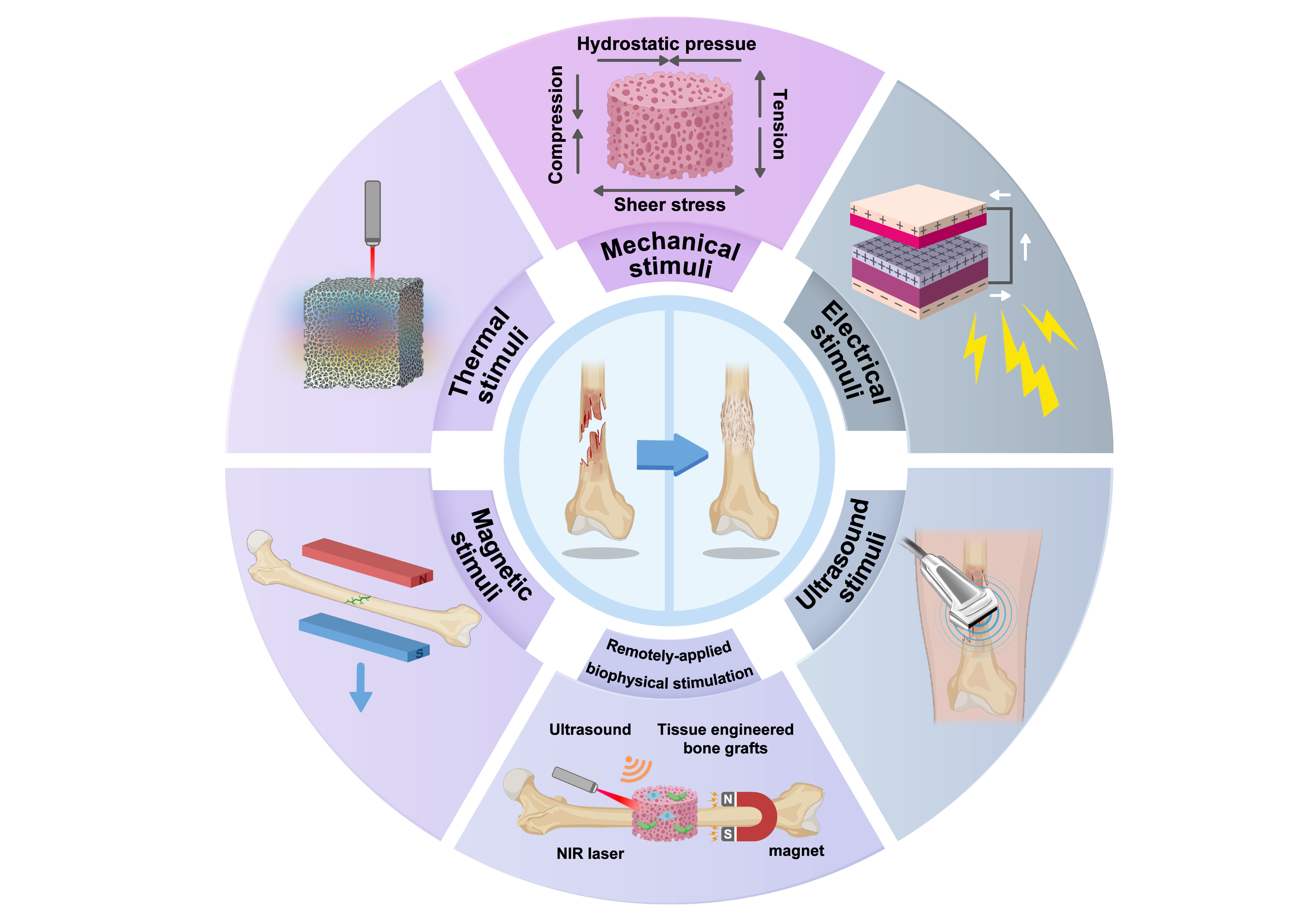
Articles in the same Issue
- Frontmatter
- Reviews
- Biophysical stimuli for promoting bone repair and regeneration
- A stepwise approach to deriving functional β-cells from human embryonic or induced pluripotent stem cells
- Eco-fertility: examining the climate change-total fertility rate nexus in the context of sustainable developmental goals in a systematic review approach
- Redefining chronic mountain sickness: insights from high-altitude research and clinical experience
- Review of organ damage from COVID and Long COVID: a disease with a spectrum of pathology
- Perspective
- Combining transcriptomic and metabolomic insights to guide the clinical application of adipose- and bone marrow-derived mesenchymal stem cells
- Research Highlight
- Precision phototherapy and imaging with aggregation-induced emission-based nanoparticles cloaked in macrophage membrane