
Giant granules in white blood cells
-
Peter Schuff-Werner
 and
Christian Junghanss
and
Christian Junghanss

A 33 years old female suffering from recurrent bacterial and fungal infections was moved from a local nursing home to our palliative care unit because of increasingly inadequate food and fluid intake due to severe painful fungal and herpes stomatitis. The evident immune-deficient patient presented ocular albinism, congenital nystagmus and mental retardation. Laboratory analyses showed slight anemia (hemoglobin (7.6 mmol/L)), leukocytopenia (2.77 × 10⁹/L) due to absolute neutropenia (0.5 × 10⁹/L), decreased electrolytes, highly elevated C-reactive protein (CRP) (128 mg/L), elevated IgG (18.5 g/L), markedly increased gamma-glutamyltransferase (286 U/L) and alkaline phosphatase (220 U/L). Liver enzymes and renal function were inconspicuous. Platelets were within the lower normal range (179 × 109/L).
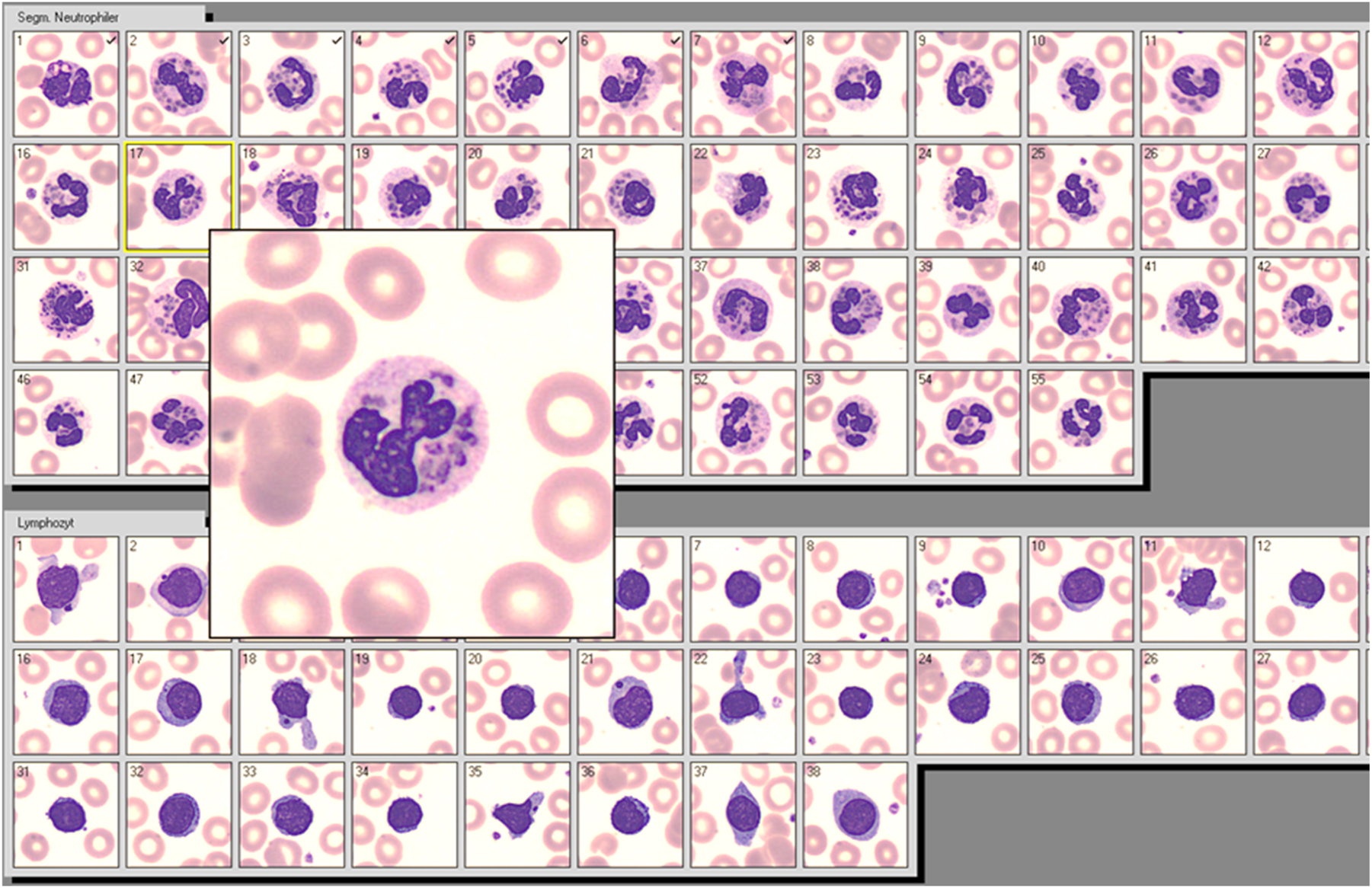
Neutrophils and lymphocytes from a patient with Chediak-Higashi-Syndrome showing cytoplasmatic aggregates of dysfunctional lysosomes.
The routine WBC differential was performed by automated digitalized microscopy (Cellavision™, Sysmex Europe, Norderstedt, Germany), showing a series of leukocytes with multiple characteristic cytoplasmic inclusions and lymphocytes with giant azurophil granules.
This autosomal recessive inherited syndrome of deficient innate immunity due to misdistribution of myeloperoxidase was first observed by Beguez-Cesar [1] and further described by Chediak, a Cuban hematologist [2], and Higashi, a Japanese pediatrician [3]. This explains the designation of the disease as Chediak-Higashi-Syndrome (CHS).
The molecular basis is the mutation of the LYST/CHS1 gene located at chromosome 1 [4]. The gene product, a highly conserved cytosolic protein (“lysosomal trafficking regulator”), is differently expressed in all cell types [5].
There is no specific treatment, except allogeneic hematological stem cell transplantation (HSCT). The here presented case of the syndrome, which normally leads to premature death within the first two decades of life [6] was far accelerated at admission, receiving palliative care until death 17 days later.
According to the literature, the giant WBC inclusions are the most reliable clinical diagnostic criterion of CHS (see Figure 1), which might be overlooked in conventional blood films of CHS patients with typical neutropenia [7]. Automated digital microscopy allows the careful review of more than hundred neutrophils displayed simultaneously; thus facilitating the detection of characteristic cellular inclusions. Digital microscopy might be therefore advantageous for rapid diagnosis of CHS in the hematological routine.
-
Research ethics: Not applicable.
-
Informed consent: Not applicable.
-
Author contributions: All authors have accepted responsibility for the entire content of this manuscript and approved its submission.
-
Use of Large Language Models, AI and Machine Learning Tools: None declared.
-
Conflict of interest: The authors state no conflict of interest.
-
Research funding: None declared.
-
Data availability: Not applicable.
References
1. Beguez-Cesar, A. Neutropenia cronica maligna familiar con granulaciones atipicas de los leucocitos. Bol Soc Cubana Pediatr 1943;15:900–22.Search in Google Scholar
2. Chediak, MM. New leukocyte anomaly of constitutional and familial character. Rev Hematol 1952;7:362–7.Search in Google Scholar
3. Higashi, O. Congenital gigantism of peroxidase granules; the first case ever reported of qualitative abnormality of peroxidase. Tohoku J Exp Med 1954;59:315–32. https://doi.org/10.1620/tjem.59.315.Search in Google Scholar PubMed
4. Kuptanon, C, Morimoto, M, Nicoli, E-R, Stephen, J, Yarnell, DS, Dorward, H, et al.. cDNA sequencing increases the molecular diagnostic yield in Chediak-Higashi syndrome. Front Genet 2023;14:1072784. https://doi.org/10.3389/fgene.2023.1072784.Search in Google Scholar PubMed PubMed Central
5. Talbert, ML, Malicdan, MCV, Introne, WJ. Chediak-Higashi syndrome. Curr Opin Hematol 2023;30:144–51. https://doi.org/10.1097/moh.0000000000000766.Search in Google Scholar
6. Kaplan, J, de Domenico, I, Ward, DM. Chediak-Higashi syndrome. Curr Opin Hematol 2008;15:22–9. https://doi.org/10.1097/MOH.0b013e3282f2bcce.Search in Google Scholar PubMed
7. Dotta, L, Parolini, S, Prandini, A, Tabellini, G, Antolini, M, Kingsmore, SF, et al.. Clinical, laboratory and molecular signs of immunodeficiency in patients with partial oculo-cutaneous albinism. Orphanet J Rare Dis 2013;8:168. https://doi.org/10.1186/1750-1172-8-168.Search in Google Scholar PubMed PubMed Central
© 2025 the author(s), published by De Gruyter, Berlin/Boston
This work is licensed under the Creative Commons Attribution 4.0 International License.
Articles in the same Issue
- Frontmatter
- Editorial
- Can a digital smear review be helpful in the routine haematology laboratory?
- Original Articles
- The impact of mutational burden, spliceosome and epigenetic regulator mutations on transfusion dependency in dysplastic neoplasms
- Improving turn-around times in low-throughput distributed hematology laboratory settings with the CellaVision® DC-1 instrument
- Hematology instruments don’t speak the same language: a comparison study between flagging messages of sysmex XN-1000 and alinity H
- Platelet clump assessment using the Cellavision peripherical blood application – do we need manual microscopy?
- Short Communication
- Development of a peripheral blood morphology proficiency assessment program using the CellaVision® Proficiency Software
- Images From the Medical Laboratory
- Giant granules in white blood cells