Enlargement of hepatic hemangioma in successive pregnancies
-
Paula E. Zozzaro-Smith
 ,
Elizabeth Fountaine
,
Elizabeth Fountaine


Abstract
Hemangiomas are the most common benign tumor of the liver and are commonly seen as incidental findings on abdominal imaging. Little is known regarding the natural course and pathogenesis of hepatic hemangiomas during pregnancy. It is believed estrogen may play a role in their growth; however, the precise mechanism remains undefined. Here we describe a patient with hepatic hemangioma enlargement in consecutive pregnancies suggesting a hormonal role in their growth.
Introduction
Hepatic hemangiomas, also known as cavernous hemangiomas, are common benign tumors found in the liver. They are typically solitary; however, they can be present as multiple lesions throughout the liver. Most are small and asymptomatic; however, larger lesions (>4 cm) can cause significant abdominal pain. Hemangiomas can present at any age but are most commonly seen between the ages of 30 and 50 years and most frequently in women [1].
There are few available cases to guide an optimal approach to obstetrical management and intervention during pregnancy. The natural progression of hepatic hemangiomas is also not completely well understood. The majority appear to remain stable over time. Insufficient evidence exists to conclusively link estrogens to the development or growth of hemangiomas. Enlargement has been noted to occur during pregnancy and during estrogen and progesterone therapy with regression following withdrawal of therapy [2, 3]. Since estrogen may predispose the growth of these tumors, concern for rupture prevents definitive conclusions on the optimal timing and route of delivery in these patients. Here we report an individual case of hepatic hemangioma enlargement in consecutive pregnancies and management during pregnancy.
Presentation of the case
A 30-year-old White female, with a known 2.7×3.7 cm hepatic hemangioma detected by abdominal ultrasound and magnetic resonance imaging (MRI) 5 years earlier was referred during her third pregnancy for worsening right upper quadrant pain. An MRI demonstrated an increase in size of the hemangioma to 6.5×4.3×6.0 cm. Her pain slightly improved with narcotics, and she underwent an uncomplicated repeat cesarean section at 37 weeks’ gestation following a mature amniocentesis secondary to continued epigastric and right upper quadrant pain. The right hepatic hemangioma was noted to measure 6.5×7.1×4.9 cm 1-month postpartum.
She then represented early in her fourth pregnancy for prenatal care 3 years later. At 8 weeks’ gestation, a baseline abdominal ultrasound demonstrated a 7.3×8.9×8.0 cm asymptomatic liver hemangioma. At 28 weeks’ gestation, she again complained of severe right upper quadrant pain where an abdominal ultrasound showed no interval change in size of the lesion. The patient continued to have abdominal pain requiring increasing narcotic use and frequent hospitalizations for pain control. She received a course of antenatal corticosteroids for fetal lung maturity during admission in which an abdominal ultrasound performed at 32 weeks’ gestation noted expansion of the hemangioma to 10 cm in width (Figure 1). A follow-up MRI confirmed hemangioma growth compared with the size noted on her previous study without evidence of hemorrhage (Figure 2).
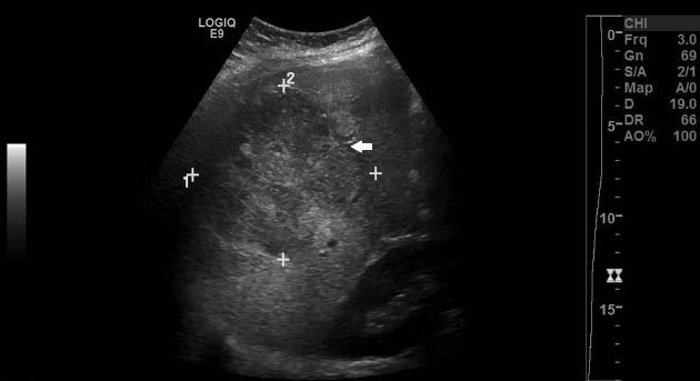
Abdominal ultrasound of the maternal liver noting interval increase in size of known right hepatic lobe hemangioma (arrow).

Axial and coronal T2-weighted magnetic resonance images. Enlarging hemangioma (arrows) occupying the right hepatic lobe.
She continued to have progressively worsening right upper quadrant pain. Secondary to the patient’s intractable abdominal pain and transplant surgery’s concern for impending rupture given the hemangioma’s size >10 cm, the patient underwent repeat cesarean delivery and bilateral tubal ligation at 34 weeks’ gestation. Her right upper quadrant pain persisted postpartum, and a follow-up abdominal ultrasound showed the liver hemangioma size to be unchanged from 10 cm noted on ultrasound performed earlier in pregnancy. She presented for a postpartum visit 6 weeks later and noted a significant decrease in her abdominal discomfort for which she no longer required medication with a plan to follow-up with her hepatologist for further management.
Discussion
Hepatic masses in pregnancy are rare; however, they can pose a significant challenge to obstetrical care. Their etiology can range from infectious to malignancies, and their progression and prognosis is dependent on the underlying disorder. Identification of a liver mass can be challenging during pregnancy as hepatic abnormalities during pregnancy require diagnosis in the context of anticipated physiologic changes. In addition, diagnostic and therapeutic strategies have to consider the implications for both the mother and the fetus. The clinical presentation of hepatic masses during pregnancy depends on the nature of the mass and does not significantly vary from the usual presentation in a non-pregnant woman [4]. An individual may be completely asymptomatic or present with pain or distension related to pressure from an expanding uterus [5]. They may also present in hemorrhagic shock from rupture of the mass [5]. The mechanism of enlargement is unclear. Hepatic hemangioma enlargement appears to be affected by both endogenous and exogenous female sex hormones. A female-male ratio of 5:1 has been observed and affected women tend to be multiparous with lesions enlarging during gestation [5]. In one study, a 2-fold increase in size was noted in women exposed to hormone therapy compared to controls [6]. Enlargement of these tumors has also been reported during pregnancy further suggesting a possible relationship between hemangiomas and estrogen [3, 7]. However, a subsequent study produced contradictory conclusions noting no significant association between oral contraceptive use and the development of hepatic hemangiomas [8]. In our patient, hepatic hemangioma enlargement was demonstrated in not only one but subsequent pregnancies. This suggests a relationship between pregnancy and hemangioma enlargement and further implies a relationship between hemangiomas and hormone exposure.
The precise mechanism linking estrogen to hemangioma enlargement is incompletely understood. In other hormonally active tumors with growth facilitated by estrogen, there has been demonstrated estrogen and progesterone receptors within these tumors. However, this has not been demonstrated on hepatic hemangiomas. For example, it has become evident that several different human breast cancers are dependent upon estrogen with growth facilitated through estrogen receptors [9]. When tested, approximately 70% of breast cancers have positive estrogen or progesterone receptors, and these tumors show progression during hormone exposure [9]. However, despite the progression of hepatic hemangiomas in the presence of estrogen, there has been no report of estrogen receptors specifically in hepatic hemangiomas. Saegusa et al. reported surgical resection of multiple hemangiomas that were negative for both estrogen and progesterone receptors [7]. An alternative premise to explain this hemangioma enlargement in the presence of hormones has been proposed – suggesting that as hemangiomas are considered to be vascular malformations that enlarge by dilation of tissue, estrogen plays an indirect role through vascular dilatation [10]. However, the exact role and interaction between estrogen and hepatic hemangioma enlargement remains unclear.
The lack of ample literature on pregnancies complicated by hepatic hemangiomas precludes definitive conclusions regarding the precise timing and route of delivery. Due to the capacity for enlargement with estrogen exposure in pregnancy, concern for potential complications can influence obstetrical management. One of the largest reviews included pregnancies with hepatic hemangiomas ranging in size from 2 to 60 cm. Spontaneous rupture was reported in one case with a hemangioma that was 10 cm in size [5]. A symptomatic liver hemangioma with intratumor hemorrhage has also been described requiring embolization during pregnancy [11]. Liver hemangiomas have also been known to cause thrombocytopenia and consumptive coagulopathy due to tumor enlargement and rupture [12]. The potential risks involved in these pregnancies clearly detail the need for multidisciplinary care. Hemangiomas can generally be managed conservatively during pregnancy and vaginal delivery is appropriate. When complications occur, they are more frequent in lesions >10 cm. However, even large lesions can result in only mild symptoms and have an uneventful course during pregnancy [13]. Each case must be individualized, and potential indications for early intervention can include worsening symptoms or rapidly enlarging lesions, as in our patient’s case.
References
[1] Gandolfi L, Leo P, Solmi L, Vitelli E, Verros G, Colecchia A. Natural history of hepatic hemangiomas: clinical and ultrasound study. Gut. 1991;32:677–80.10.1136/gut.32.6.677Search in Google Scholar
[2] Conter RL, Longmire WP. Recurrent hepatic hemangiomas: possible association with estrogen therapy. Ann Surg. 1988;207:115–9.10.1097/00000658-198802000-00001Search in Google Scholar
[3] Au W, Liu C. Growth of giant hepatic hemangioma after triplet pregnancy. J Hepatol. 2005;42:781.10.1016/j.jhep.2004.08.028Search in Google Scholar
[4] Athanassiou AM, Craigo SD. Liver masses in pregnancy. Semin Perinatol. 1998;22:166–77.10.1016/S0146-0005(98)80049-9Search in Google Scholar
[5] Cobey FC, Salem RR. A review of liver masses in pregnancy and a proposed algorithm for their diagnosis and management. Am J Surg. 2004;187:181–91.10.1016/j.amjsurg.2003.11.016Search in Google Scholar PubMed
[6] Glinkova V, Shevah O, Boaz M, Levine A, Shirin H. Hepatic hemangiomas: possible association with female sex hormones. Gut. 2004;53:1352–5.10.1136/gut.2003.038646Search in Google Scholar PubMed PubMed Central
[7] Saegusa T, Ito K, Oba N, Matsuda M, Kojima K, Tohyama K, et al. Enlargement of multiple cavernous hemangioma of the liver in association with pregnancy. Intern Med. 1995;34:207–11.10.2169/internalmedicine.34.207Search in Google Scholar PubMed
[8] Gemer O, Moscovici O, Dosoretz Ben-Horin CL, Linov L, Peled R, Shmuel S. Oral contraceptives and liver hemangioma: a case-control study. Acta Obstet Gynecol Scand. 2004; 83:1199–201.10.1111/j.0001-6349.2004.00551.xSearch in Google Scholar PubMed
[9] Mehta A, Tripathy D. Co-targeting estrogen receptor and HER2 pathways in breast cancer. Breast. 2014;23:2–9.10.1016/j.breast.2013.09.006Search in Google Scholar PubMed
[10] Van Malenstein H, Maleux G, Monbaliu D, Verslype C, Komuta M, Roskams T, et al. Giant liver hemangioma: the role of female sex hormone and treatment. Eur J Gastroenterol Hepatol. 2011;23:438–43.10.1097/MEG.0b013e328345c87dSearch in Google Scholar PubMed
[11] Graham E, Cohen A, Soulen M, Faye R. Symptomatic liver hemangioma with intra-tumor hemorrhage treated by angiography and embolization during pregnancy. Obstet Gynecol. 1993; 81:813–6.Search in Google Scholar
[12] Ebina Y, Hazama R, Nishimoto M, Tanimura K, Miyahara Y, Morizane M, et al. Resection of giant liver hemangioma in a pregnant woman with coagulopathy: case report and literature review. J Prenat Med. 2011;5:93–6.Search in Google Scholar
[13] Marques R, Taborda F, Jorge C, Areias J, Rodrigues A. Successful outcome in a pregnancy complicated by large hepatic hemangioma. Acta Obstet Gynecol Scand. 1997;76:606–7.10.3109/00016349709024595Search in Google Scholar PubMed
-
The authors stated that there are no conflicts of interest regarding the publication of this article.
©2015 by De Gruyter
Articles in the same Issue
- Frontmatter
- Case reports – Obstetrics
- Minimally invasive procedure for type II canal defect caesarean scar pregnancy with cardiac activity and high hCG titres at 8+2 weeks of gestation
- Rare causes of acute abdomen in pregnancy: “ultrasound to the rescue”. A review of two cases
- Enlargement of hepatic hemangioma in successive pregnancies
- Misdiagnosis of macroamylasemia in pregnancy as pancreatitis
- An advanced cervical ectopic pregnancy
- Multidisciplinary management of giant genital tract venous malformations during pregnancy: case report and review of the literature
- Acute uterine rupture in spontaneous term labour in a healthy primigravida: case report and review of the literature
- Massive ascites in a patient with preeclampsia
- Loeys-Dietz syndrome in pregnancy
- Prenatal diagnosis of periventricular venous infarction in utero: a case with hereditary protein C deficiency
- Case reports – Fetus
- Placental chorioangioma presenting prenatal hemolytic anemia and consumption coagulopathy: a case report
- Management of fetal ovarian cyst using in utero aspiration
- A case of fetal cardiac rupture diagnosed by postmortem magnetic resonance image
- Unusual presentation of fetus in fetu in triplet pregnancy mimicking abdominal wall defect
- Acral necrosis and upper brachial plexus palsy after prenatal fetal thrombosis
- Prenatal diagnosis of a giant fetal hepatic hemangioma: a case report
- Prenatal diagnosis and outcomes of fetal cardiac rhabdomyomas: evaluation of seven cases
- Case reports – Newborn
- Polythelia and associated hydronephrosis: a case report in neonatal age
- Necrotizing enterocolitis following intensive phototherapy in full-term newborns – is there a possible association?
- A case of neonatal toxic shock syndrome-like exanthematous disease concurrent with maternal toxic shock syndrome