A history of urine microscopy
-
J. Stewart Cameron


Abstract
The naked-eye appearance of the urine must have been studied by shamans and healers since the Stone Age, and an elaborate interpretation of so-called Uroscopy began around 600 AD as a form of divination. A 1000 years later, the first primitive monocular and compound microscopes appeared in the Netherlands, and along with many other objects and liquids, urine was studied from around 1680 onwards as the enlightenment evolved. However, the crude early instruments did not permit fine study because of chromatic and linear/spherical blurring. Only after complex multi-glass lenses which avoided these problems had been made and used in the 1820s in London by Lister, and in Paris by Chevalier and Amici, could urinary microscopy become a practical, clinically useful tool in the 1830s. Clinical urinary microscopy was pioneered by Rayer and his pupils in Paris (especially Vigla), in the late 1830s, and spread to UK and Germany in the 1840s, with detailed descriptions and interpretations of cells and formed elements of the urinary sediment by Nasse, Henle, Robinson and Golding Bird. Classes were held, most notably by Donné in Paris. After another 50 years, optical microscopy had reached its apogee, with magnifications of over 1000 times obtainable free of aberration, using immersion techniques. Atlases of the urinary sediment were published in all major European countries and in the US. Polarised light and phase contrast was used also after 1900 to study urine, and by the early 20th century, photomicroscopy (pioneered by Donné and Daguerre 50 years previously, but then ignored) became usual for teaching and recording. In the 1940s electron microscopy began, followed by detection of specific proteins and cells using immunofluorescent antibodies. All this had been using handheld methodology. Around 1980, machine-assisted observations began, and have dominated progress since.
“When the patient dies the kidneys may go to the pathologist, but while he lives the urine is ours. It can provide us day by day, month by month, and year by year with a serial story of the major events within the kidney. The examination of the urine is the most essential part of the physical examination of any patient...” (Thomas Addis, 1948 [1]).
Urine examination before microscopy and chemistry: the stone age to 1580
From the earliest days of shamans and healers in the Palaeolithic era, perhaps 30–40,000 years or more ago, the urine coming out of the body has been examined by the naked eye to see what it might tell us about what was going on inside the body. Egyptian and Babylonian texts both mention and describe the appearances of urine, its volume and colour. From one and a half millennia ago [2, 3] we have descriptions of the urine from the Greek and Roman corpus of medical writings: the examination of the urine was already part of the Hippocratic system 500 years before the beginning of the Christian era.
Thus for a 1000 years or more in the post-Classical era, from the 7th century AD in the Byzantine period, the ideas of Theophilus on “Uroscopy” held sway in an increasingly elaborate form [4–7], despite the fact that the clinical return from this system of relating urine appearance and the sediment it threw on cooling must have given information of negligible use. It was essentially diagnosis by divination.
Then, as the influence of science began in the early 17th century in the early days of the enlightenment, one of the crucial drivers of innovation was a new technology: the development of useful lenses which could magnify the microscopic as well as the stellar world. In parallel, Chemistry was born from Alchemy, and at first hesitantly to be applied to body fluids, including urine.
Glass is of course a naturally occurring substance, and on occasion must have formed droplets useful for magnification. However, there is evidence of polishing also in some ancient lenses. Several hoards from ancient sites have been found [8], from Egypt from the IVth and Vth dynasties (ca 2500 BC) (but not the VIth and later), Minoan Crete and Troy, and Carthage. A provoking object is a pre-dynastic carving from Abydos in Egypt also, which can be seen only using magnification, on an ivory handle from 1300 BC. The “Nimrud lens” from the palace of Sargon, discovered in Assyria by Layard in 1853 and now in the British Museum in London, is one of the best known ancient lenses dating from around 700–1000 BC. Certainly the Greeks of the classical period, as well as the Romans knew the magnifying properties of glass well, and Aristophanes (430 BC) (In “The clouds”), Pliny and Seneca actually describe the use of lenses for magnification – sometimes called “reading stones”. Magnifications were generally 2–3 at best, but one lens from Crete achieves ×7.
Spectacles have been suggested on Egyptian mummies, but the convex crystals were most probably for decoration of empty eye sockets – missing parts were often added post mortem to mummies. The first recorded spectacles we know of were described in Italy from 1253 onwards by Salvino d’Armate (1258–1302) and were in widespread use by 1400, but these were heavy, carried and used in the hand rather than worn on the nose.
The birth of the microscope and microscopy: 1580–1700
There remains debate as to who first conceived the idea of using two lenses in series perhaps mounted in a tube to view objects [9–11], but in the case of the microscope credit is usually given to the Janssen family of Middleburgh in the Netherlands, father Hans (d.1590) and son Zacharias (1580–1638), who made spectacles and were skilled in lens making. Other Dutch lens-makers, such as Hans Lippershey (1570–1619), were also involved [12]. Rapidly, simple tube handheld microscopes (and telescopes) became available throughout Europe, as lens-making for spectacles improved. We have reviewed the subsequent history of urine microscopy previously [13], and this article can be consulted for many unreferenced statements below.
Thus urine was examined as early as 1630 by the Provençal astronomer and polymath Nicolas-Claude Fabricius de Peiresc (1580–1637) in 1630, who described urinary crystals resembling “a heap of rhomboidal bricks”[1] [14, 15]. This description was taken up by the English polymath, Robert Hooke (1635–1703) later in the century [16], who used a three-lensed microscope giving up to ×50 magnification, and whose book Micrographia opened up the world of the very small to the world. These crystals were in all probability uric acid, possibly calcium oxalate. In contrast the famous Dutch microscopist of the late 17th century, Antoni van Leeuwenhoek (1632–1723) used only exquisitely ground droplet single lenses to achieve magnifications up to an amazing ×270, but because his specimens were mounted on to a metal point for examination, he made almost no observations on urine, although seeing crystals in a droplet of urine [12]. Magnifications were consistently much lower using two lenses, but the increased focal length was a huge practical advantage in examining specimens.
Stasis in the 18th century: 1700–1810
The images produced by all microscopes from the 18th century (of which we have a considerable number extant) showed poor resolution, with severe spherical and particularly chromatic aberrations; images were fuzzy and multicoloured, with multiple overlapping images. Although as early as 1730 the use of separate crown and flint glass lenses to diminish this by exploiting their differing refractive properties had been described, this manoeuver made only a small difference in practice. For science, better instruments were required but remained lacking, thus limiting use of microscopy in medicine right throughout the 18th century. Even so, Hermann Boerhaave (1668–1738), the famous Dutch teacher and clinician (Figure 1) almost uniquely for the period performed careful experiments in 1704 [17] watching urine from a normal healthy individual cool under the microscope to show that crystals gradually appeared, proving that on occasion constituents of normal urine could give rise, eventually, to stone formation.

Hermann Boerhaave (1668–1738) and his de Calculo of 1704.
Although bloody urine had been noted in scarlatinal nephritis throughout the 17th and 18th centuries [18–20], most notably by the astute Swedish paediatrician Rosén von Rosenstein (1706–1773) [21], I have been unable to find any record during this period of the confirmation of the presence of red cells in the urine by microscopy, although Leeuwenhoek and others had described corpuscles in the blood; that may come as a result of further enquiry, and it would be surprising if none of the enquiring 18th century minds did not think to look for Leuwenhoek’s corpuscles in urine which appeared to contain blood. However, neither Domenico Cotugno (1736–1780) [22], who discovered and named albuminuria in the 1770s, nor even Richard Bright (1789–1858) and John Bostock (1773–1846) (although they made many chemical observations on their patients’ body fluids [23]) actually used a microscope on their patient’s urine in the 1820s.
New lenses, improved microscopes and clinical microscopy is born: 1810–1840
But help was at hand. During this and the previous decade huge advances were made in lens making, and microscopy rapidly became a useful then essential tool in biology, pathology and the medical clinic. These advances were contributed from a large number of individual lens-makers in several countries, who were able eventually to form compound lenses of flint and crown glass bonded together, and so hugely improve resolution and combat aberration. Notable amongst these were, first, the London Quaker and wine merchant Joseph Jackson Lister (1786–1868) (Figure 2A), father of the noted surgeon Lord Lister [24]. The elder Lister had himself no skill in lens making, but employed skilled craftsmen to make lenses to his designs. He also described and developed optical theory considerably. In 1827 he published with the Guy’s hospital physician and pathologist Thomas Hodgkin (1798–1866) a paper [25] employing the microscope to examine organs and tissues, which founded modern microscopic anatomy.

Important creators of compound achromatic lenses: (A) Joseph Jackson Lister with one of his achromatic telescopes, (B) Giovanni Battista Amici, (C) Charles Chevalier (see text).
All images courtesy of Wikimedia Commons.
Another pioneer was the master lens-maker Italian Giovanni Battista Amici (1786–1863) of Modena [26, 27] (Figure 2B), who worked also in Paris selling his products. He was amongst the first to study compound achromatic lenses, in 1814, but separately mounted. Both he and Lister made telescopes as well as microscopes, and studied astronomy. Amici’s work in Paris helped improve the products of a number of master microscope makers already there, particularly the Chevalier family, father Vincent (1770–1841) and his son Charles (1804–1859) (Figure 2C) with whom he collaborated extensively, and later the Nachets, father Camille (1799–1881) and son Jean (1831–1908). Chevalier’s microscope had a resolution of 1.7 μm and a magnification of ×280–540, but as early as 1835 Johann Georg (Georges) Oberhauser (1798–1868), a Bavarian settled in Paris, had made a microscope capable of magnifications up to 1000 diameters with a resolution of 0.7 μm – figures scarcely surpassed since then, although distortion has been reduced dramatically[2] [28, 29]. In the late 1820s and 1830s new greatly improved objectives were developed also.
It seems credit must be given to the founder of French nephrology, Pierre Rayer (1793–1867) (Figure 3A), and his young associate (“Interne”) Eugène Napoléon Vigla (1813–1872) (Figure 3B) for the introduction of regular urine microscopy to clinical practice, described first in 1837 [30–32]. Vigla describes how impressed Rayer was by the results of microscopic examination of urine from one of his patients, supposedly full of pus, but which in fact contained abundant crystals by a visiting microscopist, the young Gottlieb Gluge (1812–1898) of Berlin, later based in Brussels. He was probably the first physician to examine renal tissue microscopically in 1839 (see below). Rayer vowed to make microscopic examination of the urine a regular feature of his clinical practice. In the preface of his magnificent “Traité des Maladies des Reins” (Treatise of Diseases of the Kidneys) published in three volumes between 1839 and 1841 [30], he writes:
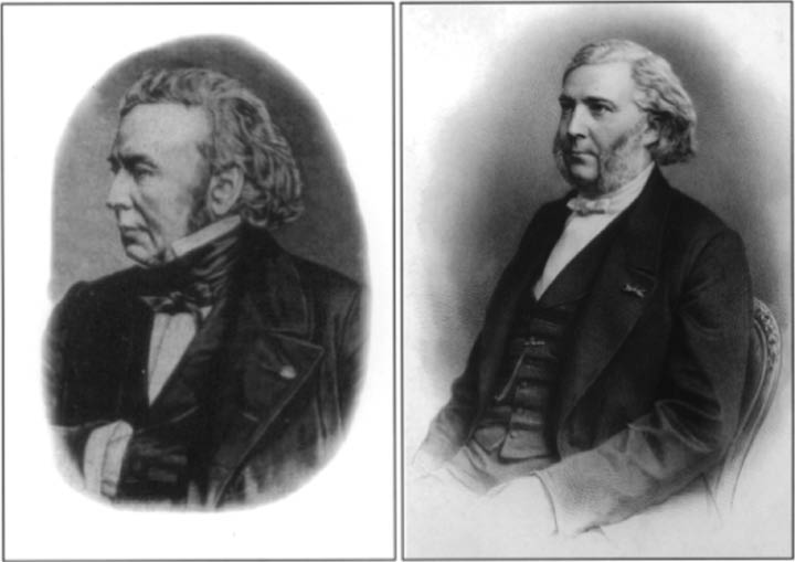
Pierre Rayer (left) and Eugène Vigla (right).
These authors were the first to record most of the formed elements of the urinary sediment (red cells, white cells epithelial cells etc., but probably not casts) in 1837–1838 (see text). Courtesy of the Wellcome Library, London.
“It is to be regretted that another method of investigation, microscopical examination, is not yet generally employed to examine the matter suspended in the urine, thrown down by cooling, or which one may precipitate by various reagents... I cannot thus understand the lack of urgency in the majority of physicians to avail themselves of the microscope in the examination of the urine”.
Gabriel Richet wrote [33] “la microscopie de l’urine, en revanche, fut crée par Rayer lui-même” (the microscopy of the urine was created by Rayer himself) and noted that Rayer made a microscope available at all hours for his associates to microscope the urine (it was probably one made by Chevalier himself, or perhaps the German Oberhauser who worked in Paris). Rayer and Vigla examined and analysed the crystals present in the urine, as many had before him, but – and this was new – he also noted the red cells, the pus cells, the epithelial cells, fatty bodies and sperm. They realised that otherwise normal (clear) urine might nevertheless contain an excess of red cells – the first description I can find, surprisingly late, of microscopic haematuria. Vigla never published another paper.
We must remember, however, that Rayer and Vigla’s priority in introducing clinical urine microscopy was hotly disputed by their colleague at the Charité Hospital Alfred Donné (1801–1878)[3] [34] (Figure 4, left), who ran courses in microscopy, not only for doctors but for the general public. The first course, begun in 1838, was published in 1844. It was illustrated by engravings of the first ever photomicrographs, taken together with his pupil Léon Foucault (1819–1869) using the new technique of Louis Daguerre (1789–1851) in the same year as it was described (1840) (Figure 4, centre), although these pictures were only published 5 years later [12, 34–36]. They included several plates of urinary crystals (Figure 4, right). Donné and Foucault went on to use artificial light (an electrically driven carbon arc) to shorten the exposure, another major innovation. Although a few British and other microscopists took up photomicrography, its complexity resulted in it being forgotten for more than 40 years. Donné, unlike Vigla and Rayer, was a general microscopist and did no further studies on urine, and is best remembered today for his advocacy of the thermometer in medicine (1835), the appearance of the blood in leukaemia (1839), descriptions of Trichomanas vaginalis (1837), and the composition of milks (1842). He also, (and not Bizzozero in 1882 as usually stated) first described platelets in 1842, although he did not study their function.
![Figure 4: (Left) Alfred Donné. From: (Thorburn AL. Alfred François Donné, 1801–1878, discoverer of Trichomonas vaginalis and of leukaemia. Br J Vener Dis 1974;50:377–80 [35]). Surprisingly, this sketch seems to be the only image of Donné, and was obtained from a descendant by Thorburn. It probably dates from 1820 to 1825. (Centre) Donné and Foucault’s photomicrograph. Illumination was supplied by a battery-powered electric carbon arc (g′) with rods vertical to the plane of the page. This supplied reflected light from the parabolic mirror on the right, to the specimen. On the left is the microscope (m) from which the image was projected on to the photographic plate, off left (not shown). From Pouillet M, Eléments de Physique Expérimentale et de Météorologie. Paris, Béchet Jeune, 1844. See pp 746–50 and Figure 30a. (Right) Engraving of a photomicrograph of uric acid crystals in urine from (Donné A, Foucault L. Cours de microscopie complémentaire des études médicales. Paris, J-B Ballière 1844–5. Donné’s atlas of 20 plates and 80 illustrations of daguerrotypes includes the urinary sediment. Donné appears to have published also a “Tableau des différens dépôts de matières salines et de substances organisées qui se font dans les urines” (also listed as: Tableau des sédiments des urines) in 1838, perhaps including the same material, but I have been unable to locate copy of this so far. Vigla quotes Donné’s work on sperm in his 1837 paper. [34]) Some authors have stated, mistakenly, that these pictures are actual photomicrographs.](/document/doi/10.1515/cclm-2015-0479/asset/graphic/j_cclm-2015-0479_fig_004.jpg)
(Left) Alfred Donné. From: (Thorburn AL. Alfred François Donné, 1801–1878, discoverer of Trichomonas vaginalis and of leukaemia. Br J Vener Dis 1974;50:377–80 [35]). Surprisingly, this sketch seems to be the only image of Donné, and was obtained from a descendant by Thorburn. It probably dates from 1820 to 1825. (Centre) Donné and Foucault’s photomicrograph. Illumination was supplied by a battery-powered electric carbon arc (g′) with rods vertical to the plane of the page. This supplied reflected light from the parabolic mirror on the right, to the specimen. On the left is the microscope (m) from which the image was projected on to the photographic plate, off left (not shown). From Pouillet M, Eléments de Physique Expérimentale et de Météorologie. Paris, Béchet Jeune, 1844. See pp 746–50 and Figure 30a. (Right) Engraving of a photomicrograph of uric acid crystals in urine from (Donné A, Foucault L. Cours de microscopie complémentaire des études médicales. Paris, J-B Ballière 1844–5. Donné’s atlas of 20 plates and 80 illustrations of daguerrotypes includes the urinary sediment. Donné appears to have published also a “Tableau des différens dépôts de matières salines et de substances organisées qui se font dans les urines” (also listed as: Tableau des sédiments des urines) in 1838, perhaps including the same material, but I have been unable to locate copy of this so far. Vigla quotes Donné’s work on sperm in his 1837 paper. [34]) Some authors have stated, mistakenly, that these pictures are actual photomicrographs.
The young Gottlieb Gluge[4] [37] was only one pioneer of renal histology in the 1830s, publishing in 1839. Another was Gabriel Gustav Valentin (1810–1883), also a Jewish German, working in Bern[5] [38], whose double-bladed knife introduced in 1838 allowed thickish sections of fresh tissue to be cut which could be trans-illuminated on the microscope stage; it was used for 4–5 decades until paraffin-embedding made microtomes more efficient. They both noted what we would today recognise as casts within the distal renal tubules, but did not examine urine.
In 1840 Bright and his student Joseph Toynbee (1815–1866) were examining normal and diseased kidneys microscopically also [39, 40] – although they used microdissection of the tissue rather than sections. Interestingly, William Bowman (1816–1892), although he made no microscopical observations on the urine, clearly appreciated that red cells could pass through the Malpighian corpuscles in disease into the urine. He describes, in a footnote to his 1842 paper, kidneys from patients with Bright’s disease that he examined under the microscope [41]:
“It is well known that blood is often passed in the urine during the course of the disease, especially at the earlier periods of it, when many circumstances contribute to prove that the kidneys are in a state of sanguineous turgescence. How does this blood escape into the ducts of the gland? The organ examined at this time presents on its surface and throughout its cortical substance, scattered red dots, of somewhat irregular shape, not accurately rounded, and generally as large as pins’ heads, that is very many times larger than the Malpighian bodies... they are nothing less than the convolutions of the tube filled with blood that has burst into it from the gorged Malpighian tuft at its extremity”.
The urine sediment described: 1840–1900
Thus as early as 1841, in his classic book on the analysis of urine [42], Alfred Becquerel (1814–1866) could refer casually to “examen micro-scopique” (microscopic examination) of the urine: and he notes again that in perfectly clear urine, one sees only sheets of epithelium, in urine with mucus, globules of this substance, closely resembling globules of pus, and also red blood cells “plus souvent déformés et irreguliers” (more often distorted and irregular) – perhaps the first description of dysmorphic red cells? He noted also sperm, and of course crystals: calcium and magnesium carbonate, and phosphates, including ammonium magnesium phosphate. Animal chemistry had developed rapidly since the beginning of the 19th century [43–45] and the chemical approach to disease was being applied in many centres throughout Europe, so Rayer and his pupils applied both approaches in parallel.
The years 1842–1844 saw a number of workers in Germany describing casts in the urine almost at the same time. Jacob Henle[6] (1809–1885) [46], using a German microscope made by Schiek, crucially recognised the tubular casts seen in histological sections of the kidney as identical to those found in the urine in 1842, supposing them to be made of coagulated fibrin; Theodor Frerichs (1819–1885), writing his influential book on “Die Bright’sche Nierenkrankheit” in Breslau (now Wroclaw) in 1851, also credits Hermann Nasse (1807–1892) [47] as reporting them at about the same time. The following year, Johann Joseph Scherer (1814–1869) [48], Julius Vogel (1814–1880) [49] and most importantly Johann Franz Simon (1807–1843) [50, 51] (Figure 5) all described casts in the urine: Simon had published data in 1842, and in 1844 writes:
![Figure 5: Franz Simon’s illustration of all the features of the urinary sediment, including casts (middle right, arrow) from his book of 1843 Beiträge zur physiologischen und pathologischen Chemie und Microskopie, Berlin, 1843 [50].](/document/doi/10.1515/cclm-2015-0479/asset/graphic/j_cclm-2015-0479_fig_005.jpg)
Franz Simon’s illustration of all the features of the urinary sediment, including casts (middle right, arrow) from his book of 1843 Beiträge zur physiologischen und pathologischen Chemie und Microskopie, Berlin, 1843 [50].
“ [they are] composed of an amorphous matter, resembling coagulated albumin….they are derived from the epithelium investing the ducts of Bellini”.
These observations were so striking that the leading British medical microscopist, Golding Bird, again of Guy’s Hospital, reproduced Simon’s illustration of them in his own book [52–54]. This was published in 1844, a year after Simon’s premature death, following Bird’s paper on the subject in Guy’s Hospital Reports of 1842 [55], which indicates Bird also had been studying the urinary sediment at the same time as the German workers.
This book of Bird’s was the first comprehensive description of the presence and significance of urinary crystals and sediments, which ran to five English and two American editions over the next decade, and placed urine microscopy firmly in the realm of routine clinical examination in the Anglo-Saxon world, as Rayer’s had previously in France.
Bird notes that his copy of Simon’s picture is “the common appearance of deposits in the urine of morbus Brightii” and that “a tubular mass of coagulated albumen, probably the cast of a uriniferous tubule, entangling granules and blood discs, occupies the centre of the figure”. Present also are red cells, epithelial cells and “large organic globules” which contain “nuclei” (although not cell nuclei). I am unsure what these particles may represent in contemporary terms. Golding Bird also illustrates fresh blood in the urine, with round cells and rouleaux, which he contrasts with older bleeding, some of the dispersed cells showing clear evidence of membrane “spikes” and “hooks” of the type illustrated using phase contrast microscopy recently (see below). Bird shows also epithelial cells and pus cells.
Bird tells us that he ordinarily used “a good achromatic objective of a quarter of an inch (6 mm) focus”, but that one of one-seventh (3.6 mm) or one-eighth (3.2 mm) focus was used occasionally.
In the later editions of his book, Bird refers to the important work of George Johnson (1818–1896) [56] on fatty deposits in the urine, published in 1846. Although it was already known that the kidneys of nephrotic patients contain an excess of fat [37, 38], Johnson was unaware of these papers. When he demonstrated fat in nephrotic kidneys as well as an excess of fat both microscopically and chemically in both epithelial cells and casts contained in the urine [56], he claimed this as an original observation; when he presented this work to the Medical Chirurgical Society in London in 1845, he was tartly reprimanded by an anonymous correspondent in the Lancet [57], who used the pseudonym “One who reads before he discovers”, drawing his attention to the prior publications from Europe [37, 38]. The idea of a fatty type of degenerative parenchymatous nephritis later gained strength from Fritz Munk’s (1879–1945) use of polarised light on the urinary sediment for the first time in 1911 (below [58]), showing the beautiful and now familiar “Achsenkreuz” or “Maltese” (St John’s) cross of the fatty casts in the urine; these observations led to the term “lipoid nephrosis” which remained current until the 1970s, especially in Paediatrics.
Thus, during the second half of the 19th century, urine microscopy became standard practice worldwide, through the writings of Golding Bird, and also of Lionel Beale (1829–1906), who although English in origin was particularly influential through the American editions of his books[7] [59–61], along with those of the American James Tyson (1841–1919) [62]. Other manuals and atlases were published in English during the second half of the nineteenth century by William Roberts (1830–1899) of Manchester, and others. One technical advance of importance was the introduction around 1850 of manual centrifuges which allowed the preparation of concentrated pellets of the urinary sediment, thus increasing ease and number of examinations. Stains also came into use during the second half of the 19th century, e.g. Sudan black and Oil red O for fat. It is important to remember that all medical microscopic observations prior to the middle 1850s were made on unstained specimens, and this has generally remained the case.
A notable series of fine Atlases illustrating the urinary sediment appeared from German speaking countries during the second half of the 19th century. In all these works, great care was taken with the preparation and printing of the illustrations, which took advantage of advances in printing technology about that time, especially chromolithography. The first of these German language Atlases was that of surgeon Robert Ultzmann (1842–1889) and his colleague, professor of medical chemistry Karl Berthold Hofmann (1842–1922) [63] of Vienna, published in 1871 [64], which to some extent set the model for subsequent publications including those in other countries.
In the same period, another source which contributed to the diffusion of the practice of urine microscopy was the publication by several authors from different countries of books on general clinical microscopy. Some of these books were largely based on images, and contained detailed microscopical descriptions of various body components, such as blood, exudates, liquor, pus, sputum, genital secretions, faeces, milk, and urine both in health and disease. They include most notably Giulio Bizzozero’s Manuale di microscopia clinica (Torino, Vallardi 1879), Hermann Lenharz’s Mikroscopie und Chemie am Krankebett. Leifaden bei der klinischen Untersuchung und Diagnose (Berlin, Springer 1893), Alexander Peyer’s Atlas der Mikroskopie am Krankenbette. Vierte Auflage (Stuttgart, Henke 1897) and Maxence Deguy and André Guillaumin‘s Traité de microscopie clinique (Paris, Masson 1906).
During this time, the significance of the “fragmented” red cells in the urine was clearly worked out, first in Germany [64–67] and in the UK [68], although forgotten for another century. Thus in 1898 in one of the finest of these German Atlases, Hermann Rieder (1858–1932) [69] published beautiful illustrations of casts in his Atlas der klinischen Mikroscopie des Harnes, and noted also:
“in renal bleeding.... (the erythrocytes) differ greatly in both size and shape, some being small and contracted whilst others look like a thorn apple. At times (the erythrocytes) are swollen, deprived of their pigment... or they are dismantled in granules or spheres containing haemoglobin”.
In Italy, Carlo Leopoldo Rovida (1844–1877) [70] of Pavia, working in the Ospedale Maggiore of Milan during the 1860s and 1870s undertook painstaking microchemical studies on the nature of urinary casts [70, 71]. He noted that especially that the colourless casts contained as their major component not albumin (Simon) fibrin (Henle) or mucus (Beale) as had been supposed hitherto, but an unknown protein which he called “cilindrina” which was only identified a century later (see below).
New technology, new ideas: 1900–2000
Optical microscopes and their use had been perfected by 1900, but the introduction of mechanical electric centrifuges in the early 1900s made specimen preparation easier. The classical study of the urinary sediment perhaps reached its apogee in the studies of Scottish-American Thomas Addis (1881–1949) [72] (Figure 6, left), quoted at the beginning of this introduction. For more than two decades from 1920, he examined the urine of countless patients with all varieties of renal diseases and noted their appearances, year in and year out over decades, correlating these data with the inevitable post mortem findings in some patients. He noted the appearance of broad “renal failure” casts [73] for the first time in uremia from any cause (Figure 6, right). All this work was first expounded in detail in his joint book with Jean Oliver (1889–1976) published in 1931[8] [74], which was based on a core study of 72 patients in whom Addis had examined serial urinary samples and in whom they also had also subsequently performed post mortem examination of the kidneys. For the first time since Donné’s pioneer efforts 80 years before, photomicrographs in black and white of the urinary sediment are used to illustrate his articles (Figure 6, right). Addis remained suspicious of functional analysis of the kidney, preferring, as he put it, the tradition epitomised by the book of Volhard and Fahr [75]; nevertheless, he advocated both urinary sediment quantitation in concentrated acid overnight urine, together with measurement of the magnitude of proteinuria.
![Figure 6: (Left) Thomas Addis. Photograph from B. Scribner. (Right) Photomicrograph of broad “renal failure” tubular casts illustrated by microphotography in the urine as shown and also in situ in the kidney (from Addis T. Renal failure casts. J Am Med Assoc 1925;84:1013–5 [73]).](/document/doi/10.1515/cclm-2015-0479/asset/graphic/j_cclm-2015-0479_fig_006.jpg)
(Left) Thomas Addis. Photograph from B. Scribner. (Right) Photomicrograph of broad “renal failure” tubular casts illustrated by microphotography in the urine as shown and also in situ in the kidney (from Addis T. Renal failure casts. J Am Med Assoc 1925;84:1013–5 [73]).
A lifetime’s study was summarised in his book of 1948, quoted at the beginning of this article [1], one of the very few books devoted exclusively to the study of kidney disease at that time. However, after his death the following year it proved that his method of quantitating 12- or 24-h cell and cast excretion rates had fallen on deaf ears. The practical problems of obtaining such samples in busy hospital or office practice, and the undoubted fact that even in acid concentrated urines, the majority of the cells lysed overnight in a variable fashion in the bladder, militated against its use. Although the “Addis count” was known by this time everywhere, and although I saw it in use during the 1950s in England, by 1960 it was performed regularly almost nowhere, and died out completely by the end of the following decade.
Addis wrote the epigraph at the beginning of this article just after Nils Alwall (1904–1986) had attempted, unsuccessfully and unknown, to add renal biopsy to autopsy examination [76, 77], and just before its later successful introduction by Claus Brun (1914–2014) [78] (who died last year aged 100). It was popularised in particular by Bob Muehrcke (1921–2004) together with his mentor Robert Kark (1910–2003) [79]. In the excitement of actually following renal histology during life, many clinicians partially forgot the powerful lessons the urine can teach us. Just how far this neglect was allowed to progress has been demonstrated by one of the authors of this supplement [80].
In the US at least, however, perhaps as a result of Addis’ influence, urine microscopy of fresh urine continued in use, as again I can testify from experience in New York in the early 1960s, when in the Renal Unit at the New York Hospital a centrifuge and a microscope was available on each ward and in out-patients – and were used regularly by all members of staff, young fellows as well as attending physicians, at each consultation.
A major influence determining continuity of examination of the urinary sediment in the US throughout the 20th century was the colour atlas created by Richard W. Lippman (1916–1959) [81], about whom however almost nothing has been written. Lippman trained in New York at Columbia, but after army service was a pupil of Addis’ for 2 years (and also of Addis’ collaborator Jean Oliver [1889–1976]) who both shared Addis’ left-wing views. Lippman worked in the Cedars of Lebanon hospital in Los Angeles, and managed to achieve good colour photography of the urine sediment for the first time, which is the major achievement of his monograph first published in 1952 [81]. Unfortunately, he says little about how these images were achieved: in the introduction he writes simply “these photographs of material in an ordinary hemocytometer chamber were taken with simple equipment and with the high dry (4 mm) objective.” It is sad to record that Lippman was dismissed from his post, almost certainly as a result of his political views, unacceptable in the McCarthyite era of the 1950s when “blacklisting” was common. He died shortly afterwards in 1959 aged only 43, and we have no portrait of him as yet[9].
Colour microphotography had been realised early in the first decade of the 20th century, but usually involved multiple exposures using filters and combining of the images. However, in 1935 colour film became generally available in 35 mm format and led to an explosion in this type of photography. Even so, until well after the Second World War most photomicrographic slides and almost all book illustrations were in black and white – paintings and drawings were used when colour was wanted for books, as in Volhard and Fahr’s magnificent textbook of 1914.
The problem of using colour in printed papers remained for several decades the cost of the printing, and Lippman notes he had grants for this from the John Simon Guggenheim Memorial Foundation and from CIBA Pharmaceutical products, who first published some of this material in a November 1950 number (vol 2 no 9) of their “CIBA Clinical Symposia” series (pp 287–98). The problem was solved eventually by advances in print technology which made the process cheaper. The second edition of Lippman’s book was as late as 1977, long after Lippman had died in 1959, and was prompted solely by the publisher but still sold 1400 copies! This monograph must have been widely available in most US laboratories and renal units in the 1960s–1980s.
Between 1952 and 1977 many technical advances took place. Two important examples are the introduction of phase contrast microscopy, pioneered by Frits Zernike (1888–1966) in the 1930s, whose advantages over open bright field microscopy for urine sediment examination was first described in the 1940s, and emphasised in 1967 by Paul Michielsen and in 1968 by the group of Kark in Chicago [82] (Figure 7, left). Edwin Spencer (born 1934) and his assistant Ib Pedersen in Aarhus, Denmark also published a book on the subject in 1971 [83] (Figure 7, right). Another advance was the identification of the nature of the matrix protein of casts as Tamm-Horsfall glycoprotein, also known as uromodulin [84], and the composition of granules in casts [85], both achieved by using immunofluorescence-antibody techniques.
![Figure 7: The advantages of phase contrast over bright field microscopy.(Left) From (Brody I, Webster MC, Kark RM. Identification of the elements of urinary sediment by phase contrast microscopy. J Am Med Assoc 1968;206:1777–81 [82])– bright field on left, phase contrast on right; and (right) (Spencer E, Pedersen I. Hand atlas of the urinary sediment. Bright field, contrast and polarized light. Copenhagen: Munksgaard, 1971 [83]) – bright field above and phase contrast below.](/document/doi/10.1515/cclm-2015-0479/asset/graphic/j_cclm-2015-0479_fig_007.jpg)
The advantages of phase contrast over bright field microscopy.
(Left) From (Brody I, Webster MC, Kark RM. Identification of the elements of urinary sediment by phase contrast microscopy. J Am Med Assoc 1968;206:1777–81 [82])– bright field on left, phase contrast on right; and (right) (Spencer E, Pedersen I. Hand atlas of the urinary sediment. Bright field, contrast and polarized light. Copenhagen: Munksgaard, 1971 [83]) – bright field above and phase contrast below.
In 1985, the first automated analysis of the urinary sediment was performed [86] – but this is the subject of the rest of this symposium. If problems of storage – and delay – can be solved, this should give the urinary sediment its central place in clinical Nephrology again. Why optical microscopy of fresh urine is undoubtedly less commonly practiced today than half a century ago is a complex subject [80]. From the 1950s onwards, renal biopsy seemed to provide a court of appeal more precise than urinary microscopy could ever provide, but is much more expensive, cannot be done daily or be available at midnight immediately when an anuric or other acutely ill patient presents. The renewed debate on red cell morphology in relation to the origin of the bleeding [13], begun in the early 1980s by work in Australia [87, 88] did bring urine microscopy back to centre stage to some extent, but most nephrologists today still do not know how to exploit urine microscopy, and are not today taught to perform it adequately on fresh specimens of urine in the clinic or bedside.
However, we can return always to the simple optical microscopy of our ancestors for most of our work, allowing the urine to present to us day-by-day, free and easily accessible, a sample of what is happening in the kidney and the urinary tract.
Acknowledgments
I am grateful to Professor Leon Fine for supplying information about Lippman, and to the late Gabriel Richet for help with information on Donné and especially of his photo-microscope with Foucault.
Author contributions:All the authors have accepted responsibility for the entire content of this submitted manuscript and approved submission.
Financial support: None declared.
Employment or leadership:None declared.
Honorarium: None declared.
Competing interests:The funding organisation(s) played no role in the study design; in the collection, analysis, and interpretation of data; in the writing of the report; or in the decision to submit the report for publication.
References
1. Addis T. Glomerular nephritis. Diagnosis and treatment. New York: Macmillan, 1948:2.Search in Google Scholar
2. Marketos SG. Hippocratic medicine and nephrology. Am J Nephrol 1994;14:264–9.10.1159/000168733Search in Google Scholar
3. Scarborough J. Galen’s investigations of the kidney. Clio Medica 1976;11:171–7.Search in Google Scholar
4. Fine L. Circle of urine glasses: art of uroscopy. Am J Nephrol 1986;6:307–11.10.1159/000167180Search in Google Scholar
5. Haber MH. Pisse prophecy: a brief history of urinalysis. Clin Lab Med 1988;8:415–30.10.1016/S0272-2712(18)30665-6Search in Google Scholar
6. Dal Canton A, Castellano M. Theory of urine formation and uroscopic diagnosis in the medical school of Salerno. Kidney Int 1988;34:273–7.10.1038/ki.1988.176Search in Google Scholar
7. Voswinckel P. From uroscopy to urinalysis. Clin Chim Acta 2000;297:5–16.10.1016/S0009-8981(00)00229-1Search in Google Scholar
8. Ancient lenses. Available from: http://www.ancient-wisdom.co.uk/optics.htm.Search in Google Scholar
9. Disney AN, Hill CF, Baker WE. Origin and development of the microscope. London: Microscopical Society, 1928.Search in Google Scholar
10. Bradbury S. The evolution of the microscope. Oxford: Pergamon, 1967.Search in Google Scholar
11. Croft WJ. Under the microscope: a brief history of microscopy. Boston: Harvard University Press, 2006.10.1142/4034Search in Google Scholar
12. Bradbury S, Turner GL. Historical aspects of microscopy. Cambridge: Heffer & sons, 1967.Search in Google Scholar
13. Fogazzi GB, Cameron JS. Urinary microscopy from the seventeenth century to the present day. Kidney Int 1996;50:1058–68.10.1038/ki.1996.409Search in Google Scholar PubMed
14. Van Swieten J. Commentarius. Edinburgh: 1776; xvi, 81.Search in Google Scholar
15. Brown H. Peiresc NC. In: Gillespie CC, editor. Dictionary of scientific biography, vol 10. New York: Scribner’s, 1974:488–92.Search in Google Scholar
16. Hooke R. Micrographia: or some physiological descriptions of minute bodies made by magnifying glasses with observations and inquiries thereupon. London: Royal Society, Printed by Jo. Martin & Ja. Allestry, 1665:81–2.Search in Google Scholar
17. Boerhaave H. Praelections publicae de calculo.1704. In: Consultationes medicae: sive sylloge epistolarum cum responsis Hermanni Boerhaave in Britania primum editae nunc aliquot exemplis auctiores. Gottingae: Vandenhoeck, 1744:33–7.Search in Google Scholar
18. Storch J. Praktischer und theoretischer Traktat vom Scharlachfieber. Gotha: C Mevius, 1742:238–42.Search in Google Scholar
19. Navier PT. Dissertation en forme de lettres sur plusieurs maladies popularies qui ont regné à Chalons sur Marne. Paris: Cavelier, 1753:308, 438.Search in Google Scholar
20. Reil JC. Űber die Erkentniss und cur der Fieber, book 5. Halle: Cur‘sche Buchhandlung, 1799–1815:123–5.Search in Google Scholar
21. Rosén von Rosenstein N. Underratleser on bjarnsjukdomar och deras botmedel. Stockholm, 1765 [translated into English by Sparrman A]. London: T Cadel, 1776:158–9.Search in Google Scholar
22. Cotugno D. De ischiade nervosa commentarius. Vienna: Graeffer, 1778.Search in Google Scholar
23. Bright R. Reports of medical cases. London: Longman, Green Orme, etc. 1827.Search in Google Scholar
24. Lister J. (Baron Lister). JJ Lister. In: Lee S, editor. Dictionary of national biography, 33. London: Smith Elder, 1893.Search in Google Scholar
25. Hodgkin T, Lister JJ. Notice of some microscopic observations of the blood and animal tissues. Phil Mag 1827;32(n.s.):130–8.10.1080/14786442708674422Search in Google Scholar
26. Amici GB. The biography of Giovanni Battista Amici. Available from: http://www.gbamici.sns.it/eng/biografia/biografia_pt5.htm.Search in Google Scholar
27. Chevalier C. De microscopes et leur usage etc. Paris: self-published, 1839.Search in Google Scholar
28. Bracegirdle B. The microscopical tradition. In: Bynum WF, Porter R, editors. Companion encyclopaedia of the history of medicine. London: Routeledge, 1993:102–19.Search in Google Scholar
29. Hughes A. Studies in the history of microscopy. I – the influence of achromatism. J R Microscop Soc March 1955;1–21.10.1111/j.1365-2818.1955.tb00403.xSearch in Google Scholar PubMed
30. Rayer PF. Traité des maladies des reins, etc. 3 vols and atlas. Paris: JB Ballière, 1839–41. Preface, viii-ix; and 58, 99, 105, 114, 116–7, 122, 207 for comments on urine microscopy.Search in Google Scholar
31. Vigla EN. Ētude microscopique de l’urine, éclairée par l’analyse chimique etc. l’Expérience 1837;12:177–90; 1838;1:193–204.Search in Google Scholar
32. Gurlt E, Wernich A, Hirsch A, editors. Biographisches lexicon der hervorragenden artze aller ziet und volker. Zweite auflage. Berlin und Wien: Urban and Schwartzenberg, 1934; band v, pp. 756–7.Search in Google Scholar
33. Richet G. Pierre Rayer, createur de la méthodologie néphrologique. Histoire Sci Méd 1991;25:285–92.Search in Google Scholar
34. Donné A, Foucault L. Cours de microscopie complémentaire des études médicales. Paris, J-B Ballière 1844–5.Search in Google Scholar
35. Thorburn AL. Alfred François Donné, 1801–1878, discoverer of Trichomonas vaginalis and of leukaemia. Br J Vener Dis 1974;50:377–80.10.1136/sti.50.5.377Search in Google Scholar PubMed PubMed Central
36. Degos L. Un pionnier de la médecine scientifique, Alfred Donné. Hist Méd Sci 1995;11:1478–81.10.4267/10608/2334Search in Google Scholar
37. Gluge G. Anatomisch-microskopische Untersuchung zur allgemeinen und speziellen Pathologie. Jena 1842:ii:126–31.Search in Google Scholar
38. Valentin G. Repertorium für anatomie und physiologie. Bern und St Gall: Huber, 1837;11:290–1.Search in Google Scholar
39. Toynbee J. On the intimate structure of the human kidney and on the changes which its several component parts undergo in “Bright’s disease”. Med Chir Trans 1843;29:304–26.Search in Google Scholar
40. Cameron JS, Becker EL. Richard Bright and observations in renal histology. Guy’s Hosp Rep 1964;114:159–71. Toynbee later became one of the founders of ophthalmology.Search in Google Scholar
41. Bowman J. On the structure and use of the Malpighian bodies of the kidney with observations on the circulation through that gland. Phil Trans R Soc Lond 1842;132:57–80 (see pp 67–8 in particular).10.1098/rstl.1842.0005Search in Google Scholar
42. Becquerel A. Séméiotique des urines; ou traité des altérations des urines dans les maladies; suivi d’un traité de la maladie de Bright au divers ages de la vie. Paris: Fortin et Masson, 1841:172.Search in Google Scholar
43. Foster WD. The early history of chemical pathology in Great Britain. Med Hist 1959;3:173–87.10.1017/S0025727300024595Search in Google Scholar
44. Brock WH. The life and work of Wiliam Prout. Med Hist 1965;9:101–26.10.1017/S0025727300030386Search in Google Scholar PubMed PubMed Central
45. Richet G. The chemistry of urinary stones around 1800; a first in clinical chemistry. Kidney Int 1995;48:876–86.10.1038/ki.1995.364Search in Google Scholar PubMed
46. Henle FG. In: Pfeufer C. Morbus Bright. Klinische Mitteilungen. Z Nat Med 1844;1:57–60.Search in Google Scholar
47. Nasse H. Schmidt’s Jahrbucher 1843, 356. Quoted in: Frerichs FT. Die Bright’sche nierenkrankheit und deren behandlung. Braunschweig: Bieweg, 1851:9.Search in Google Scholar
48. Scherer JJ. Chemische und mikroskopische Untersuchungen zur Pathologie. Heidelberg: C.F. Winter, 1843. quoted ibid. p. 9Search in Google Scholar
49. Vogel J. Icones histologiae pathologicae. Tabulae histologiam pathologicam illustrantes. Lipsiae: Voss, 1843:108–9.Search in Google Scholar
50. Simon JF. Beiträge zur physiologischen und pathologischen Chemie und Mikroskopie. Berlin: August Hirschwald, 1843. b.s. 190.Search in Google Scholar
51. Ueber eigentümliche Formen in Harnsediment bei Morbus Bright. Arch Anat Physiol Wissenschaft Med 1843:28–30.Search in Google Scholar
52. Bird G. Urinary deposits. Their diagnosis, pathology and therapeutical indications. London: Churchill, 1844.10.1097/00000441-184510000-00013Search in Google Scholar
53. Wilks S, Bettany GT. A biographical history of Guy‘s Hospital. London, Ward Lock Bowden and co., 1892:245–50.Search in Google Scholar
54. Stephen S, Lee S, editors. Dictionary of National Biography, vol II. London: Smith, Elder & co., 1908:536–7.Search in Google Scholar
55. Bird G. Note on the microscopic globules found in the urine. Guy’s Hosp Rep 1842;7:336–40.Search in Google Scholar
56. Johnson G. On the minute anatomy and pathology of Bright’s disease of the kidney and on the relation of the renal disease to those diseases of the liver, heart and arteries with which it is associated. Med Chir Trans 1846;29:1–43.10.1177/095952874602900102Search in Google Scholar PubMed PubMed Central
57. One who reads before he “discovers”. On granular degeneration of the kidneys. Lancet 1846;i:239 (letter).Search in Google Scholar
58. Munk F. Klinische Diagnostik der degenerativen Nierenkrankungen. Klin Med 1913;78:1–52.Search in Google Scholar
59. Beale LS. On casts composed of mucus formed in the straight portion of the uriniferous tubules. Arch Med 1867;iv:56.Search in Google Scholar
60. Beale LS. Tables for the clinical and microscopical examination of urine in health and disease. London: Churchill, 1856.Search in Google Scholar
61. Beale LS. Kidney diseases, urinary deposits and calculous disorders. Their nature and treatment, 3rd ed. London: Churchill, 1869.Search in Google Scholar
62. Tyson J. A guide to the practical examination of the urine for the use of physicians and students. Philadelphia: Lindsay & Blakiston, 1875.10.1097/00000441-187504000-00042Search in Google Scholar
63. Fogazzi GB. An atlas on urinary sediment written by a surgeon and a chemist still of interest today. Nephrol Dialysis Transplant 1999;14:2038–40.10.1093/ndt/14.8.2038Search in Google Scholar PubMed
64. Ultzmann R, Hofmann KR. Atlas der physiologische und pathologische Harndsedimente. Braumüller: Vienna, 1871.Search in Google Scholar
65. Küster S, Ritz E. Fragmentocytes in the diagnosis of renal hematuria – observations in the 19th century. Nephrol Dialysis Transplant 1994;9:569–70.10.1093/ndt/9.5.569Search in Google Scholar PubMed
66. Friedrich F. Eine Beitrag zur Lebensgeschichte der rothen Blutkörperchen. Virchows Arch 1867;41:395–411.10.1007/BF01932582Search in Google Scholar
67. Gumprecht K. Die Fragmentation der rothen Blutkörperchen und ihre Bedeutung fur die Diagnose der Hämaturie. Dtsch Arch Klin Med 1894;11:53–5.Search in Google Scholar
68. Harley G. The urine and its derangements etc. London: Churchill, 1872:178–9.Search in Google Scholar
69. Rieder H. Atlas der klinischen Mikroscopie des Harnes. Leipzig: Vogel, 1898.Search in Google Scholar
70. Fogazzi GB, Testanera G. The farsighted studies of the Italian Carlo L. Rovida (1844–1877) on the nature of urinary casts. Am J Nephrol 2002;22:300–8.10.1159/000063778Search in Google Scholar
71. Rovida CL. I cilindri e loro rapporti colle lesioni delle reni. Arch Sci Med 1876–7;1:279–314;365–419.Search in Google Scholar
72. Peitzman SJ. Thomas Addis (1881–1949): mixing patients, rats and politics. Kidney Int 1990;37:833–40.10.1038/ki.1990.53Search in Google Scholar
73. Addis T. Renal failure casts. J Am Med Assoc 1925;84:1013–5.10.1001/jama.1925.02660400001001Search in Google Scholar
74. Addis T, Oliver J. The renal lesion in Bright’s disease. New York: Paul Hoeber, 1931.10.1097/00007611-193110000-00030Search in Google Scholar
75. Alwall N. On the organization of treatment with the artificial kidney and clinical nephrology in the 1940s and following decades. A contribution to the history of medicine. 1. The nineteen-forties. Dial Transplant 1980;9:307–11.Search in Google Scholar
76. Cameron JS, Hicks JA. The introduction of renal biopsy into nephrology from 1901 to 1961: a paradigm of the forming of nephrology by technology. Am J Nephrol 1997;17:347–59.10.1159/000169122Search in Google Scholar
77. Iversen P, Brun C. Aspiration biopsy of the kidney. Am J Med 1951:11:324–30.10.1016/0002-9343(51)90169-6Search in Google Scholar
78. Muercke RC, Kark RM, Pirani CL. Technique of percutaneous kidney biopsy in the prone position. J Urol 1955;74:267–77.10.1016/S0022-5347(17)67279-3Search in Google Scholar
79. Fogazzi GB, Grignani S. Urine microscopic analysis – an art abandoned by nephrologists? Nephrol Dial Transplant 1998;13:2485–7.10.1093/ndt/13.10.2485Search in Google Scholar PubMed
80. Lippman RW. Urine and the urinary sediment, 2nd ed. Springfield IL: Charles C Thomas, 1952. 1957–1977.Search in Google Scholar
81. Die Brightsche Nierenkrankheit [The Bright’s Renal Disease]. Berlin: Julius Springer, 1914.Search in Google Scholar
82. Brody I, Webster MC, Kark RM. Identification of the elements of urinary sediment by phase contrast microscopy. J Am Med Assoc 1968;206:1777–81.10.1001/jama.1968.03150080057014Search in Google Scholar
83. Spencer E, Pedersen I. Hand atlas of the urinary sediment. Bright field, contrast and polarized light. Copenhagen: Munksgaard, 1971.Search in Google Scholar
84. McQueen IG. Composition of urinary casts. Lancet 1966;i:397–8.10.1016/S0140-6736(66)91392-4Search in Google Scholar
85. Rutecky GJ, Goldsmith CG, Schreiner GE. Characterization of proteins in urinary casts. N Engl J Med 1971;284:1049–52.10.1056/NEJM197105132841901Search in Google Scholar
86. Deinhoerfer FH, Gangwer JR, Laird CW, Ringold RR. “The yellow iris tm” urinalysis workstation – the first commercial application of “automated intelligent microscopy”. Clin Chem 1985;31:1491–9.10.1093/clinchem/31.9.1491Search in Google Scholar
87. Fairley K, Birch DF. Hematuria: a simple method for identifying glomerular bleeding. Kidney Int 1982;21:105–8.10.1038/ki.1982.16Search in Google Scholar
88. Fasset RG, Horgan BA, Mathew TH. Detection of glomerular bleeding by phase contrast microscopy. Lancet 1982;i:1432–4.10.1016/S0140-6736(82)92451-5Search in Google Scholar
©2015 by De Gruyter
Articles in the same Issue
- Frontmatter
- Editorial
- Urinary sediment: still an important diagnostic tool
- Introduction
- A history of urine microscopy
- The modern view on some urinary sediment particles
- Lipiduria – with special relevance to Fabry disease
- Cylindruria
- Crystalluria
- External quality control programs on urinary sediment
- External quality assessment of urine particle identification: a Northern European experience
- The Italian External Quality Assessment (EQA) program on urinary sediment: results of the period 2012–2015
- Recent advances in the automation of urinary sediment analysis
- Screening of presumptive urinary tract infections by the automated urine sediment analyser sediMAX
- Automated urine screening devices make urine sediment microscopy in diagnostic laboratories economically viable