Scalp Sarcoidosis Presenting as Cicatricial Alopecia
-
Joseph Prohaska

Abstract
Sarcoidosis is a granulomatous condition that has a highly variable presentation. One rare presentation of sarcoidosis is cutaneous scalp sarcoidosis. Usually scalp sarcoid presents as a scarring alopecia, but it can be nonscarring. The presence of sarcoidal lesions on the scalp is associated with systemic disease, as are other cutaneous manifestations of sarcoidosis. The authors present the case of a 64-year-old woman with a history of sarcoidosis who presented with alopecia and hypopigmented patches on her scalp. She also had papular sarcoid lesions on her upper back and a history of pulmonary involvement, which is consistent with previous reports in the literature. The condition subsequently improved with topical clobetasol propionate.
Sarcoidosis is a disease characterized by a wide array of cutaneous and extracutaneous clinical presentations with distinctive histopathologic findings of noncaseating epithelioid granulomas, also called naked granulomas because of a sparse lymphocytic infiltrate surrounding the granuloma.1 Sarcoidosis has been reported to affect all body systems, but it has a predilection for the lungs, lymphatic system, and skin. The disease most commonly presents in black women aged 20 to 80 years. Approximately 20% of patients with sarcoidosis have cutaneous lesions.2 These lesions can be divided into specific and nonspecific types. Specific lesions have the characteristic histopathologic findings associated with the disease but can vary widely in clinical morphology and location. Papular sarcoidosis is the most common cutaneous presentation, marked by numerous nonscaled papules often located on the nasolabial folds and eyelids, but it can also involve other areas, such as the upper back.2,3 Scalp sarcoidosis, although rare, presents with important associations that are relevant to diagnosis and treatment, as seen in the following case of a woman with alopecia and rash.
Report of Case
A 64-year-old black woman presented to the dermatology clinic with a 2-month history of alopecia and an asymptomatic rash on her scalp, chest, and back. She was referred by her primary care physician, who had initiated treatment 2 weeks earlier with cephalexin, topical clobetasol propionate, and fluocinonide. She had an extensive medical history, including a 15-year history of pulmonary sarcoidosis and subacute lupus erythematous. She had been taking long-term low-dose prednisone (4 mg daily) and methotrexate (10 mg weekly) for more than 10 years.
Examination of the patient's scalp revealed patches of mottled hypopigmentation associated with loss of follicular ostia and thinning of the hair (Figure 1). She also had hyperpigmented to violaceous flat-topped grouped papules coalescing into plaques on her chest and back (Figure 2). She denied fever, shortness of breath, abdominal pain, myalgia, or arthralgia. A punch biopsy of her scalp was performed. The differential diagnosis at the time of biopsy was discoid lupus, sarcoidosis, and lichen planopilaris. The biopsy specimen revealed epithelioid granulomas with minimal inflammatory infiltrate. Under polarized light, no foreign material was observed in the granulomas. Fungal and acid-fast bacilli stains were negative. The patient was given a diagnosis of a relapse of sarcoidosis presenting as hypopigmented cicatricial alopecia of the scalp with papular sarcoidosis on her upper back. She was advised to continue taking prednisone and methotrexate, as well as topical clobetasol propionate. Topical minoxidil foam was also recommended. At follow-up several months later, the patient reported that her hair loss had stabilized.
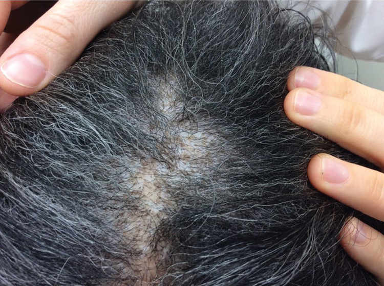
Patches with mottled hypopigmentation, loss of follicular ostia, and thinning of the hair in a patient with scalp sarcoidosis.

Hyperpigmented to violaceous flat-topped grouped papules coalescing into plaques in a patient with scalp sarcoidosis.
Discussion
Involvement of the scalp is rare in sarcoidosis.2,4 However, when the scalp is involved, it presents more frequently as a scarring alopecia and less frequently as a nonscarring alopecia. Clinical presentations of scalp sarcoidosis are protean and can range from discoid lupus-like lesions, lichen planopilaris–like lesions, or indurated red-orange nodules or plaques.4,5 Patients with long-standing sarcoidosis may present with end-stage scarring alopecia. One diagnostic clue for scalp sarcoidosis is the presence of other sarcoidal lesions, such as on the head4-6 or upper back, as in the current report. Thus, for patients with alopecia, a thorough examination of the scalp and skin should be performed. Once a diagnosis of scalp sarcoidosis is confirmed, a systemic workup should be undertaken, as most cases are associated with systemic disease, usually pulmonary or lymph node involvement.4,5
As in other forms of sarcoidosis, sarcoid-induced alopecia has numerous treatment strategies, but most are anecdotal with mixed results.4-8 The most widely suggested treatment regimen includes topical or intralesional steroids for small areas of active alopecia. For widespread or recalcitrant disease, antimalarials, oral corticosteroids, methotrexate, and tetracycline antibiotics have been used.9,10 In one case, a prednisone taper of 60 mg cleared active lesions, with results persisting after 2 years of follow-up.7 Another small case series demonstrated success in treating 2 cases of recalcitrant scalp sarcoidosis with 5 mg/kg of infliximab dosed at 0, 2, and 6 weeks followed by infusions every 8 weeks. One patient had no progression of her scalp sarcoidosis for 4 years, but reactivation occurred after she stopped taking infliximab. She subsequently restarted infliximab treatment, with clearance of active lesions again. The other case in the series remained stable without adverse effects for more than a year.8
Conclusion
Scalp sarcoidosis may be associated with other cutaneous manifestations of sarcoidosis. The cutaneous finding in our patient was papular sarcoid lesions on her back. Because of this association, suspicion of scalp sarcoidosis should prompt a complete skin examination. Systemic disease is also commonly found in patients with scalp sarcoidosis. Our patient reaffirmed this finding; she had a long-standing history of pulmonary sarcoidosis. If scalp sarcoidosis is identified in a patient, he or she should receive a workup for systemic involvement to rule out systemic sarcoidosis.
Acknowledgment
We thank Grace D. Brannan, PhD, for her guidance.
References
1. Mangas C , Fernández-FiguerasMT, FitéE, Fernández-ChicoN, SàbatM, FerrándizC. Clinical spectrum and histological analysis of 32 cases of specific cutaneous sarcoidosis. J Cutan Pathol. 2006;33(12):772-777. doi:10.1111/j.1600-0560.2006.00563.xSearch in Google Scholar
2. Haimovic A , SanchezM, JudsonMA, PrystowskyS. Sarcoidosis: a comprehensive review and update for the dermatologist: part I. cutaneous disease. J Am Acad Dermatol. 2012;66(5):699-e1. doi:10.1016/j.jaad.2011.11.965Search in Google Scholar
3. Elgart ML . Cutaneous sarcoidosis: definitions and types of lesions. Clin Dermatol. 1986;4(4):35-45.10.1016/0738-081X(86)90032-5Search in Google Scholar
4. House NS , WelshJP, EnglishJCIII. Sarcoidosis-induced alopecia. Dermatol Online J. 2012;18(8):4.10.5070/D30FP92370Search in Google Scholar
5. Katta R , NelsonB, ChenD, RoenigkH. Sarcoidosis of the scalp: a case series and review of the literature. J Am Acad Dermatol. 2000;42(4):690-692.10.1067/mjd.2000.104679Search in Google Scholar
6. Henderson CL , LafleurL, SontheimerRD. Sarcoidal alopecia as a mimic of discoid lupus erythematosus. J Am Acad Dermatol. 2008;59(1):143-145. doi:10.1016/j.jaad.2008.02.014Search in Google Scholar PubMed
7. Yazdani Abyaneh MA , RaghuP, KircherK, KutznerH, KortzA, CarlsonJA. Circumscribed cicatricial alopecia due to localized sarcoidal granulomas and single-organ granulomatous arteritis: a case report and systematic review of sarcoidal vasculitis. J Cutan Pathol. 2015;42(10):746-756. doi:10.1111/cup.12530Search in Google Scholar PubMed
8. Tu J , ChanJ. Cutaneous sarcoidosis and infliximab: evidence for efficacy in refractory disease. Australas J Dermatol. 2014;55(4):279-281. doi:10.1111/ajd.12056Search in Google Scholar PubMed
9. Badgwell C , RosenT. Cutaneous sarcoidosis therapy updated. J Am Acad Dermatol. 2007;56(1):69-83. doi:10.1016/j.jaad.2006.06.019Search in Google Scholar PubMed
10. Mosam A , MorarN. Recalcitrant cutaneous sarcoidosis: an evidence-based sequential approach. J Dermatolog Treat. 2004;15(6):353-359. doi:10.1080/09546630410023584Search in Google Scholar PubMed
© 2018 American Osteopathic Association
This work is licensed under the Creative Commons Attribution-NonCommercial-NoDerivatives 4.0 International License.
Articles in the same Issue
- OMT MINUTE
- An Osteopathic Approach to Stasis Dermatitis and Chronic Venous Insufficiency
- LETTERS TO THE EDITOR
- Investigating the Influence of Vitamin D Replacement Therapy on Magnesium Status
- Response
- HEALTH POLICY
- Guns and Older Adults: The Physician's Role
- ORIGINAL CONTRIBUTION
- Industry Payments in Cardiology: A Cross-sectional Analysis of Open Payments Data
- Utility of 4-Factor Prothrombin Complex Concentrate in Trauma and Acute-Care Surgical Patients
- BRIEF REPORT
- Adjuvant Lymphatic Osteopathic Manipulative Treatment in Patients With Lower-Extremity Ulcers: Effects on Wound Healing and Edema
- JAOA/AACOM MEDICAL EDUCATION
- Moving Toward Milestone-Based Assessment in Osteopathic Manipulative Medicine
- Exodus From the Classroom: Student Perceptions, Lecture Capture Technology, and the Inception of On-Demand Preclinical Medical Education
- CASE REPORT
- Scalp Sarcoidosis Presenting as Cicatricial Alopecia
- Spontaneous Infrarenal Aortic Dissection in an Athlete Managed Emergently With Endovascular Stent Grafts, Occluders, and Femoral-Femoral Artery Bypass
- CLINICAL IMAGES
- Bullous Diabeticorum
- Posterior Cortical Atrophy