Posterior Reversible Encephalopathy Syndrome
-
Brian J. Lewis

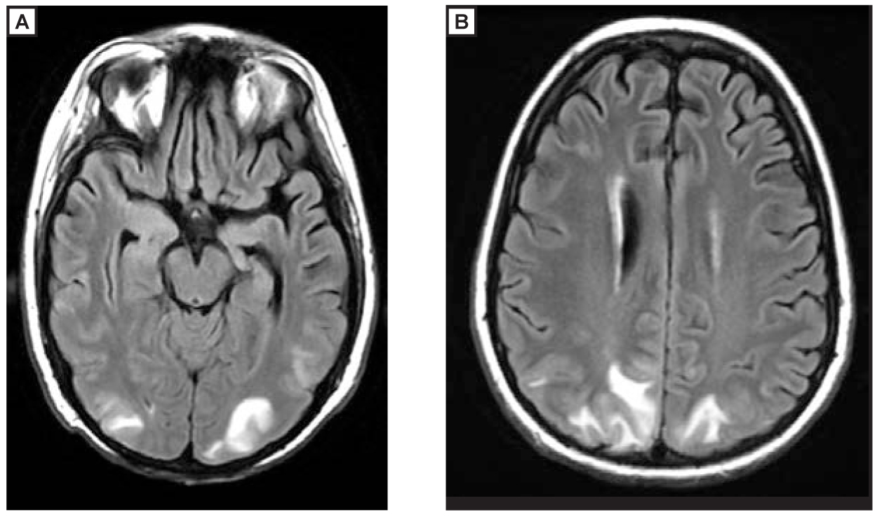
A 15-year-old boy undergoing chemotherapy for acute lymphoblastic leukemia presented with new-onset headaches and visual disturbances. Clinical evaluation revealed moderate hypertension, and no focal neurologic deficits were identified. Magnetic resonance imaging demonstrated bilateral regions of cortical and subcortical signal abnormality predominantly within the occipital (image A) and posterior parietal (image B) lobes, consistent with posterior reversible encephalopathy syndrome. After successful treatment for hypertension, the patient’s symptoms completely resolved.
In posterior reversible encephalopathy syndrome, autoregulation of the intracerebral perfusion is thought to be hindered during a hypertensive episode, resulting in vasogenic edema.1 Owing to a relative decrease in sympathetic innervation, the posterior circulation (posterior temporal, parietal, and occipital lobes) is most often affected.1-3 Common causes include preeclampsia or eclampsia, renal failure, autoimmune disorders, and chemotherapeutic drug toxicities. Patients frequently present with headache, nausea and vomiting, visual disturbances, or seizures.3 Symptoms and imaging findings typically resolve after management of the hypertensive episode. However, complications, including superimposed infarct, necessitate early detection and treatment.
References
1. Stevens CJ ,HeranMK. The many faces of posterior reversible encephalopathy syndrome.Br J Radiol.2012;85(1020):1566-1575. doi:10.1259/bjr/25273221.10.1259/bjr/25273221Search in Google Scholar PubMed PubMed Central
2. Bartynski WS. Posterior reversible encephalopathy syndrome, part 1: fundamental imaging and clinical features.AJNR Am J Neuroradiol.2008;29(6):1036-1042. doi:10.3174/ajnr.A0928.10.3174/ajnr.A0928Search in Google Scholar PubMed PubMed Central
3. Fugate JE ,ClaassenDO,CloftHJ,KallmesDF,KozakOS,RabinsteinAA. Posterior reversible encephalopathy syndrome: associated clinical and radiologic findings.Mayo Clin Proc.2010;85(5):427-432. doi:10.4065/mcp.2009.0590.10.4065/mcp.2009.0590Search in Google Scholar PubMed PubMed Central
© 2015 American Osteopathic Association
This work is licensed under the Creative Commons Attribution-NonCommercial-NoDerivatives 4.0 International License.
Articles in the same Issue
- EDITORIAL
- Health Issues Among Military Populations: Are We Providing Health Care or Health and Care?
- LETTERS TO THE EDITOR
- Effectiveness of OMT for Carpal Tunnel Syndrome
- Response
- AOA COMMUNICATION
- Official Call: 2015 Annual Business Meeting of the American Osteopathic Association
- Proposed Amendments to the AOA Constitution
- ORIGINAL CONTRIBUTION
- Perceptions of Physicians in Civilian Medical Practice on Veterans’ Issues Related to Health Care
- Relationships Between Polypharmacy and the Sleep Cycle Among Active-Duty Service Members
- Screening for At-Risk Drinking Behavior in Trauma Patients
- Age-Related Decline in Chest Wall Mobility: A Cross-Sectional Study Among Community-Dwelling Elderly Women
- MEDICAL EDUCATION
- The Doctors Hospital and Nationwide Children’s Hospital Dually Accredited Pediatric Residency Program: A Potential Best Model for Pediatric Osteopathic GME Training
- CASE REPORT
- Role of Osteopathic Manipulative Treatment in the Management of Stiff Person Syndrome
- CLINICAL IMAGES
- Posterior Reversible Encephalopathy Syndrome
- IN YOUR WORDS
- Law #1: Don’t Panic!