Evaluation of the Surface Contamination by Cytotoxic Agents of a Chemotherapy Reconstitution Unit: From Analysing Work Practices to Preventive and Corrective Actions
-
Florent Drapeau is a hospital pharmacist in the University Hospital of La Réunion Saint Pierre, particulary in charge of the reconstitution of the cytotoxic drugs ; he is the Quality Manager according to ISO 9001.
 ,
,
Caroline Rossard is a hospital pharmacist since 2014 in the University Hospital of La Réunion Saint Pierre. She’s in charge of the reconstitution of the cytotoxic drugs and of the sterilization of surgical materials.
Nicolas Bouscaren is a public health physician and an epidemiologist in the Clinical Center of Investigation/University Hospital of La Reunion since 2015. He conducts and manages research in collaboration with clinicians.

Abstract
Introduction:
The staff handling the cytotoxics are exposed to contamination as an occupational hazard. The unit of the Centre Hospitalier Universitaire (C.H.U) of Saint Pierre is ISO9001 certified. In a constant effort to improve the quality of our work, samples of the work surface were collected and allowed us, in a first step, to evaluate the contamination in a CRU in Reunion Island. As a result, corrective and preventive actions were implemented in the work routines.
Material and method:
53 samples of the work surface, collected in December 2015 and in March and April 2016 in seven different spots, were treated in this observational study. The 5-FU was chosen as example and analysed and quantified by using liquid chromatography associated with a mass spectrometer with a detection limit of 2 ng/pf.
Results:
We observed a great difference in contamination; 29 samples were positive, the isolator container was the location with the highest level of contamination, including manipulator gloves under the isolator, work surface and the inside of the isolator bin. The contamination was reduced after a first clean-up with water only.
The overwrapping bags and the manipulator gloves are only a little or not at all contaminated.
Conclusions
In order to reduce the exposure to cytotoxic residuals, corrective actions have been implemented: more frequent changes of manipulator gloves, the use of closed systems, and cleaning of the isolator with water. The efficiency of these modifications of work practices will be evaluated again in a few months.
Introduction
The offer of anti-cancer treatments is vastly growing in order to provide optimal care for patients, but those treatments are also widely known for their proven and potential toxicities [1, 2]: that is why special attention is being paid not only to the patients’ risk of exposure, but also to occupational exposure [3]. For a few fears, numerous studies have been dealing with teratogenic, mutagenic or carcinogenic occupational exposure [4–6]. Consequently, a vast number of guidelines and regulatory texts have since been published, [7–9] A centralised space for preparation, under a laminar flow or an isolator, and individual safety equipment reduce occupational exposure [10, 11]. These actions seem to be indispensable since one of the primary routes of entry is the dermal route [12, 13].
The pharmaceutical unit of the University Hospital (U.H) of Saint Pierre in Reunion Island is ISO9001 certified. Within this continuous improvement environment, the chemical contamination was analysed in the controlled atmosphere area (Z.A.C.) [14]. The aim of this study is to evaluate the chemical contamination of the hospital’s cytotoxic reconstitution unit (U.R.C.) by using different sampling points. We can so specify the potential risk of exposure in order to consider modifying of our work practices through preventive and corrective actions.
Material and methods
The work consisted of measuring, at different times and in different places of the unit, the residual presence of 5-FU. Only 5-FU was analysed. It was chosen as a proof of the contamination levels of the unit since it is the most used cytotoxic. Also, its stability at ambient temperatures was ideal to withstand the transport of the samples to continental France.
The sampling kits were provided by the Centre de Lutte Contre le Cancer Léon Bérard in Lyon: a filter paper saturated with 0.3 ml water for injection was used to perform the sampling on a frame of 15 cm on 15 cm. The analysis and the quantification of the 5-FU contamination was carried out in the pharma-toxicological laboratory of the U.H in Lyon: analysis of 5-FU was performed by LC-MS/MS using a Quantum-Ultra (ThermoElectron, San Jose, USA). Samples (10 µL injection volume) were separated on a Hypercarb column, (ThermoElectron), 5 µm, (100 mm×2.1 mm i. d.). The mobile phase consisted of water and acetonitrile using isocratic mode and was delivered through the column at a flow rate of 200 µL/min. The triple quadrupole mass spectrometer operated in negative ion mode with electrospray (ESI) source. [15N2]-5-FU was used as internal standard (IS). 5-FU and IS were detected using transitions (m/z 129 42 and m/z 131 43) respectively. We will present the results in graphic form by sampling site and will express the results as median [min, max] given the low numbers. The level is expressed in nanogram/filter paper(ng/fp) this quantity corresponds to the quantity found in the surface of a 15 cm×15 cm frame. A positive contamination threshold is set at 2 ng/fp (Low control: quantity of 5-FUdeposited on wipe sample (ng)=2, standard deviation=0.2, accuracy (%)=103).
A few clarifications on the sampling points: the isolator bin is a zipper bag in which only the paper wrappings issued from the isolator container. The “wastebin cell” corresponds to the joint system of the device for the tight transfer linked to a health-care waste bag that assures the evacuation of contaminated waste from the container. The SIEVE® spray is a desinfectant virucide, the WIP’ANIOS® wipes are biocide.
Results
Description of sampling points
The 53 samples were collected within two periods: first in December 2015, then in Mars and April 2016; the different sampling points are listed in Table 1.
Description of sampling points by place and date.
| Date | Type of sample | Number N=53 | Comments |
|---|---|---|---|
| December 2015 | Manipulator glove | n=5 | 2h00 after end of production |
| Manipulator glove | n=5 | at the end of production | |
| Manipulator gloves in the isolator | n=5 | ||
| Isolator bin | n=5 | before taking out, carried out by the manipulator with new gloves | |
| Overwrapping bags | n=5 | within the control area at the opening of a 5-FU preparation (except one case) | |
| March 2016 | Work surface isolator cleaned with SIEVE® spray only | n=4 | |
| Waste bin cell cleaned with wipes | n=5 | Interior surface of the cell, i. e. the most exposed surface | |
| Waste bin cell cleaned with water and wipes | n=5 | ||
| March and April 2016 | Preparations in the isolator at the end of production | n=10 | No 5-FU in the preparations; the manipulator changes gloves after two preparations |
| April 2016 | Work surface isolator cleaned with water+SIEVE® spray | n=4 |
Sampling sites: gloves manipulator, manipulator gloves in the isolator, isolator bin, overwrapping bags, work surface isolator cleaned with SIEVE® spray only, wastebin cell cleaned with wipes, waste bin cell cleaned with water and wipes, preparations in the isolator at the end of production, work surface isolator cleaned with water + SIEVE® spray.
Concerning the work surface isolator and the wastebin cell, the samples were collected following two different cleaning methods: spray cleaning SIEVE® single and water cleaning and the spray SIEVE® for the work surface; cleaning wipe only and cleaning with water then wipe for wastebin cell.
Contamination level by sampling point
In total, 53 samplings were collected: 29 of them were returned positive, 24 below quantification limit.
The Figures 1 to 9 illustrate the surface contamination with 5-FU, showing the different sampling points numbered 1 to 53:
Figure 1 shows a near-absence of contamination on the manipulator gloves: the samples 1 to 5 were collected two hours after the beginning of the activity, the other five show the samples collected at the end of the activity. Nine samples were below quantification limit, only sample 7 was positive at 3 ng/fp.
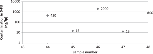
Figure 2 illustrates the manipulator glove’s contamination under isolator: samples 11 to 15, collected juste before changing gloves, all indicate a high contamination with a median of 155 ng/fp [62–1500]. Samples 12 and 46 show the highest contamination, with 1,500 et 2,000 ng/fp respectively (cf. Figure 8).
Figure 3 indicates a partial contamination of the isolator bin, plastic zipper bag in which the paper wrappings are disposed of, with a median of 12 ng/fp for the five samples [2–65].
The contamination level of the isolator bags’ surface is very low (Figure 4): Six samples out of ten are undetectable, sample 37 shows a high contamination with 600 ng/fp. The median value is 2 ng/fp [2–600].
The overwrapping bags for the preparations do not show any contamination with 5-FU: the five samples are below detection limit as is shown in Figure 5. The samples were collected outside the production zone, in the discharge control area.
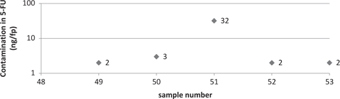
The contamination level of the isolator work surface after cleaning with Sieve® spray is sensitive with a median value of the five samples of 22 ng/fp [6–44] (Figure 6). However, after being cleaned with water first, then with the spray (Figure 7), the work surface shows a low contamination with a median of 5 ng/fp [3–6].
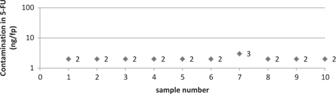
ontamination level of manipulator gloves.
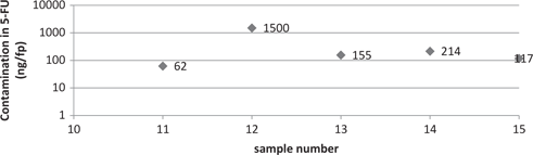
Contamination level of manipulator gloves (under isolator).
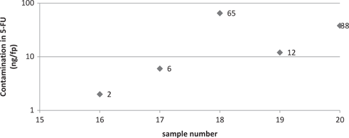
Contamination level of the isolator bin.
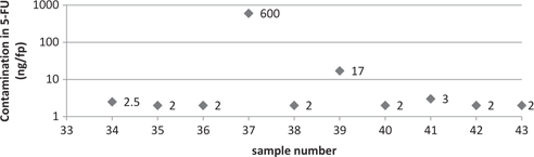
Contamination level of isolator bags at the end of production.
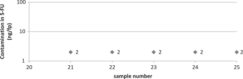
Contamination of overwrapping bags.
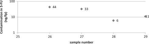
Contamination level of isolator work surface cleaned with spray only.
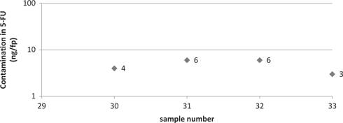
Contamination level of isolator work surface cleaned with water and spray.
Contamination level of the wastebin cell after cleaning with wipes.
Contamination level of wastebin cell after cleaning with water and wipes.
The samples 26 to 29 and the samples 44 to 48 do all indicate a residual contamination within the isolator after cleaning with a biocide cleaner, the median value of contamination is at 33 ng/fp, versus a median value of 3 ng/fp for the nine samples collected after cleaning with water first, then with a biocide cleaner (Tables 2 and 3).
Samples collected in the isolator after cleaning with biocide cleaner; (n=9); median=33 ng/fp.
| Sample number | 26 | 27 | 28 | 29 | 44 | 45 | 46 | 47 | 48 |
|---|---|---|---|---|---|---|---|---|---|
| Contamination in 5-FU (ng/fp) | 44 | 33 | 6 | 11 | 450 | 15 | 2000 | 13 | 800 |
Samples collected in the isolator after cleaning with water first, then with biocide cleaner; (n=9) median=3 ng/fp.
| Sample number | 30 | 31 | 32 | 33 | 49 | 50 | 51 | 52 | 53 |
|---|---|---|---|---|---|---|---|---|---|
| Contamination in 5-FU (ng/fp) | 4 | 6 | 6 | 3 | 2 | 3 | 32 | 2 | 2 |
Discussion
The diversity of sampling points has shown a great disparity in contamination levels within pharmaceutics. The highest contamination levels can obviously be found within the container of the isolator, the manipulator gloves, the work surface, and the wastebin cell DASRI. Several remedial actions have been introduced.
Changing gloves more frequently, after every other preparation, in order to limit the contamination by hand. Note that since 2002, B. Favier has been interested in contamination by hand due to the manipulation of cytotoxics, insisting on the contamination of the manipulator gloves [15]; more recently, in 2015, Paul J.M. Sessink affirms the systematic contamination of manipulator gloves [16].
Provision of Wastebag® disposable bin bags that limit the exposure of the agent responsible for cleaning the cells.
The use of bags with nozzle tips (Luerlock type) allows to limit the use of needles, to reduce the risk of contamination by micro droplets and aerosols as well as the risk of accidental pricking. Several teams have demonstrated the advantages of using closed systems in order to reduce contamination: M. Berruyer in 2015 confirmed in a multicentric follow-up study in Canada a significant diminution in contamination in the units using closed systems, as did Paul J.M. Sessink who compared the contamination levels within a six-year interval in units using a method including syringes and closed systems [17, 18].
The near non-existence of contamination on the manipulator gloves, nine samples out of ten being undetectable, suggest the non-contamination of his individual safety equipment: it was therefore decided that the waste elimination circuit was to be altered: thus, mask, cap and coat of the mani pulator now follow the waste circuit assimilated to domestic waste: thus, we were able to diminuish the volume of waste in healthcare activities bearing a risk of infection.
However, it seems delicate for now to extrapolate these data to the whole of our U.R.C. (furniture, handles, computers...) even if S. Crauste-Manciet et al. confirmed in 2005 that exterior exposure can be limited if the contamination remains within the isolator [19].
Note that the isolator bins present a significant contamination level (Figure 3); therefore, they continue to be removed with the contaminated waste.
The near non-existence of contamination on overwrapping bags and on the surface of the bags, shown by 13 undetectable samples out of 15, is reassuring, while there remains a risk of occupational exposure of the pharmacist within the discharge control area and of the courier in charge of transport.
In fact, F. Sadeghipour emphases the importance of a specific manipulation training for the preparers of cytotoxics, [20] as it has been the custom within the U.R.C of the U.H of Saint Pierre for years now. Recently, R. Vazquez shows that the manual process can lead to errors of re-use of medical devices and can potentially have consequences for the patient [21].
Concerning the sample 37: this outlying result is probably due to a micro droplet. Sample size allow no generalization of our results. More samples would have been necessary. On the other hand we were able to lead a reflection on our professional practices.
Conclusion
This first observational study, evaluating the contamination by cytotoxic and conducted in a U.R.C in Reunion Island, allowed confirming the surface contamination of one part of the unit and thus the necessity of individual and collective safety measures. In spite of these safety measures, we found a residual contamination of the work environment. That is why the U.R.C. of the U.H in Saint Pierre has introduced an internal training that has to be passed by all new preparers.
The analysis of our work practices and of occupational risks are at the source of corrective and preventive actions implemented in our quality management system according to ISO9001.
Clearly, a sampling conducted several months after those actions would allow evaluating their efficiency. Furthermore, we are instantly thinking about the possibility of organising a regular follow-up to evaluate the contamination of the work environment by “relying on adapted environment controls” as suggested in the “Bonnes Pratiques de Préparations” (“good habits of preparation”).
About the authors

Florent Drapeau is a hospital pharmacist in the University Hospital of La Réunion Saint Pierre, particulary in charge of the reconstitution of the cytotoxic drugs ; he is the Quality Manager according to ISO 9001.

Caroline Rossard is a hospital pharmacist since 2014 in the University Hospital of La Réunion Saint Pierre. She’s in charge of the reconstitution of the cytotoxic drugs and of the sterilization of surgical materials.
Nicolas Bouscaren is a public health physician and an epidemiologist in the Clinical Center of Investigation/University Hospital of La Reunion since 2015. He conducts and manages research in collaboration with clinicians.
Acknowledgments
We would like to thank Dr. Jean-François Latour, pharmacist at the Centre Léon Berard in Lyon, and Professor Jérôme Guitton, laboratory biologists at the pharmaceutic laboratory of the U.H in Lyon for their advice.
Conflicts of interest statement: Authors state no conflict of interest. All authors have read the journal’s Publication ethics and publication malpractice statement available at the journal’s website and hereby confirm that they comply with all its parts applicable to the present scientific work.
References
1. Dode X. Dossiers du CNHIM, revue d’évaluation thérapeutique, anticancéreux: utilisation pratique. 7ème éd. Paris: CNHIM;2013. 599 p.Search in Google Scholar
2. Remesh A. Toxicities of anticancer drugs and its management. Int J Basic Clin Pharmacol [Internet]. 2012 [cited 2016 Sept 01];1(1):2. Available at: http://www.ijbcp.com/?mno=2762910.5455/2319-2003.ijbcp000812Search in Google Scholar
3. Ministère du travail, de l’emploi et de la santé Arrêté du 6 avril 2011 relatif au management de la qualité de la prise en charge médicamenteuse et aux médicaments dans les établissements de santé. JORF n°0090 du 16 avril 2011. 6687. Disponible sur https://www.legifrance.gouv.fr/eli/arrete/2011/4/6/ETSH1109848A/joSearch in Google Scholar
4. Lawson CC, Rocheleau CM, Whelan EA, Hibert EN, Grajewski B, Spiegelman D, et al. Occupational exposures among nurses and risk of spontaneous abortion. Am J Obstet Gynecol [Internet] 2012 Apr 1 [cited 2016 Aug 26];206(4):327.e1–327.e8. Available at: http://www.ajog.org/article/S0002-9378(11)02470-710.1016/j.ajog.2011.12.030Search in Google Scholar PubMed PubMed Central
5. Kusnetz E, Condon M. Acute effects from occupational exposure to antineoplastic drugs in a para-professional health care worker. Am J Ind Med. 2003; 44:107–109.10.1002/ajim.10230Search in Google Scholar PubMed
6. Fransman W, Kager H, Meijster T, Heederik D, Kromhout H, Portengen L, et al. Leukemia from dermal exposure to cyclophosphamide among nurses in the Netherlands: quantitative assessment of the risk. Ann Occup Hyg [Internet] 2014 4–1 [cited 2016 Oct 26];58(3):271–82. Available at: http://annhyg.oxfordjournals.org/content/58/3/271Search in Google Scholar
7. NIOSH [2014]. NIOSH list of antineoplastic and other hazardous drugs in healthcare settings 2014. By Connor TH, MacKenzie BA, DeBord DG, Trout DB, O’Callaghan JP. Cincinnati, OH: U.S. Department of Health and Human Services, Centers for Disease Control and Prevention, National Institute for Occupational Safety and Health, DHHS (NIOSH) Publication No. 2014–138 (Supersedes 2012–150).Search in Google Scholar
8. Green E, Johnston M, Trudeau M, Schwartz L, Poirier S, Macartney G, et al. Safe handling of parenteral cytotoxics: recommendations for Ontario. J Oncol Pract [Internet] 2009 Sep [cited 2016 Sept 10];5(5):245–9. Available at: http://www.ncbi.nlm.nih.gov/pmc/articles/PMC2790670/10.1200/JOP.091014Search in Google Scholar PubMed PubMed Central
9. Bonnes Pratiques de Préparations JORF n°270 du 21 novembre 2007. Disponible sur http://ansm.sante.fr/var/ansm_site/storage/original/application/a5d6ae4b3d5fdee013ca463462b7b296.pdfSearch in Google Scholar
10. Balty I, Chapouthier A. Equipements de Protections Individuelles règles d’utilisation 2ème éd. Paris: INRS; 2013. 27 p.Search in Google Scholar
11. Kopp B, Crauste-Manciet S, Guibert A, Mourier W, Guerrault-Moro M-N, Ferrari S, et al. Environmental and biological monitoring of platinum-containing drugs in two hospital pharmacies using positive air pressure isolators. Ann Occup Hyg [Internet] 2013 4–1 [cited 2016 Sept 12];57(3):374–83. Available at: http://annhyg.oxfordjournals.org/content/57/3/374Search in Google Scholar
12. Fransman W, Vermeulen R, Kromhout H. Occupational dermal exposure to cyclophosphamide in Dutch hospitals: a pilot study. Ann Occup Hyg. 2004 Apr;48(3):237–44.Search in Google Scholar
13. Sessink PJ, Van de Kerkhof MC, Anzion RB, Noordhoek J, Bos RP. Environmental contamination and assessment of exposure to antineoplastic agents by determination of cyclophosphamide in urine of exposed pharmacy technicians: is skin absorption an important exposure route? Arch Environ Health 1994 May–Jun;49(3):165–9.10.1080/00039896.1994.9940377Search in Google Scholar PubMed
14. ISO editor. ISO9001:2015 Système de management de la qualité. Paris: AFNOR; 2015. 31 pSearch in Google Scholar
15. Favier B, Latour JF, Ardiet C, Voloch A. Évaluation de la contamination des gants et des mains du personnel infirmier avant et après formation à la manipulation des anticancéreux./data/revues/12503274/00630001/20/[Internet]. 2008 15–2 [cited 2016 Aug 29]; Available at: http://www.em-consulte.com/en/article/72327Search in Google Scholar
16. Sessink PJ, Leclercq GM, Wouters D-M, Halbardier L, Hammad C, Kassoul N. Environmental contamination, product contamination and workers exposure using a robotic system for antineoplastic drug preparation. J Oncol Pharm Pract. 2015 Apr;21(2):118–27.10.1177/1078155214522840Search in Google Scholar PubMed
17. Berruyer M, Tanguay C, Caron NJ, Lefebvre M, Bussières JF. Multicenter study of environmental contamination with antineoplastic drugs in 36 Canadian hospitals: a 2013 follow-up study. J Occup Environ Hyg. 2015;12(2):87–94.10.1080/15459624.2014.949725Search in Google Scholar PubMed
18. Sessink PJM, Trahan J, Coyne JW. Reduction in surface contamination with cyclophosphamide in 30 US hospital pharmacies following implementation of a closed-system drug transfer device. Hosp Pharm [Internet]. 2013 Mar [cited 2016 Sept 10];48(3):204–12. Available at: http://www.ncbi.nlm.nih.gov/pmc/articles/PMC3839517/10.1310/hpj4803-204Search in Google Scholar PubMed PubMed Central
19. Crauste-Manciet S, Sessink PJM, Ferrari S, Jomier, J-Y, Brossard D. Environmental contamination with cytotoxic drugs in healthcare using positive air pressure isolators. Ann Occup Hyg [Internet]. 2005 10–1 [cited 2016 Sept 10];49(7):619–28. Available at: http://annhyg.oxfordjournals.org/content/49/7/619Search in Google Scholar
20. Sadeghipour F, Lorenzini KI, Ziewitz C, Dobrinas M, Fleury M, Bonnabry P. Chemical contamination during the preparation of cytotoxics: validation protocol for operators in hospital pharmacies. J Oncol Pharm Pract. 2013 Mar;19(1):57–64.10.1177/1078155212452764Search in Google Scholar PubMed
21. Vazquez R, Boubet K, Guerrault-Moro, M-N, Crauste-Manciet S. Impact of handling errors for chemical cross-contamination risk for the preparation of parenteral cytotoxic drugs. Pharma Technol Hosp Pharmacy [Internet]. 2016 [cited 2016 Nov 30];1(3):115–22. Available at: https://www.degruyter.com/view/j/pthp.2016.1.issue-3/pthp-2015-0009/pthp-2015-0009.xml?format=INT10.1515/pthp-2015-0009Search in Google Scholar
©2016 Walter de Gruyter GmbH, Berlin/Boston
Articles in the same Issue
- Frontmatter
- graphical-abstract
- Editorial
- Quality and Safety in the Hospital: The Pharmacist is the Key Person
- Research Articles
- Rubber Coring of Injectable Medication Vial Stoppers: An Evaluation of Causal Factors
- Prospective Descriptive Study of RFID Tag Detection Rates based on Various Exploratory Scenarios Aimed at Identifying Optimal Conditions of Use
- Surface Contamination in a Teaching Hospital: A 6 Year Perspective
- Short Communications
- Evaluation of the Surface Contamination by Cytotoxic Agents of a Chemotherapy Reconstitution Unit: From Analysing Work Practices to Preventive and Corrective Actions
- Simultaneous Determination of Sufentanil and Ziconotide in Combination for Intrathecal Analgesia by UPLC-UV
- Reviewer Acknowledgment