Green synthesis and characterization of zero-valent iron nanoparticles using stinging nettle (Urtica dioica) leaf extract
-
Alireza Ebrahiminezhad
Alireza Ebrahiminezhad received both his BS and MS in microbiology at Islamic Azad University, Jahrom Branch, Iran. He also obtained a PhD degree in pharmaceutical biotechnology at Tabriz University of Medical Sciences, Tabriz, Iran. He now works as an assistant professor of nanobiotechnology at Fasa University of Medical Sciences.
 ,
Alireza Zare-Hoseinabadi
,
Alireza Zare-Hoseinabadi
Alireza Zare-Hoseinabadi obtained his BS degree in genetics. He is now an MS student of medical biotechnology at Fasa University of Medical Sciences.
Aydin Berenjian has BE, ME and PhD degrees in biochemical engineering. He is interested in developing cutting edge bioprocess technologies to generate high value products. He serves on the editorial board of
Molecular Biotechnology and is an associate editor ofAmerican Journal of Biochemistry and Biotechnology . Aydin Berenjian has published more than 50 peer-reviewed articles and has received several prestigious international research awards.Younes Ghasemi is a professor of pharmaceutical biotechnology. He is the head of Department of Pharmaceutical Biotechnology, School of Pharmacy and Pharmaceutical Sciences Research Center, Shiraz University of Medical Sciences. He is also the head of School of Advanced Medical Sciences and Technologies.

Abstract
For the first time, aqueous leaf extract of Urtica dioica was used as a sustainable source of reducing and capping agents to synthesize iron nanoparticles (INPs). In this regard, zero-valent INPs were produced and entrapped in a biologic coating. INPs were characterized by transmission electron microscopy (TEM), particle size analysis, Fourier transform infrared (FTIR) spectroscopy, X-ray diffractometer, vibrating sample magnetometer, thermogravimetric analysis (TGA), and differential TGA. Based on the results, the prepared INPs were completely composed of zero-valent iron atoms without any iron oxide impurities. Nanoparticles were spherical in shape with diameters ranging from 21 nm to 71 nm with a mean particle size of 46 nm. Particles were entrapped in a biologic coating which resulted in formation of complexes ranging from 117 nm to 605 nm. High zeta potential value of these complexes (−82.6 mV) and the presence of hydrophilic groups on the biologic coating provided a stable colloid system. Prepared INPs were non-crystalline (amorphous) having a low magnetization value of 0.14 emu/g. The prepared particles are of significant interest in a wide range of scientific and technical applications.
1 Introduction
Iron nanoparticles (INPs) are one of the most important and useful nanostructures in various science and technology fields due to unique properties such as biocompatibility, surface modifiability, and high surface to volume ratio [1], [2], [3], [4], [5]. The high surface to volume ratio of nanomaterials causes a large portion of the atoms to reside on the surface as compared to those in the bulk materials. The presence of a large percentage of exposed atoms greatly increases the surface activity. This unique surface structure and large surface area are required for inducing and catalyzing chemical reactions. The applications of nanoparticles in chemical industries are so well developed that the size and shape dependent catalytic properties of nanoparticles have been widely studied [6], [7].
In addition to catalytic applications, INPs are now used in a broad range of environmental clean-up technologies as nanosorbents and photocatalysts [8]. INPs have applications in detoxification and removal of compounds such as carbon monoxide, arsenic, chromium, and mercury [4], [9], [10], [11], [12], [13], [14]. Water and wastewater treatment is another field for application of INPs [8], [15]. Clean water, free of toxic compounds and pathogens, is a critical issue for the world’s health and considerable attention has been paid to water treatment techniques. Application of INPs can resolve or improve many of the current problems in the water treatment industries. INPs are used to remove toxic ions or organic pollutants from water with a higher removal capacity than bulk materials [15].
The considerable scientific and technical applications of INPs increase the demand for their economic and sustainable synthesis. There are many physical and chemical methods for synthesis of INPs [16], [17], [18], [19], however they are energy consuming, employ toxic chemicals, and require harsh process conditions. Green synthesis has emerged as a novel method to develop and implement chemical processes that reduce or eliminate the use of hazardous substances [20], [21], [22], [23].
Plant extracts are among the most used and abundant compounds for green synthesis in nanochemistry. Metal nanoparticles have been synthesized by using plant leaf extracts such as black tea, Lippia citriodora (Lemon Verbena), maple (Acer sp.), Lantana camara, Artemisia annua, and eucalyptus [24], [25], [26], [27], [28], [29]. Other parts of plants have also been used such as Medicago sativa and Sterculia foetida seed exudate, orange peel extract, sorghum bran extract, Piper longum, Crataegus douglasii fruit extract, coffee extract, Chrysanthemum morifolium Ramat extract, and Cinnamon zeylanicum bark extract [23], [25], [30], [31], [32], [33], [34], [35], [36], [37]. Prepared nanoparticles are usually coated with biologic compounds from plants. This coating makes the prepared particles more stable and biocompatible [21], [22], [38].
Urtica dioica is the most studied and known species of the genus Urtica which is also called common nettle or stinging nettle. This is a very widely distributed herbaceous perennial plant found in Europe, America, Asia, and north Africa [39]. Medical applications of U. dioica as anti-inflammatory, anti-hyperglycaemic, antioxidant, diuretic, natriuretic, hypotensive, antimicrobial, antiulcer, and analgesic are well studied [40], [41], [42], [43]. This herb also has traditional applications as a food additive in some countries [41]. U. dioica contains large amounts of phenolic and flavonoid compounds which are reported to be effective in bioreduction and green synthesis of metal nanoparticles [25], [44]. In the present study, U. dioica leaf extract was investigated as a natural source of reducing and capping agent for INPs synthesis. Physical and chemical properties of obtained particles were also characterized.
2 Materials and methods
2.1 Materials
Dried leaves of U. dioica were purchased from a local shop (Shiraz, Fars, Iran). Ferric chloride (FeCl3·6H2O) was purchased from Merck Chemicals (Darmstadt, Hessen, Germany) and used as received. All glassware was acid washed and then rinsed with deionised water. Chemical reactions were conducted using Millipore water (Millipore Corp., Bedford, MA, USA, conductivity range=0.055−0.294 lS/cm).
2.2 Leaf extract preparation
Dried leaves were washed with deionized water to remove any mud and dust and were dried at room temperature. Using about a 5% (w/v) mixture of dried leaves in deionized water has been the most common ratio for leaf extract preparation in previous experiments [45], [46], [47], [48], [49], [50], [51], [52]. Therefore, in the current experiment, 5 g dried leaves were boiled in 100 ml deionized water for 15 min by using a heater mantel under reflux. The mixture was cooled to room temperature and filtered through a Whatman filter paper (Reeve angel, Grade 201). In order to remove leaf microparticles, the prepared extract was centrifuged at 2000 rpm for 5 min. The resulting clear solution was sealed in polypropylene tubes and refrigerated.
2.3 Synthesis of INPs
Synthesis of INPs was conducted using a 10 ml reaction liquid. In practice, 1 ml iron precursor (FeCl3·6H2O, 0.2 m) was added to 9 ml leaf extract while stirring vigorously at room temperature. After 12 h, the reaction mixture was centrifuged and the resulting black pellet was washed with deionized water and dried in an oven at 50°C.
2.4 Characterization of INPs
The visual appearance and morphology of prepared INPs were evaluated by transmission electron microscopy (TEM). A drop of nanoparticles dispersion in distilled water was dripped on to a carbon-coated copper grid and dried at room temperature. Micrographs were obtained using a Philips CM 10, TEM, operated at high voltage (HT) 100 kV [53], [54], [55]. Particle size analyses were conducted using ImageJ software version 1.47v, an image analysis software developed by the NIH (http://imagejnihgov/ij/). The hydrodynamic diameter and zeta potential of particles were measured using a Microtrac S3500 particle size analyser. The Fourier transform infrared (FTIR) spectroscopy analyses were done using KBr pellets. Prepared INPs were mixed and pressed with 150 mg KBr and the spectra were taken using a Bruker, Vertex 70, FTIR spectrometer. The IR absorption analyses were done from 4000 cm−1 to 400 cm−1 [20], [56]. The crystallinity and composition of the INPs were analysed via X-ray powder diffractometry (Siemens D5000). The X-ray diffraction pattern of the sample was collected over a 2-θ range from 20° to 90° at a scan rate of 2° min−1 [57], [58]. Thermogravimetric analysis (TGA) and derivative thermogravimetric (DTG) analyses were done to determine the presence and quantification of organic compounds from U. dioica leaf extract in the final INPs product. Magnetic characteristics of nanoparticles were evaluated using a vibration sample magnetometer. The analysis was conducted at room temperature with increasing magnetic field up to 10 kOe and field sweeping from −10 to +10 kOe [17], [59].
3 Results and discussion
3.1 Synthesis of INPs
By addition of iron precursor to the leaf extract a sudden change in the color of the reaction mixture was observed and a dark black suspension was formed. Instantaneous change in the reaction color and appearance of deep black suspension is a common phenomenon in green synthesis of INPs [48], [49], [60], [61], [62], [63] and considered as an indicator for reduction of iron ions and formation of INPs [46], [64], [65]. This phenomenon was also observed in semi-green synthesis [50], [66], [67], [68], [69] and chemical synthesis of INPs [18], [19]. Various parameters such as reaction time, metal precursor concentration, amount of leaf extract, and reaction temperature were reported to be the key parameters for the synthesis of nanoparticles in a green approach. The significance of these parameters does vary in different plant extracts [21], [38], [70], [71], [72]. While using a particular plant extract, various investigations should be done to evaluate the effects of each parameter on the characteristic features of the obtained particles. Finally, an optimization study would determine the optimal state of each parameter toward determining the best result.
3.2 Characterization of INPs
Visual appearance of the prepared INPs was evaluated by TEM analysis (Figure 1A). The obtained particles were dominantly spherical while some oval particles were also observed. Particle size distribution analyses were conducted and results are shown in Figure 1B. Diameters of INPs were measured in the range of 21 nm to 71 nm with a mean particle size of 46 nm. Green synthesized INPs with similar particle size distribution were also reported by using Adean blackberry, green tea, and eucalyptus leaf extract [46], [64], [67], [73]. Very small INPs (i.e. about 5 nm) were also synthesized by green methodology using mulberry, pomegranate, and Syzygium jambos leaf extract and soybean sprouts [61], [74], [75]. By contrast, Yuvakkumar and Hong [76] reported that using rambutan peel waste extract for the synthesis of INPs resulted in large spinel particles with 200 nm diameter. Based on the TEM micrograph, nanoparticles were surrounded by a biologic matrix from leaf extract. Particles were surrounded by a matrix and formed a complex. The sizes of INPs complexes with surrounding materials were measured in the range of 117–605 nm. Formation of nanoclusters with phytochemical capping was also reported for Andean blackberry leaf extract mediated synthesized INPs. The authors believed that various polyphenols in the leaf extract can be responsible for the formation of INP clusters [67].

Transmission electron microscopy (TEM) micrographs of the green synthesized iron nanoparticles (INPs) (A) which indicate that nanoparticles are surrounded by a biologic matrix from leaf extract, and particle size distribution diagram of the INPs (B).
Precise particle size analyses were done to evaluate the hydrodynamic diameter of INP complexes. According to the results, the diameter of complexes was measured from 121.5 nm up to 2.75 μm. The particle size distribution curve (Figure 2) shows two distribution peaks at 243 nm (volume per cent: 13.4%) and 1.94 μm (volume per cent: 4.0%) which can be an indication for some agglomeration of INPs complexes. Muthukumar and Matheswaran have shown that biologic coating can reduce the rate of INPs agglomeration. They measured the size of biogenic synthesized INPs to be from 58 nm to 530 nm using a particle size analyser. However, chemically synthesized nanoparticles were agglomerated up to 2.3 μm [69]. Similar agglomeration was also reported by Niraimathee et al. [45] for the green synthesized INPs using Mimosa pudica root extract. They revealed particle size distribution of prepared particles ranging from 32 nm to 600 nm. The INPs complexes had a negative zeta potential of −82.6 mV. Biologic coatings from various plants can provide different zeta potentials ranging from −35 mV for green tea to about zero for Hordeum vulgare, and +71 mV for Amaranthus spinosus [60], [65], [69]. This variation is mainly due to different phytochemicals in various plants. Generally, the surface charge values above −30 mV are considered to be sufficient for suppressing particles agglomeration and providing a stable colloidal system [77].

Particle size distribution curve of iron nanoparticles (INPs) complexes which were obtained by particle size analyser.
Figure 3 shows the FTIR absorption spectra of the synthesized INPs. The OH groups induce a broad indicative peak which appeared at 3467 cm−1 [56]. The carbonyl group stretching vibration can be seen at 1636 cm−1 and the peak at about 1070 cm−1 produced by C–O bonds [56]. The peaks of aliphatic C–H appeared around 2800 cm−1 [53], [54], [55]. Usually, presence of these peaks in the FTIR spectra of green synthesized nanoparticles is indicative of nanoparticles functionalization with organic compounds [20], [21], [22], [38]. It has been shown that exposure of INPs to aqueous leaf extract resulted in the coating and stabilization of INPs with herbal biochemical compounds [78]. Functionalization of nanostructures with hydrophilic groups such as hydroxyl, carbonyl, and C–O increases the interactions with water molecules and results in more stable colloids.

Fourier transform infrared (FTIR) spectra of green synthesized iron nanoparticles (INPs); peaks at 1070 cm−1, 1636 cm−1, 2800 cm−1, and 3467 cm−1 were due to C–O, C=O, C–H, and OH bonds, respectively.
Iron oxide nanoparticles (IONs) exhibit two intense and sharp peaks at about 640 cm−1 and 450 cm−1 due to the presence of Fe–O bonds [17], [18], [19], [57], [58], [59]. These peaks were not observed in the FTIR spectrum of the prepared particles (Figure 3), which is indicative of zero-valent INPs. Similar spectra were also reported for chemically synthesized zero-valent INPs [79], [80]. Zero-valent INPs exhibit dual characteristics of iron oxide/hydroxide and of zero-valent iron [81]. There is a core-shell structure, zero-valent iron atoms forming the core which is surrounded by an iron oxide shell [80], [82], [83]. This feature is due to oxidation of exposed iron atom to iron oxide/hydroxide. The thin iron oxide shell resulted in the appearance of Fe–O absorption bonds, but in lower intensity than that usually observed for IONs [17], [59], [84]. Absence of these peaks in FTIR spectra of the prepared INPs indicated the absence of iron oxide shell. Organic coating from leaf extract seems to protect the surface of Fe0 atoms from oxidation.
The crystallinity of the prepared INPs was evaluated by X-ray powder diffraction analysis and results were evaluated by using PANalytical X’Pert HighScore software. As shown in Figure 4, there is no distinctive diffraction peak in the whole pattern, suggesting that the green synthesis approach resulted in the production of amorphous INPs. Fabrication of amorphous zero-valent INPs was also reported by using leaf extract of different plants and food industry wastes [46], [52], [75]. In addition, green synthesis of INPs by using extracts of green tea, eucalyptus, S. jambos (L.) Alston (Myrtaceae), and sorghum bran was reported to result in amorphous IONs [30], [46], [64], [74]. Some researchers also considered a small peak at 2θ=44.9° as an indicative peak for zero-valent INPs [48], [49], [51]. The broad shoulder peak from 10° to 20° of 2θ values was proposed to be due to the presence of organic materials from leaf extract which are responsible for capping and stabilizing nanoparticles [46].

X-ray powder diffractometry (XRD) pattern of the green synthesized iron nanoparticles (INPs); no characteristic peak was observed which indicates the amorphous structure of nanoparticles.
TGA and DTG curves of green synthesized zero-valent INPs are provided in Figure 5. There is a significant weight loss in the TGA curve of the synthesized INPs. The initial weight loss below 200°C is usually attributed to evaporation of residual and adsorbed water [85]. Organic compounds are reported to exhibit a considerable thermal stability up to 200°C, whereas at higher temperatures, breakdown of the organic skeleton takes place [86], [87]. Decomposition of organic capping materials makes the main peak in the DTG curve as reported for dextran-coated magnetite nanoparticles [87]. About 30% weight loss was recorded due to decomposition of INPs biologic coating.

Thermogravimetric analysis (TGA) and derivative thermogravimetric (DTG) curves of iron nanoparticles (INPs); the weight loss below 200°C was due to water evaporation and decomposition of biologic matrix resulted in 30% weight loss which makes the main peak in the DTG curve.
Magnetic characteristics of the prepared INPs were evaluated by a vibrating sample magnetometer at room temperature and the results are shown in Figure 6. Magnetization value of the synthesized INPs was measured to be 0.14 emu/g. Magnetization value about 130 emu/g was reported for chemically synthesized particles which contrasts with the current report [88]. High magnetization properties provide the possibility for magnetic handling of particles. However, high values of magnetization have undesirable impacts on INPs colloidal stability and formation of aggregated particles [88].
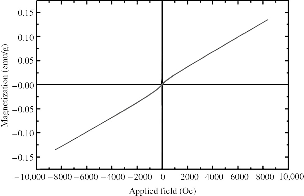
Magnetization curve of iron nanoparticles (INPs); magnetization value of the synthesized INPs was measured to be 0.14 emu/g.
4 Conclusion
Zero-valent INPs were synthesized in a green manner by using U. dioica leaf extract as a natural source of reducing agents. The biochemical compounds of U. dioica leaves also showed the ability to act as capping and protecting agents. Coating of INPs with biologic materials provides desirable characteristics which cannot be easily achieved via chemical synthesis. Green synthesized zero-valent INPs were completely protected against oxidation and iron oxide was not detected in FTIR analysis. It was found that one to several INPs were trapped in a biologic coating to fabricate a complex. These complexes formed a stable colloid system with a very low rate of agglomeration. Therefore, the prepared particles were physicochemically and colloidally stable which makes them desirable for many scientific and technical applications in future.
About the authors

Alireza Ebrahiminezhad received both his BS and MS in microbiology at Islamic Azad University, Jahrom Branch, Iran. He also obtained a PhD degree in pharmaceutical biotechnology at Tabriz University of Medical Sciences, Tabriz, Iran. He now works as an assistant professor of nanobiotechnology at Fasa University of Medical Sciences.

Alireza Zare-Hoseinabadi obtained his BS degree in genetics. He is now an MS student of medical biotechnology at Fasa University of Medical Sciences.

Aydin Berenjian has BE, ME and PhD degrees in biochemical engineering. He is interested in developing cutting edge bioprocess technologies to generate high value products. He serves on the editorial board of Molecular Biotechnology and is an associate editor of American Journal of Biochemistry and Biotechnology. Aydin Berenjian has published more than 50 peer-reviewed articles and has received several prestigious international research awards.

Younes Ghasemi is a professor of pharmaceutical biotechnology. He is the head of Department of Pharmaceutical Biotechnology, School of Pharmacy and Pharmaceutical Sciences Research Center, Shiraz University of Medical Sciences. He is also the head of School of Advanced Medical Sciences and Technologies.
Acknowledgments
This work was financially supported by Fasa University of Medical Sciences.
References
[1] Akbarzadeh A, Mikaeili H, Zarghami N, Mohammad R, Barkhordari A, Davaran S. Int. J. Nanomed. 2012, 2012, 511–526.10.2147/IJN.S24326Search in Google Scholar
[2] Gupta AK, Gupta M. Biomaterials 2005, 26, 3995–4021.10.1016/j.biomaterials.2004.10.012Search in Google Scholar
[3] Laurent S, Forge D, Port M, Roch A, Robic C, Vander Elst L, Muller RN. Chem. Rev. 2008, 108, 2064–2110.10.1021/cr068445eSearch in Google Scholar
[4] Li P, Miser DE, Rabiei S, Yadav RT, Hajaligol MR. Appl. Catal. B 2003, 43, 151–162.10.1016/S0926-3373(02)00297-7Search in Google Scholar
[5] Yu S, Chow GM. J. Mater. Chem. 2004, 14, 2781–2786.10.1039/B404964KSearch in Google Scholar
[6] Zhou X, Xu W, Liu G, Panda D, Chen P. J. Am. Chem. Soc. 2009, 132, 138–146.10.1021/ja904307nSearch in Google Scholar PubMed
[7] Narayanan R, El-Sayed MA. Nano Lett. 2004, 4, 1343–1348.10.1021/nl0495256Search in Google Scholar
[8] Xu P, Zeng GM, Huang DL, Feng CL, Hu S, Zhao MH, Lai C, Wei Z, Huang C, Xie GX, Liu ZF. Sci. Total Environ. 2012, 424, 1–10.10.1016/j.scitotenv.2012.02.023Search in Google Scholar PubMed
[9] Mak S-Y, Chen D-H. Dyes Pigm 2004, 61, 93–98.10.1016/j.dyepig.2003.10.008Search in Google Scholar
[10] Cumbal L, SenGupta AK. Environ. Sci. Technol. 2005, 39, 6508–6515.10.1021/es050175eSearch in Google Scholar PubMed
[11] Gupta V, Agarwal S, Saleh TA. Water Res. 2011, 45, 2207–2212.10.1016/j.watres.2011.01.012Search in Google Scholar PubMed
[12] Parham H, Zargar B, Shiralipour R. J. Hazard. Mater. 2012, 205, 94–100.10.1016/j.jhazmat.2011.12.026Search in Google Scholar PubMed
[13] Sylvester P, Westerhoff P, Möller T, Badruzzaman M, Boyd O. Environ. Eng. Sci. 2007, 24, 104–112.10.1089/ees.2007.24.104Search in Google Scholar
[14] Zargar B, Parham H, Hatamie A. Chemosphere 2009, 76, 554–557.10.1016/j.chemosphere.2009.02.065Search in Google Scholar PubMed
[15] Zhong LS, Hu JS, Liang HP, Cao AM, Song WG, Wan LJ. Adv. Mater. 2006, 18, 2426–2431.10.1002/adma.200600504Search in Google Scholar
[16] Hyeon T. Chem. Commun. 2003, 2003, 927–934.10.1039/b207789bSearch in Google Scholar PubMed
[17] Ebrahimi N, Rasoul-Amini S, Ebrahiminezhad A, Ghasemi Y, Gholami A, Seradj H. Acta Metall. Sin. 2016, 2016, 1–9.Search in Google Scholar
[18] Ebrahiminezhad A, Ghasemi Y, Rasoul-Amini S, Barar J, Davaran S. Bull. Korean Chem. Soc. 2012, 33, 3957–3962.10.5012/bkcs.2012.33.12.3957Search in Google Scholar
[19] Ebrahiminezhad A, Ghasemi Y, Rasoul-Amini S, Barar J, Davaran S. Colloids Surf. B. 2013, 102, 534–539.10.1016/j.colsurfb.2012.08.046Search in Google Scholar PubMed
[20] Ebrahiminezhad A, Bagheri M, Taghizadeh S, Berenjian A, Ghasemi Y. Adv. Nat. Sci. 2016, 7, 015018.10.1088/2043-6262/7/1/015018Search in Google Scholar
[21] Ebrahiminezhad A, Barzegar Y, Ghasemi Y, Berenjian A. Chem. Ind. Chem. Eng. Q. DOI: 10.2298/CICEQ150824002E.Search in Google Scholar
[22] Ebrahiminezhad A, Berenjian A, Ghasemi Y. IET Nanobiotechnol. 2016, 2016, 1–4.Search in Google Scholar
[23] He Y, Du Z, Lv H, Jia Q, Tang Z, Zheng X, Zhang K, Zhao F. Int. J. Nanomed. 2013, 8, 1809–1815.10.2147/IJN.S43289Search in Google Scholar PubMed PubMed Central
[24] Cruz D, Falé PL, Mourato A, Vaz PD, Luisa Serralheiro M, Lino ARL. Colloids Surf. B. 2010, 81, 67–73.10.1016/j.colsurfb.2010.06.025Search in Google Scholar PubMed
[25] Nadagouda MN, Varma RS. Green Chem. 2008, 10, 859–862.10.1039/b804703kSearch in Google Scholar
[26] Vivekanandhan S, Schreiber M, Mason C, Mohanty AK, Misra M. Colloids Surf. B. 2014, 113, 169–175.10.1016/j.colsurfb.2013.08.033Search in Google Scholar PubMed
[27] Mo Y-y, Tang Y-k, Wang S-y, Ling J-m, Zhang H-b, Luo D-y. Mater. Lett. 2015, 144, 165–167.10.1016/j.matlet.2015.01.004Search in Google Scholar
[28] Ajitha B, Reddy YAK, Reddy PS. Mater. Sci. Eng., C 2015, 49, 373–381.10.1016/j.msec.2015.01.035Search in Google Scholar PubMed
[29] Basavaiah K, Rao AVP. Curr Nanosci. 2012, 8, 215–220.10.2174/157341312800167722Search in Google Scholar
[30] Njagi EC, Huang H, Stafford L, Genuino H, Galindo HM, Collins JB, Hoag GE, Suib SL. Langmuir 2010, 27, 264–271.10.1021/la103190nSearch in Google Scholar PubMed
[31] Kahrilas GA, Wally LM, Fredrick SJ, Hiskey M, Prieto AL, Owens JE. ACS Sustainable Chem. Eng. 2013, 2, 367–376.10.1021/sc4003664Search in Google Scholar
[32] Ghaffari-Moghaddam M, Hadi-Dabanlou R. J. Ind. Eng. Chem. 2014, 20, 739–744.10.1016/j.jiec.2013.09.005Search in Google Scholar
[33] Ghaffari-Moghaddam M, Hadi-Dabanlou R, Khajeh M, Rakhshanipour M, Shameli K. Korean J. Chem. Eng. 2014, 31, 548–557.10.1007/s11814-014-0014-6Search in Google Scholar
[34] Sathishkumar M, Sneha K, Won S, Cho C-W, Kim S, Yun Y-S. Colloids Surf. B. 2009, 73, 332–338.10.1016/j.colsurfb.2009.06.005Search in Google Scholar
[35] Sathishkumar M, Sneha K, Yun Y-S. Bioresour. Technol. 2010, 101, 7958–7965.10.1016/j.biortech.2010.05.051Search in Google Scholar
[36] Rajasekharreddy P, Rani PU. Mater. Sci. Eng., C 2014, 39, 203–212.10.1016/j.msec.2014.03.003Search in Google Scholar
[37] Reddy NJ, Nagoor Vali D, Rani M, Rani SS. Mater. Sci. Eng., C 2014, 34, 115–122.10.1016/j.msec.2013.08.039Search in Google Scholar
[38] Ebrahiminezhad A, Taghizadeh S, Berenjiand A, Rahi A, Ghasemi Y. Adv. Mater. Lett. 2016, 7, 122–127.10.5185/amlett.2016.6458Search in Google Scholar
[39] Olsen C. J. Ecol. 1921, 9, 1–18.10.2307/2255757Search in Google Scholar
[40] Riehemann K, Behnke B, Schulze-Osthoff K. FEBS Lett. 1999, 442, 89–94.10.1016/S0014-5793(98)01622-6Search in Google Scholar
[41] Bnouham M, Merhfour F-Z, Ziyyat A, Mekhfi H, Aziz M, Legssyer A. Fitoterapia 2003, 74, 677–681.10.1016/S0367-326X(03)00182-5Search in Google Scholar
[42] Obertreis B, Giller K, Teucher T, Behnke B, Schmitz H. Arzneimittelforschung 1996, 46, 52–56.Search in Google Scholar
[43] Tahri A, Yamani S, Legssyer A, Aziz M, Mekhfi H, Bnouham M, Ziyyat A. J. Ethnopharmacol. 2000, 73, 95–100.10.1016/S0378-8741(00)00270-1Search in Google Scholar
[44] Sun Q, Cai X, Li J, Zheng M, Chen Z, Yu C-P. Colloids Surf. Physicochem. Eng. Aspects 2014, 444, 226–231.10.1016/j.colsurfa.2013.12.065Search in Google Scholar
[45] Niraimathee V, Subha V, Ravindran RE, Renganathan S. Int. J. Environ. Sustain. Dev. 2016, 15, 227–240.10.1504/IJESD.2016.077370Search in Google Scholar
[46] Wang T, Jin X, Chen Z, Megharaj M, Naidu R. Sci. Total Environ. 2014, 466, 210–213.10.1016/j.scitotenv.2013.07.022Search in Google Scholar PubMed
[47] Shahwan T, Sirriah SA, Nairat M, Boyacı E, Eroğlu AE, Scott TB, Hallam KR. Chem. Eng. J. 2011, 172, 258–266.10.1016/j.cej.2011.05.103Search in Google Scholar
[48] Huang L, Weng X, Chen Z, Megharaj M, Naidu R. Spectrochim. Acta Mol. Biomol. 2014, 117, 801–804.10.1016/j.saa.2013.09.054Search in Google Scholar PubMed
[49] Huang L, Weng X, Chen Z, Megharaj M, Naidu R. Spectrochim. Acta Mol. Biomol. 2014, 130, 295–301.10.1016/j.saa.2014.04.037Search in Google Scholar PubMed
[50] Prasad AS. Mater. Sci. Semicond. Process. 2016, 53, 79–83.10.1016/j.mssp.2016.06.009Search in Google Scholar
[51] Kuang Y, Wang Q, Chen Z, Megharaj M, Naidu R. J. Colloid Interface Sci. 2013, 410, 67–73.10.1016/j.jcis.2013.08.020Search in Google Scholar PubMed
[52] Machado S, Grosso J, Nouws H, Albergaria JT, Delerue-Matos C. Sci. Total Environ. 2014, 496, 233–240.10.1016/j.scitotenv.2014.07.058Search in Google Scholar PubMed
[53] Ebrahiminezhad A, Davaran S, Rasoul-Amini S, Barar J, Moghadam M, Ghasemi Y. Curr. Nanosci. 2012, 8, 868–874.10.2174/157341312803989178Search in Google Scholar
[54] Ebrahiminezhad A, Rasoul-Amini S, Davaran S, Barar J, Ghasemi Y. Curr. Nanosci. 2014, 10, 382–388.10.2174/15734137113096660109Search in Google Scholar
[55] Ebrahiminezhad A, Rasoul-Amini S, Kouhpayeh A, Davaran S, Barar J, Ghasemi Y. Curr. Nanosci. 2015, 11, 113–119.10.2174/1573413710666140911224743Search in Google Scholar
[56] Ebrahiminezhad A, Najafipour S, Kouhpayeh A, Berenjian A, Rasoul-Amini S, Ghasemi Y. Colloids Surf. B. 2014, 118, 249–253.10.1016/j.colsurfb.2014.03.052Search in Google Scholar PubMed
[57] Ebrahiminezhad A, Varma V, Yang S, Berenjian A. Appl. Microbiol. Biotechnol. 2016, 100, 173–180.10.1007/s00253-015-6977-3Search in Google Scholar PubMed
[58] Ebrahiminezhad A, Varma V, Yang S, Ghasemi Y, Berenjian A. Nanomaterials 2015, 6, 1–9.10.3390/nano6010001Search in Google Scholar PubMed PubMed Central
[59] Gholami A, Rasoul-amini S, Ebrahiminezhad A, Seradj SH, Ghasemi Y. J. Nanomater. 2015, 2015, Article ID: 451405.10.1155/2015/451405Search in Google Scholar
[60] Makarov VV, Makarova SS, Love AJ, Sinitsyna OV, Dudnik AO, Yaminsky IV, Taliansky ME, Kalinina NO. Langmuir 2014, 30, 5982–5988.10.1021/la5011924Search in Google Scholar PubMed
[61] Cai Y, Shen Y, Xie A, Li S, Wang X. J. Magn. Magn. Mater. 2010, 322, 2938–2943.10.1016/j.jmmm.2010.05.009Search in Google Scholar
[62] Soliemanzadeh A, Fekri M, Bakhtiary S, Mehrizi MH. Chem. Ecol. 2016, 32, 286–300.10.1080/02757540.2016.1139091Search in Google Scholar
[63] Pattanayak M, Nayak P. World J. Nano Sci. Technol. 2013, 2, 06–09.Search in Google Scholar
[64] Wang T, Lin J, Chen Z, Megharaj M, Naidu R. J. Clean. Prod. 2014, 83, 413–419.10.1016/j.jclepro.2014.07.006Search in Google Scholar
[65] Xiao L, Mertens M, Wortmann L, Kremer S, Valldor M, Lammers T, Kiessling F, Mathur S. ACS Appl. Mater. Interfaces. 2015, 7, 6530–6540.10.1021/am508404tSearch in Google Scholar PubMed
[66] Raja M. J. Biol. Info. Sci. 2015, 4, 6–8.Search in Google Scholar
[67] Kumar B, Smita K, Cumbal L, Debut A, Galeas S, Guerrero VH. Mater. Chem. Phys. 2016, 179, 310–315.10.1016/j.matchemphys.2016.05.045Search in Google Scholar
[68] Darroudi M, Hakimi M, Goodarzi E, Oskuee RK. Ceram. Int. 2014, 40, 14641–14645.10.1016/j.ceramint.2014.06.051Search in Google Scholar
[69] Muthukumar H, Matheswaran M. ACS Sustainable Chem. Eng. 2015, 3, 3149–3156.10.1021/acssuschemeng.5b00722Search in Google Scholar
[70] Dubey SP, Lahtinen M, Särkkä H, Sillanpää M. Colloids Surf. B. 2010, 80, 26–33.10.1016/j.colsurfb.2010.05.024Search in Google Scholar PubMed
[71] Dubey SP, Lahtinen M, Sillanpää M. Colloids Surf. Physicochem. Eng. Aspects 2010, 364, 34–41.10.1016/j.colsurfa.2010.04.023Search in Google Scholar
[72] Dubey SP, Lahtinen M, Sillanpää M. Process Biochem. 2010, 45, 1065–1071.10.1016/j.procbio.2010.03.024Search in Google Scholar
[73] Zhuang Z, Huang L, Wang F, Chen Z. Indus. Crops Prod. 2015, 69, 308–313.10.1016/j.indcrop.2015.02.027Search in Google Scholar
[74] Xiao Z, Yuan M, Yang B, Liu Z, Huang J, Sun D. Chemosphere 2016, 150, 357–364.10.1016/j.chemosphere.2016.02.056Search in Google Scholar PubMed
[75] Machado S, Pacheco J, Nouws H, Albergaria JT, Delerue-Matos C. Sci. Total Environ. 2015, 533, 76–81.10.1016/j.scitotenv.2015.06.091Search in Google Scholar PubMed
[76] Yuvakkumar R, Hong S. Adv. Mater. Res. 2014, 1051, 39–42.10.4028/www.scientific.net/AMR.1051.39Search in Google Scholar
[77] Li D, Mueller MB, Gilje S, Kaner RB, Wallace GG. Nat. Nanotechnol. 2008, 3, 101–105.10.1038/nnano.2007.451Search in Google Scholar PubMed
[78] Arokiyaraj S, Saravanan M, Udaya Prakash NK, Valan Arasu M, Vijayakumar B, Vincent S. Mater. Res. Bull. 2013, 48, 3323–3327.10.1016/j.materresbull.2013.05.059Search in Google Scholar
[79] Singh R, Misra V, Singh RP. J. Nanopart. Res. 2011, 13, 4063–4073.10.1007/s11051-011-0350-ySearch in Google Scholar
[80] Akbari A, Mohamadzadeh F. J. Nanostruct. 2012, 2, 175–181.Search in Google Scholar
[81] Sun Y-P, Li X-q, Cao J, Zhang W-x, Wang HP. Adv. Colloid Interface Sci. 2006, 120, 47–56.10.1016/j.cis.2006.03.001Search in Google Scholar PubMed
[82] Üzüm Ç, Shahwan T, Eroğlu AE, Hallam KR, Scott TB, Lieberwirth I. Appl. Clay Sci. 2009, 43, 172–181.10.1016/j.clay.2008.07.030Search in Google Scholar
[83] Fan M, Yuan P, Chen T, He H, Yuan A, Chen K, Zhu JX, Liu D. Chin. Sci. Bull. 2010, 55, 1092–1099.10.1007/s11434-010-0062-1Search in Google Scholar
[84] Noruzi M, Mousivand M. J. Appl. Chem. Res. 2015, 9, 37–50.Search in Google Scholar
[85] Shan Z, Yang W-S, Zhang X, Huang Q-M, Ye H. J. Braz. Chem. Soc. 2007, 18, 1329–1335.10.1590/S0103-50532007000700006Search in Google Scholar
[86] Durmus Z, Kavas H, Toprak MS, Baykal A, Altınçekiç TG, Aslan A, Bozkurt A, Coşgun S. J. Alloys Compd. 2009, 484, 371–376.10.1016/j.jallcom.2009.04.103Search in Google Scholar
[87] Juríková A, Csach K, Miškuf J, Koneracká M, Závišová V, Kubovčíková M, Kopčanský P. Acta. Phys. Pol., A 2012, 121, 1296.10.12693/APhysPolA.121.1296Search in Google Scholar
[88] Yusmartini ES, Setiabudidaya D. Adv. Mater. Res. 2015, 1112, 62–65.10.4028/www.scientific.net/AMR.1112.62Search in Google Scholar
©2017 Walter de Gruyter GmbH, Berlin/Boston
This article is distributed under the terms of the Creative Commons Attribution Non-Commercial License, which permits unrestricted non-commercial use, distribution, and reproduction in any medium, provided the original work is properly cited.
Articles in the same Issue
- Frontmatter
- In this issue
- Original articles
- A safe, efficient and environment friendly biosynthesis of silver nanoparticles using Leucaena leucocephala seed extract and its antioxidant, antimicrobial, antifungal activities and potential in sensing
- Preparation, characterization, and antioxidant activity of water-soluble oligochitosan
- Green synthesis and characterization of zero-valent iron nanoparticles using stinging nettle (Urtica dioica) leaf extract
- Green synthesis of silver nanoparticles using aqueous extract of dried Juglans regia green husk and examination of its biological properties
- Characterization and adsorption properties of La and Fe modified activated carbon for dye wastewater treatment
- Study on regeneration of spent activated carbon by using a clean technology
- Desulfurization of liquid fuel via extraction with imidazole-containing deep eutectic solvent
- Experimental optimization of microwave drying zinc oxide leach residues by response surface methodology