Green synthesis of dual-surface nanocomposite films using Tollen’s method
-
Sayed M. Badawy
Sayed M. Badawy, born in 1966, studied Chemistry at Cairo University and obtained his PhD in the field of Physical Chemistry in 2001. He was employed as a fellow of Chemistry at Cairo University from 2008. Currently, he works as an Assistant Professor of Physical Chemistry at Aljouf University. His major research interests are polymer and environmental chemistry. He is author or co-author of about 20 peer-reviewed publications.


Abstract
Nanocomposite films with dual-surface, silver (Ag) layers and silver-polyvinyl alcohol (Ag-PVA) bulk layers were prepared by Tollen’s reagent using β-D-glucose as a green reducing agent in water as an environmentally-friendly solvent and in PVA as a bio-friendly polymer. The gradual color change of Ag in PVA solutions from colorless to light brown and then to brown, and finally dark brown, indicated the formation of Ag nanoparticles in the bulk of the PVA solution which finally formed Ag layers on the inner side of the glass. Energy dispersive X-ray (EDX) analysis and X-ray diffraction (XRD) were used to characterize the nanocomposite films with two surfaces. The Ag content of the Ag layers reached 93% with a higher crystallinity. The Ag content of the Ag-PVA bulk layers reached 10.8% with a lower crystallinity due to incorporation of Ag nanoparticles in the polymer chains. The structure and morphology of the two surfaces were confirmed by scanning electron microscopy (SEM).
1 Introduction
Nanosized metal particles are attracting the attention of the science field and have numerous industrial applications because of their physical and chemical properties, which are quite dissimilar from those of bulk materials [1]. Silver (Ag) nanoparticles have been found to have applications in various fields such as intercalation of materials for electrical batteries [2], optical receptors [3], polarizing filters, catalysts in chemical reactions, biolabelling [4], sensors [5], bioactive materials [6], signal enhancers in surface-enhanced Raman scattering (SERS)-based enzyme immunoassay [7] and antimicrobial agents [8]. Ag is known for its antimicrobial properties and has been used for years in the medical field for antimicrobial applications; it has even been shown to prevent HIV binding to host cells [9–13]. Additionally, Ag has been used in water and air filtration to eliminate microorganisms [14–16].
It has been shown that polymers are suitable host material for metal nanoparticles with known characteristics [17–22]. Due to electrical, magnetic and optical properties and the ability to preclude oxidation, agglomeration and precipitation, polymer-based nanocomposites have attracted more attention [19]. The embedding of such particles in polymer matrix is also advantageous from the point of view of film casting. Polyvinyl alcohol (PVA) hydrogel is one of the well-known polymer gels that, due to its good biocompatibility, has been used in numerous biomedical applications, for example, as implants [23], artificial organs [24], contact lenses [25], drug delivery devices [26] and also wound dressings in wound management [27]. PVA is used as a stabilizer due to its optical clarity, which enables investigation of the nanoparticle formation [28, 29]. Introduction of nanosized Ag into PVA provides antibacterial activity, which is highly desired in textiles used in medicine, clothing and household products [30, 31]. Ag in the colloidal state has germicidal properties and has shown negligible toxicity even at high concentrations [32, 33].
Several methods for nanocomposite synthesis are used, e.g., gamma radiation [34], chemical reduction method [17, 18], sol-gel method [35] and laser ablation [36]. In most of the cases the polymer is prepared in the first step, then metal ions are entered into polymers, and ions are reduced to the zero valent state by a reducing agent or by heating.
In nanoparticle preparation, it is very important to control the particle size, particle shape and morphology. The particle size plays a crucial role in nanoparticle properties. The size of nanoparticles influences their antimicrobial, chemical, electronic, magnetic and mechanical properties. The melting point of nanoparticles is evidently decreased when the size reaches the nanometer scale [37, 38]. Ag nanoparticles with sizes below 10 nm have a weaker antibacterial activity than those with sizes between 40 nm and 60 nm widths, which have the best antibacterial effect [39, 40].
The aim of this work is to characterize Ag nanocomposite films with dual- surface prepared by Tollen’s reagent using β-D-glucose as a green reducing agent. X-ray diffraction (XRD), energy dispersive X-ray (EDX) analysis and scanning electron microscopy (SEM) analysis were used to confirm the formation of nanocomposite films with dual-surface. Although transmission electron microscopy (TEM) is one of the powerful techniques for crystallite size measurement, it has certain limitations. The XRD technique is free from these limitations. XRD is a simpler and easier approach for the determination of the crystallite size of powder samples [41]. XRD has a good potential for the analysis of nanostructures, because the width and shape of reflections yield information about the substructure of the materials such as crystal structure, crystallite size and strain [42]. EDX microanalysis is a technique used for identification of the elemental composition of a specimen [43].
2 Materials and methods
2.1 Method of preparation
The materials used for this study are: PVA (99–100% hydrolyzed), silver nitrate (Avonvhem, UK), β-D-glucose (Interchem, UK) and distilled water. The PVA bulk solution was prepared by dissolving PVA powder (5 wt%) in distilled water under controlled temperature at 90°C and continued stirring for 3 h. After the solution cooled down to ambient temperature, Tollen’s reagent solution (0.1 M Ag) was added with stirring and then a solution of β-D-glucose as the reducing agent was added dropwise and stirred. The final ratio of Ag to glucose is 1/2 mole. The blend solution was poured into Petri dishes, 10 ml/dish, and allowed to dry to form the films by casting under ambient temperature for 1 day in a dark room. Tollen’s reagent was prepared by adding drops of NH4OH to a solution of AgNO3 until a brown precipitate formed and continuing to add NH4OH until the solution became clear.
2.2 X-ray analysis
The XRD pattern was recorded in a continuous scanning mode at room temperature on an Empyrean diffractometer system operated at 45 kV and a current of 30 mA using a Cu tube with Cu Kα radiation (λ=1.5406 Å). The diffraction intensities were recorded from 4° to 80°, in 2θ angles in a step of 0.026° with a scan step time of 19 s. The diffraction intensity was compared with the standard inorganic crystal structure database (ICSD) collection files. SEM for samples was performed using a Quanta 250 field emission gun scanning electron microscope with an EDX unit attached, with an accelerating voltage of 30 kV and magnification 14× up to 1,000,000.
3 Results and discussion
3.1 Mechanism for the formation of nanocomposite films with two surface layers
The basic method for synthesis of nanoparticles in PVA is to disperse the metal ion solution in the polymer and reduce them to a zero valent state [44].
In the case of using Tollen’s reagent, Ag(NH3)2+ ions are reduced by β-D-glucose in the presence of ammonia, yielding Ag nanoparticles. Here, excess ammonia and Ag nitrate are reacted in the presence of the aldehyde, which oxidizes to form carboxylic acid and release Ag, which deposits on the inner surface of the glass container wall to give an Ag mirror [45]. The Ag mirror test is commonly used to detect the presence of an aldehyde group in organic compounds. The general reaction can be written as:
Following this reaction, the process was modified to get Ag nanoparticles. The proposed reaction mechanism for this reaction is very similar to that of the Ag mirror test. When the ammonia is dissolved in water, it withdraws hydrogen ions by leaving hydroxyl ions in solution. The ammonium ion further reacts with Ag nitrate to form a complex of Ag. The remaining hydroxyl ions oxidize the aldehyde group here in glucose to form gluconic acid and an electron is released in the process. This electron reduces the Ag complex to get metallic Ag [45]:
The Tollen’s synthesis method gives Ag nanoparticles with a controlled size in a one-step process [31]. PVA is appropriate as a stabilizer and polymeric medium for reducing the AgNO3 using β-D-glucose as a reducing agent. PVA is a bio-friendly polymer since it is water soluble and has extremely low cytotoxicity [46].
The blend solution was poured into Petri dishes. The gradual color change of Ag in PVA solutions from colorless to light brown and then to brown and finally dark brown, indicated the formation of Ag nanoparticles in the bulk of the PVA solution which finally formed Ag layers on the inner side of the glass of the Petri dish. The blend solution was allowed to dry to form the nanocomposite films with dual-surface, (a) Ag nanoparticles layers and (b) Ag-PVA nanocomposite bulk layers, by casting under ambient temperature as shown in Scheme 1. The new in this technique in comparison to other techniques [44] is the formation of Ag nanocomposite films with dual-surface.

Nanocomposite films with dual-surface.
3.2 XRD
XRD has a good potential for the analysis of nanostructures, because the width and shape of reflections yield information about the substructure of the materials such as crystal structure, crystallite size and strain [42]. The XRD of a pure PVA sample shown in Figure 1 exhibits a broad diffraction peak located at 19.66°. The broadening of the peaks is due to the amorphous nature of the polymer.

X-ray diffraction (XRD) of polyvinyl alcohol (PVA).
The XRD patterns nanocomposite films with dual-surface, Ag layers and Ag-PVA bulk layers, prepared by Tollen’s reagent are presented in Figures 2 and 3, respectively. XRD shows the prominent peaks at 2θ values of about 38°, 44°, 64° and 77°. This observation is a confirmation of reduction of Ag+ ions to metallic Ag [47]. No peaks of other impurity crystalline phases were detected. There was no diffraction peak for Ag2O suggesting that no oxidation occurred during synthesis. The XRD peaks exactly matched 111, 200, 220 and 331 crystal planes of Ag with a face-centered cubic (FCC) crystal structure in the ICSD collection (code: 53762) as shown in Table 1.
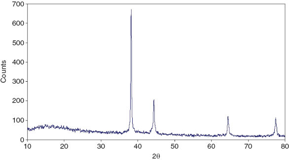
X-ray diffraction (XRD) of silver layers of the nanocomposite films.

X-ray diffraction (XRD) of Ag-polyvinyl alcohol (PVA) bulk layers of the nanocomposite films.
Peak list of Ag [Inorganic Crystal Structure Database (ICSD) collection code: 53762].
| No. | h k l | d-spacing (Å) | Position (°2θ) | Relative intensities (%) |
|---|---|---|---|---|
| 1 | 1 1 1 | 2.35905 | 38.117 | 100.00 |
| 2 | 0 0 2 | 2.04300 | 44.301 | 47.5 |
| 3 | 0 2 2 | 1.44462 | 64.447 | 27.8 |
| 4 | 1 1 3 | 1.23198 | 77.402 | 30.9 |
| 5 | 2 2 2 | 1.17953 | 81.545 | 8.9 |
The degree of crystallinity determines the amount of crystal phase (Ag nanoparticles) in the amorphous polymer (PVA), which can be calculated by dividing the total area of crystalline peaks by the total area of all peaks. The crystallinity of Ag layers was found to be 82%, indicating a higher concentration of Ag nanoparticles on the Ag layers. The crystallinity of Ag-PVA bulk layers was 59%, which indicates a lower concentration of Ag nanoparticles on the Ag-PVA bulk layers.
The Sherrer diffraction formula was used to estimate the crystallite domain size (D):
where D is the crystallite size in (nm), k is the so-called shape factor which usually takes a value of about 0.94, λ is the X-ray wavelength, (CuKa´=1.5406 Å), β is the full width half maximum (FWHM) of the main diffraction peak and θ is the diffraction angle corresponding to the maximum intensity peak in the XRD pattern. The crystallite size calculation from the FWHM of the (111) peak using Scherrer’s equation resulted in average crystallite sizes of about 43 nm for the Ag layers and about 29 nm for the Ag-PVA bulk layers.
3.3 EDX analysis
EDX microanalysis is a technique used for identification of the elemental composition of a specimen. The “fingerprint” energies of the emitted X-rays can then be used to identify an element. Also, EDX microanalysis is capable of generating a map of one or more chemical elements of interest [43]. Analysis of nanocomposite films with dual-surface with an EDX spectrometer confirmed the presence of elemental Ag signals of the Ag nanoparticles at 2.6 keV, 3.0 keV, 3.2 keV and 3.4 keV, which correspond to the binding energies of Ag lines. The EDX analysis of Ag layers represents the element percentage (%) of Ag, C and O as 93.06, 0.003 and 6.93, respectively, (Table 2 and Figure 4B), and indicates a higher concentration of Ag nanoparticles on the Ag layers. EDX analysis of the Ag-PVA bulk layers represents the element percentage (%) of Ag, C and O as 10.80, 24.11 and 65.06, respectively, (Table 2 and Figure 4A), which indicates a lower concentration of Ag nanoparticles on the Ag-PVA bulk layers. The carbon and oxygen in the examined samples are attributed to the matrix of PVA. The structure and morphology of the two surfaces were confirmed by SEM.

Energy dispersive X-ray (EDX) analysis of (A) Ag-polyvinyl alcohol (PVA) bulk layers and (B) silver layers.
Atomic % and element % for the two surfaces of polyvinyl alcohol (PVA)-silver (Ag) nanocomposite films.
| Elements | Atomic % | Element % | ||
|---|---|---|---|---|
| Ag-PVA bulk layers | Ag layers | Ag-PVA bulk layers | Ag layers | |
| C K | 32.50 | 0.02 | 24.11 | 0.003 |
| Ag L | 1.62 | 66.19 | 10.80 | 93.06 |
| O K | 65.86 | 33.25 | 65.06 | 6.93 |
3.4 SEM
The SEM images of the nanocomposite films with dual-surface are presented in Figures 5 and 6, respectively. SEM of the Ag layers showed concentrated and agglomerated nanoparticles of spherical shape with a diameter range of 30 nm-60 nm. SEM of Ag-PVA bulk layers showed that the Ag nanoparticles formed were predominantly cubical, uniform shape with no agglomeration and well-defined morphology observed in the micrograph. The average diameter of the Ag nanoparticles was found to be approximately 200 nm–350 nm on the Ag-PVA bulk layers. The function of PVA in the Ag-PVA nanocomposites prevents the process of agglomeration of Ag nanoparticles. The growth of the nanoparticles distributed uniformly between the networks of the PVA hydrogel.

Scanning electron microscopy (SEM) of silver layers of the nanocomposite films.

Scanning electron microscopy (SEM) of Ag-polyvinyl alcohol (PVA) bulk layers of the nanocomposite films.
XRD, EDX and SEM analysis confirmed the formation of nanocomposite films with dual-surface, Ag layers and Ag-PVA bulk layers shown in Scheme 1. Formation of Ag layers could be explained by an increase in reaction time, particle size and aggregation of Ag nanocrystals gradually increased together. Inside the beaker or the inner surface of the glass of a Petri dish finally turned into an Ag mirror. Ag particles deposited on a glass substrate to be Ag layers as shown in Scheme 1. Ag layers were formed very easily because the wetting-contact angle between two phases (substrate and solution) was greater than zero and reduced the energy needed. The Tollen’s reaction occurred as a heterogeneous nucleation [48].
4 Conclusion
Nanocomposite films with dual-surface, Ag layers and Ag-PVA bulk layers were prepared by Tollen’s reagent at ambient temperature using β-D-glucose as a green reducing agent, in comparison to other techniques. EDX microanalysis identified the elemental composition of the two surfaces. The Ag content of the Ag layers reached 93%. The Ag content of the Ag-PVA bulk layers reached 10.8%. XRD revealed the formation of an FCC phase of Ag nanoparticles on Ag layers and the formation of an amorphous phase of PVA matrix incorporated Ag nanoparticles on Ag-PVA bulk layers. SEM of the Ag layers showed concentrated and agglomerated small nanoparticles. SEM of Ag-PVA bulk layers showed that the Ag nanoparticles formed were predominantly a cubical, uniform shape with no agglomeration and well-defined morphology was observed in the micrograph.
About the author

Sayed M. Badawy, born in 1966, studied Chemistry at Cairo University and obtained his PhD in the field of Physical Chemistry in 2001. He was employed as a fellow of Chemistry at Cairo University from 2008. Currently, he works as an Assistant Professor of Physical Chemistry at Aljouf University. His major research interests are polymer and environmental chemistry. He is author or co-author of about 20 peer-reviewed publications.
Acknowledgments
Financial support from Aljouf University (Research Project Number 232/34) is gratefully acknowledged.
References
[1] Wang X, Zhuang J, Peng Q, Li Y. Nature 2005, 437, 121–124.10.1038/nature03968Search in Google Scholar
[2] Joerger R, Joerger TK, Olsson E, Granqvi CG. Solar Energy 2001, 69, 27–33.10.1016/S0038-092X(01)00002-0Search in Google Scholar
[3] Schultz S, Smith DR, Mock JJ, Schultz DA. Proc. Nat. Acad. Sci. USA 2000, 97, 996–1001.10.1073/pnas.97.3.996Search in Google Scholar PubMed PubMed Central
[4] Handley DA. Colloidal Gold: Principles, Methods and Applications, Hayat MA, Ed., Academic; San Diego, CA, 1989, Vol. 1.Search in Google Scholar
[5] Dubas ST, Pimpan V. Talanta 2008, 76, 29–33.10.1016/j.talanta.2008.01.062Search in Google Scholar PubMed
[6] Blaker JJ, Nazhat SN, Boccaccini AR. Biomaterials 2004, 25, 1319–1329.10.1016/j.biomaterials.2003.08.007Search in Google Scholar PubMed
[7] Chen J, Lo Y, Liang Y, Jiang J, Shen G, Yu R. Anal. Sci. 2009, 25, 347–352.Search in Google Scholar
[8] Morones JR, Elechiguerra JL, Camacho A, Holt K, Kouri JB, Ramirez JT, Yacama JM. Nanotechnology 2005, 16, 2346–2353.10.1088/0957-4484/16/10/059Search in Google Scholar PubMed
[9] Nino-Martinez N, Martinez-Castanon GA, Aragon-Pina A, Martinez-Gutierrez F, Martinez-Mendoza JR, Ruiz F. Nanotechnology 2008, 19, 065711/1-065711/8.10.1088/0957-4484/19/6/065711Search in Google Scholar PubMed
[10] Alt V, Bechert T, Steinrücke P, Wagener M, Seidel P, Dingeldein E, Schnettler R. Biomat. 2004, 25, 4383–4391.Search in Google Scholar
[11] Russell AD, Hugo WB. Prog. Med. Chem. 1994, 31, 351–370.Search in Google Scholar
[12] Lee HY, Park HK, Lee YM, Kim K, Park SB. Chem. Commun. 2007, 28, 2959–2961.Search in Google Scholar
[13] Jeong SH, Yeo SY, Yi SC. J. Mater. Sci. 2005, 40, 5407–5411.Search in Google Scholar
[14] Chou W-L, Yu D-G, Yang M-C. Polym. Adv. Technol. 2005, 16, 600–607.Search in Google Scholar
[15] Jin M, Zhang XT, Nishimoto S, Liu ZY, Tryk DA, Emeline AV, Murakami T, Fujishima A. J. Phys. Chem. C 2007, 111, 658–665.10.1021/jp065590nSearch in Google Scholar
[16] Chen Q, Yue L, Xie F, Zhou M, Fu Y, Zhang Y, Weng J. J. Phys. Chem. C 2008, 112, 10004–10007.10.1021/jp800306cSearch in Google Scholar
[17] Khanna PK, Singh N, Charan S, Subbarao V, Gokhale R, Mulik UP. Mater. Chem. Phys. 2005, 93, 117–121.Search in Google Scholar
[18] Mbhele ZH, Salemane MG, van Sittert CG, Nedeljkovi JM, Djokovi V, Luyt AS. Chem. Mater. 2003, 15, 5019–5024.Search in Google Scholar
[19] Zeng R, Rong MZ, Zhang MQ, Liang HC, Zeng HM. Appl. Surf. Sci. 2002, 187, 239–247.Search in Google Scholar
[20] Zhang Z, Han M. J. Mater. Chem. 2003, 13, 641–643.Search in Google Scholar
[21] Liu H, Ge X, Zhu Y, Xu X, Zhang Z, Zhang M. Mater. Lett. 2000, 46, 205–208.Search in Google Scholar
[22] Faraji N, Yunus WM, Kharazmi A, Saion E, Shahmiri M, Tamchek N. J. Eur. Opt. Soc.: Rapid 2012, 7, 12040–12047.10.2971/jeos.2012.12040Search in Google Scholar
[23] Juang JH, Bonner WS, Ogawa YJ, Vacanti P, Weir GC. Transplantation 1996, 61, 1557–1561.10.1097/00007890-199606150-00001Search in Google Scholar
[24] Chen DH, Leu JC, Huang TC. J. Chem. Technol. Biotechnol. 1994, 61, 351–357.Search in Google Scholar
[25] Hyon SH, Cha WI, Ikada Y, Kita M, Ogura Y, Honda Y. J. Biomater. Sci. Polym. Ed. 1994, 5, 397–406.Search in Google Scholar
[26] Li JK, Wang N, Wu XS. J. Controlled Release 1998, 56, 117–126.10.1016/S0168-3659(98)00089-3Search in Google Scholar
[27] Yoshii F, Makuuchi K, Darwis D, Iriawan T, Razzak MT, Rosiak JM. Radiat. Phys. Chem. 1995, 46, 169–174.Search in Google Scholar
[28] Stepanov AL, Popok VN, Khaobullin IB, Kreibig U. Nucl. Instrum. Methods Phys. Res. Sect. B 2002, 191, 473–477.10.1016/S0168-583X(02)00595-5Search in Google Scholar
[29] Badr Y, Mahmoud MA. J. Appl. Polym. Sci. 2006, 99, 3608–3614.Search in Google Scholar
[30] Hong KH, Park JL, Sul IH, Youk JH, Kang TJ. J. Polym. Sci. Part B Polym. Phys. 2006, 44, 2468–2474.Search in Google Scholar
[31] Sharma VK, Yngard RA, Lin Y. Adv. Colloid Interface Sci. 2009, 145, 83–96.Search in Google Scholar
[32] Searle AB. The Use of Colloids in Health and Disease: Germicides and Disinfectants, Toronto Collection, London Constable and Co; Gerstein University, 1920.Search in Google Scholar
[33] Becker RO, Spardaro MD. J. Bone Joint Surg. 1978, 60, 871–881.Search in Google Scholar
[34] Liu H, Ge X, Zhu Y, Xu X, Zhang Z, Zhang M. Mater. Lett. 2000, 46, 205–208.Search in Google Scholar
[35] Bai J, Li Y, Sun L, Zhang C, Yang Q. Bull. Mater. Sci. 2009, 32, 161–164.Search in Google Scholar
[36] Zeng R, Rong MZ, Zhang MQ, Liang HC, Zeng HM. Appl. Surf. Sci. 2002, 187, 239–247.Search in Google Scholar
[37] Nalwa HS. Encyclopedia for Nanoscience and Nanotechnology, American Scientific Publishers: USA, 2004, Vol. 1.Search in Google Scholar
[38] Akbari B, Tavandashti MP, Zandrahimi M. Iran. J. Mater. Sci. Eng. 2011, 8, 48–56.Search in Google Scholar
[39] Goyal A, Kumar A, Parta P, Mahendra S, Tabatabai S, P Alvzrez P, John G, Ajayan P. Macromol. Rapid Commun. 2009, 30, 1116–1122.Search in Google Scholar
[40] Oorha C, Manea F, Pop A, Burtica G, Todea I. Rev. Chim (Bucharest) 2008, 59, 173–177.10.37358/RC.08.2.1728Search in Google Scholar
[41] Rehani B, Joshi P, Lad K, Pratap A. Indian J. Pure Appl. Phys. 2006, 44, 157–161.Search in Google Scholar
[42] Dorofeeva G, Streletskiib A., Povstugara I, Protasova A, Elsukova E. Colloid J. 2012, 74, 675–685.Search in Google Scholar
[43] Zheng J, Nagashima K, Parmiter D, de la Cruz J, Patri A. Characterization of Nanoparticles Intended for Drug Delivery, Methods in Molecular Biology, Springer Science+Business Media, LLC: Boston, MA, 2011, 697.Search in Google Scholar
[44] Badawy SM. Green Process. Synth. 2014, 3, 229–234.Search in Google Scholar
[45] Janardhanan R, Karuppaiah M, Hebalkar N, Rao T. Polyhedron 2009, 28, 2522–2530.10.1016/j.poly.2009.05.038Search in Google Scholar
[46] Razzak MT, Zainuddin E, Dewi S, Lely H, Taty S. Radiat. Phys. Chem. 1999, 55, 153–165.Search in Google Scholar
[47] Sadeghi B, Pourahmad A. Adv. Powder Technol. 2011, 22, 669–673.Search in Google Scholar
[48] Chitvoranund N, Jiemsirilers S, Kashima DP. J. Aust. Ceram. Soc. 2013, 49, 62–69.Search in Google Scholar
©2014 by De Gruyter
This article is distributed under the terms of the Creative Commons Attribution Non-Commercial License, which permits unrestricted non-commercial use, distribution, and reproduction in any medium, provided the original work is properly cited.
Articles in the same Issue
- Frontmatter
- In this issue
- Editorial
- Science Woodstock and Nobel Prize: what remains in 50 years?
- GPE 2014
- 4th International Congress on Green Process Engineering (GPE2014) continued
- Beyond biofuels: economic opportunities, recent advances and challenges in property modeling for vegetable oils
- Greener route to 4-vinyl-1-cyclohexane 1,2-epoxide synthesis using batch and continuous reactors
- Intensification of waste cooking oil transformation by transesterification and esterification reactions in oscillatory baffled and microstructured reactors for biodiesel production
- One-step processes for in situ transesterification to biodiesel and lutein extraction from microalgae Phaeodactylum using instant controlled pressure drop (DIC)
- Hydrodeoxygenation of stearic acid for the production of “green” diesel
- Original articles
- Eco-friendly conjugate hydrocyanation of 2-aroyl α,β-unsaturated ketones with potassium hexacyanoferrate(II)
- Facile and green synthesis of Hantzsch derivatives in deep eutectic solvent
- Green synthesis of dual-surface nanocomposite films using Tollen’s method
- Optimized microemulsion production of biodiesel over lipase-catalyzed transesterification of soybean oil by response surface methodology
- Adsorption of organic chemicals on graphene coated biochars and its environmental implications
- Company profile
- iX-factory GmbH: development of a microfluidic chromatography chip
- Conference announcements
- International Workshop on Process Intensification 2015 (IWPI2015): Towards Sustainable Process Technologies in the 21st Century (Canik Basari University, Samsun, Turkey, April 27–30, 2015)
- Conferences 2015–2017
- Book review
- Domino reactions: concepts for efficient organic synthesis