Thoracoschisis secondary to a mesenchymal hamartoma associated with diaphragmatic eventration
-
Colm P. Travers


Abstract
Thoracoschisis is an extremely rare congenital anomaly associated with limb body wall defect and diaphragmatic hernia. We describe a case of a female infant who was noted at birth to have tissue coming through a left sided thoracic defect next to an accessory nipple. The stomach bubble was displaced superiorly on radiographs. At surgery the tissue was attached to the left lateral lobe of the liver and was protruding through the chest wall via an intercostal defect below an eventrated diaphragm. The tissue was resected and the defect closed. Pathological examination was consistent with a mesenchymal hamartoma. The diaphragm may have formed abnormally in this case due to the presence of the mesenchymal hamartoma in this location.
Introduction
Mesenchymal hamartoma of the liver is the second most common benign liver tumor in childhood [1]. Mesenchymal hamartomas of the chest wall have also rarely been described in the literature [2]. This is the first case report of a mesenchymal hamartoma of the liver involving the left side of the chest wall and the liver. Liver hamartomas can act as space occupying lesions interfering with the growth of surrounding tissues and structures.
Presentation of the case
A female infant was delivered at 39 weeks of gestation by spontaneous vaginal delivery weighing 2665 g. Apgar scores were 8 at 1 min and 9 at 5 min. There was evidence of mild respiratory distress at birth and she required oxygen initially to maintain adequate saturations. The mother was 24 years old and medical history was significant for a previous child with dilated cardiomyopathy of unknown cause who died at 18 months of age and the use of prenatal medications including escitalopram, tramadol, topiramate, oxycodone/acetaminophen, and prenatal multivitamins. The mother denied cigarette smoking, alcohol use, or illicit drug use during pregnancy. An anomaly scan at 20 weeks of gestation was interpreted as normal. At birth she was noted to have red/purple tissue protruding from a left sided thoracic defect (Figures 1 and 2). There was an accessory nipple on the upper left side of the chest and the defect was situated between the two nipples. There was no evidence of pectoralis muscle hypoplasia or any limb anomalies. There were no other dysmorphic features or congenital anomalies noted on physical exam. She required oxygen via nasal cannula. The stomach bubble was displaced superiorly on radiographs (Figure 3). A pre-operative computerized tomography scan showed that the tissue was connected to the liver (Figure 4). Echocardiogram showed a moderate sized patent ductus arteriosus, patent foramen ovale, and dextroposition of the heart.

Digital photograph of left side of thorax showing tissue protruding from left sided chest wall defect and associated accessory nipple.

Close-up digital photograph of left sided thoracoschisis.

Chest radiograph showing dextrocardia, superiorly displaced gastric bubble, and a radiodensity within left hemi-thorax.

Reconstructed coronal CT image showing the tissue connection from the liver to the thoracic wall defect.
At surgery there was a left sided diaphragmatic eventration. The diaphragm was intact with a superiorly displaced attachment to the anterior costal margin. The left lateral segment of the liver and omentum appeared adherent to the diaphragm. There was tissue protruding through an intercostal defect below the diaphragm. This tubular tissue was attached to the omentum and to the superior extent of the left lateral lobe of the liver. There was medial rib aplasia of the two overlying ribs. The tissue was resected and the defect closed. The infant was discharged home on day 6 after birth on full feeds, breathing room air with no support.
Pathological examination showed that the lesion was composed of loose myxoid stroma with scattered stellate and spindle cells (Figure 5). Numerous lymphangiomatous channels and mixed inflammatory cells were present. Branching bile ducts as well as small, well-formed bile ducts were identified. Hepatocytes were present singly or in small groups with no significant atypia seen. These features were consistent with a mesenchymal hamartoma of the liver.
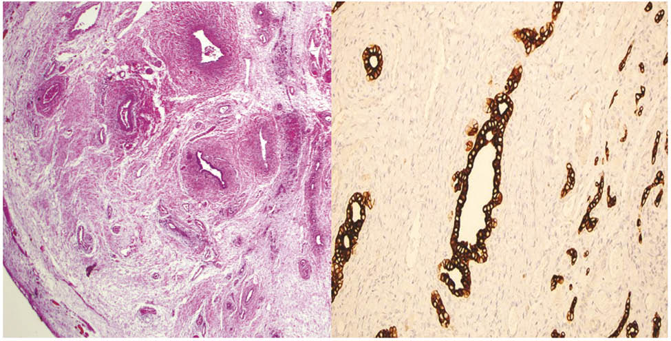
Image on left represents a hematoxylin and eosin stain at low power magnification. Image on right represents a cytokeratin immunohistochemical stain highlighting bile ducts.
Array comparative genomic hybridization of white blood cells did not demonstrate any constitutional copy number variations, but did detect a 16.99 Mb run of homozygosity on chromosome 14 that was concerning for uniparental disomy (UPD). However, subsequent testing was negative for UPD 14. Further genetic work up is pending.
Discussion
Thoracoschisis is an extremely rare congenital anomaly which has been associated with diaphragmatic hernia and limb-body wall complex [3, 4]. Early vascular disruption has been postulated as a potential cause of these cases of thoracoschisis [5]. The teratogenic effects of topiramate have been associated with orofacial clefting, pyloric stenosis and hypospadias, but not with body wall defects such as throracoschisis or mesenchymal hamartomas. It is possible that the teratogenic effects of topiramate during the period of organogenesis may have been compounded by the polypharmacy seen in this patient. Paternal UPD 14 has been associated with thoracic and abdominal wall defects but testing in this case was negative. Additionally, nine autosomal recessive genes lie within the run of homozygosity. The most notable of these is DNAAF2, abnormalities in which have been associated with cilliary dyskinesia and situs anomalies. Abnormalities of the diaphragm have been reported in each case of thoracoschisis to date [6]. This includes a case report of isolated thoracoschisis where abdominal contents including intestines and a Reidel lobe of the liver were found protruding through a thoracic wall defect below an eventrated diaphragm [7]. Although mesenchymal hamartoma of the liver is the second most common benign liver tumor in childhood [1], mesenchymal hamartomas of the chest wall are rare and are thought to have a different embryonal origin compared to mesenchymal hamartomas of the liver. Mesenchymal hamartomas are benign tumors although rare cases of malignant transformation have been reported. There has been one previous case report of a mesenchymal hamartoma involving both the chest wall and the liver [8]. This previous case also described thoracoschisis in association with diaphragmatic eventration but on the right side of the body, unlike in this case. It is known that liver hamartomas can act as space occupying lesions interfering with the growth of surrounding tissues and structures. It is possible that the abnormalities including the thoracic wall defect, eventrated diaphragm, and dextrocardia described in this case occurred due to the presence of this large mesenchymal hamartoma causing displacement thereby affecting normal growth and development of these structures.
References
[1] Siddiqui MA, McKenna BJ. Hepatic mesenchymal hamartoma: a short review. Arch Pathol Lab Med. 2006;130:1567–9.10.5858/2006-130-1567-HMHASRSearch in Google Scholar
[2] Jozaghi Y, Emil S, Albuquerque P, Klam S, Blumenkrantz M. Prenatal and postnatal features of mesenchymal hamartoma of the chest wall: case report and literature review. Pediatr Surg Int. 2013;29:735–40.10.1007/s00383-013-3280-1Search in Google Scholar
[3] Bhattacharyya NC, Gogoi M, Deuri PK. Thoracoschisis with limb agenesis. J Indian Assoc Pediatr Surg. 2012;17:78–9.10.4103/0971-9261.93972Search in Google Scholar
[4] Martínez-Frías ML. Clinical and epidemiological characteristics of infants with body wall complex with and without limb deficiency. Am J Med Genet. 1997;73:170–5.10.1002/(SICI)1096-8628(1997)73:2<170::AID-AJMG11>3.0.CO;2-RSearch in Google Scholar
[5] Van Allen MI, Curry C, Gallagher L. Limb body wall complex: I. Pathogenesis. Am J Med Genet. 1987;28:529–48.10.1002/ajmg.1320280302Search in Google Scholar
[6] McKay JD, Parker CM, Loewen J, Cundiff CA, Herman HK, Abramowsky CR, et al. Thoracoschisis: a case report and review of literature. Fetal Pediatr Pathol. 2015;34:307–14.10.3109/15513815.2015.1051254Search in Google Scholar
[7] Seleim HM, ElFiky MM, Fares AE, Elbarbary MM. Isolated thoracoschisis: case report and review of literature. European J Pediatr Surg Rep. 2015;3:40–2.10.1055/s-0034-1396013Search in Google Scholar
[8] Yesildag E, Yeker Y, Erdogan E, Yeker D. Extrathoracic liver hamartoma. Indian J Pediatr. 2004;71:265–7.10.1007/BF02724281Search in Google Scholar
-
The authors stated that there are no conflicts of interest regarding the publication of this article.
©2016 Walter de Gruyter GmbH, Berlin/Boston
Articles in the same Issue
- Frontmatter
- Case Reports – Obstetrics
- The Bakri balloon implementation during cesarean section without switching to the lithotomy position
- Recurrent large uterine fundal dehiscence during cesarean section after hysteroscopic uterine septum resection with uterine perforation
- Unexpected pregnancy during tamoxifen treatment: a case report and review of the literature
- Postpartum hemorrhage in the setting of a mechanical heart valve
- Spontaneous cord hematoma: report of two cases
- Anencephaly with placental adhesion
- Negative pressure wound treatment for uterine incision necrosis following a cesarean section
- Management of very early preterm premature rupture of membranes (PPROM) in twin pregnancies by selective feticide
- Spontaneous carotid artery dissection in pregnancy
- Spontaneous heterotopic triplet pregnancy with intrauterine monochorionic-monoamnionic twins
- Case Reports – Fetus
- Importance of perinatal care for pregnant women with severe fetal multiple limb abnormalities
- Mitoxantrone exposure in pregnancy: a new case report in a multiple sclerosis patient
- Transient iatrogenic heart block following foetal intracardiac transfusion for severe twin anaemia-polycythaemia sequence
- Vertical transmission of Zika virus (ZIKV) in early pregnancy: two cases, two different courses
- Case Reports – Newborn
- Early neonatal pyloric stenosis after exposure to maternal macrolide therapy
- Case report of neonatal near drowning associated with underwater birth
- Thoracoschisis secondary to a mesenchymal hamartoma associated with diaphragmatic eventration
- Acute myocardial infarction in a premature infant on the first day of life
- A rare case of acrocephaly: Saethre-Chotzen syndrome or Crouzon?
- Chest drain associated neonatal pneumopericardium
- Raynaud’s phenomenon in a newborn: case report and review of the literature
- Late-onset brain abscess due to group B Streptococcus