
Keratin 17 is induced in prurigo nodularis lesions
-
Li-Li Yang
 ,
Wei Zhang
and
Bo Yu
,
Wei Zhang
and
Bo Yu

Abstract
Prurigo nodularis (PN) is a highly pruritic chronic inflammatory dermatosis with unknown pathogenesis. It is characterized by the existence of many hyperkeratotic, erosive papules and nodules, and the development of lesions may be associated with hyperproliferation and aberrant differentiation of keratinocytes. Keratin 17 (K17) is overexpressed selectively in human proliferative skin diseases, promoting keratinocyte proliferation not found in normal epidermis. In this study, we investigated the mRNA levels and protein levels of K17 in lesional and perilesional skin using quantitative real-time polymerase chain reaction and western blot. We demonstrate that K17 is induced in lesional and perilesional skin in PN. The mRNA expression level of K17 was upregulated in PN lesions (P < 0.01), with multifold changes in the PN lesion (normalized to glyceraldehyde-3-phosphate dehydrogenase as the housekeeping gene) showing a median positive correlation with PRUNOSI (P < 0.05). The protein level of K17 was also markedly increased in PN lesions (P < 0.01). In conclusion, K17 is highly induced in PN lesions, which may contribute to the proliferation of keratinocytes and the pathogenesis of PN.
Graphical Abstract
1 Introduction
Prurigo nodularis (PN) is a chronic disorder of the skin characterized by multiple, pruritic, and hyperkeratotic nodules that tend to distribute symmetrically with a predilection on the extensor surfaces of the limbs [1,2]. Recently, PN was defined as a subtype of chronic prurigo and may possibly be triggered by neuronal sensitization to itch and the development of an itch–scratch cycle [3]. Various disorders have been considered to be linked to PN (e.g., atopic predisposition, systemic diseases, infections, neurological disorders, xerosis cutis, and psychiatric comorbidities) [1,4,5,6,7,8,9,10]. However, its exact pathophysiology remains unclear. Most patients with PN suffer from a high itch intensity with limited existing management, resulting in a reduced quality of life [1,11].
In the epidermis, keratinocytes, resident immune cells, and innervating somatosensory nerve fibers interact in direct and indirect ways and are involved in itch [12]. There is an aberrant expression of histamine-independent pruritogenic mediators in keratinocytes, underlining the importance of keratinocytes in the pathogenesis of PN [13]. The behavior of keratinocytes in normal and diseased skin is determined mostly by its state of activation or differentiation [14]. Histopathological studies of prurigo nodularis have shown thick epidermis, irregular epidermal hyperplasia, focal parakeratosis, and hypergranulosis in the epidermis [11,15], suggesting that hyperproliferation and aberrant differentiation of keratinocytes may be involved in the formation of PN.
The keratins are the typical intermediate filament proteins of epithelia. More than half of keratin genes are expressed in mature mammalian skin tissues and are expressed in highly specific patterns related to the epithelial type, stage of cellular differentiation, and striking modulation upon wounding, infection, or disease [16,17,18]. In the homeostatic state, only basal keratinocytes expressing keratin 5 and keratin 14 (K5/K14) can regenerate and differentiate through the spinous and granular layers of the epidermis to become corneocytes; complete terminal differential keratinocytes express keratin 1 and keratin 10 (K1/K10). In response to various stressors observed in wounding, KC activation is triggered, expressing keratin 6 and keratin 16 (K6/K16) as well as keratin 17 (K17), which leads to different phenotypes, including keratinocyte hyperproliferation, migration, and cytoskeleton alteration [14,17,19,20].
K17 was originally discovered in epidermis cells as a specific cytoskeleton in basal cell skin carcinoma [19]. It has limited expression in certain healthy epithelia such as sweat glands and the outer root sheath. However, it is strongly induced in challenged keratinocytes [21]. K17 stimulates protein synthesis and epithelial cell growth by binding to the adaptor protein 14-3-3-sigma [22]. K17 can induce epidermal hyperplasia by regulating the immune response in the skin and promoting the Th1-/Th17-dominated inflammatory response, which contributes to the development of basal skin tumor [23]. In psoriasis, K17 is overexpressed in psoriatic lesional epidermis and can act as an autogenic target for autoreactive T cells in the lesional epidermis, causing epithelial proliferation [24,25]. Hence, K17 is considered to be the hallmark of the psoriasis.
In this work, to discover the pattern of keratin gene expression in PN skin lesions and to understand dysregulated keratinocytes in PN pathogenesis, we detected the K17 expression pattern in PN lesional and perilesional skin by using quantitative real-time polymerase chain reaction (qRT-PCR), Western blot and immunohistochemistry studies.
2 Materials and methods
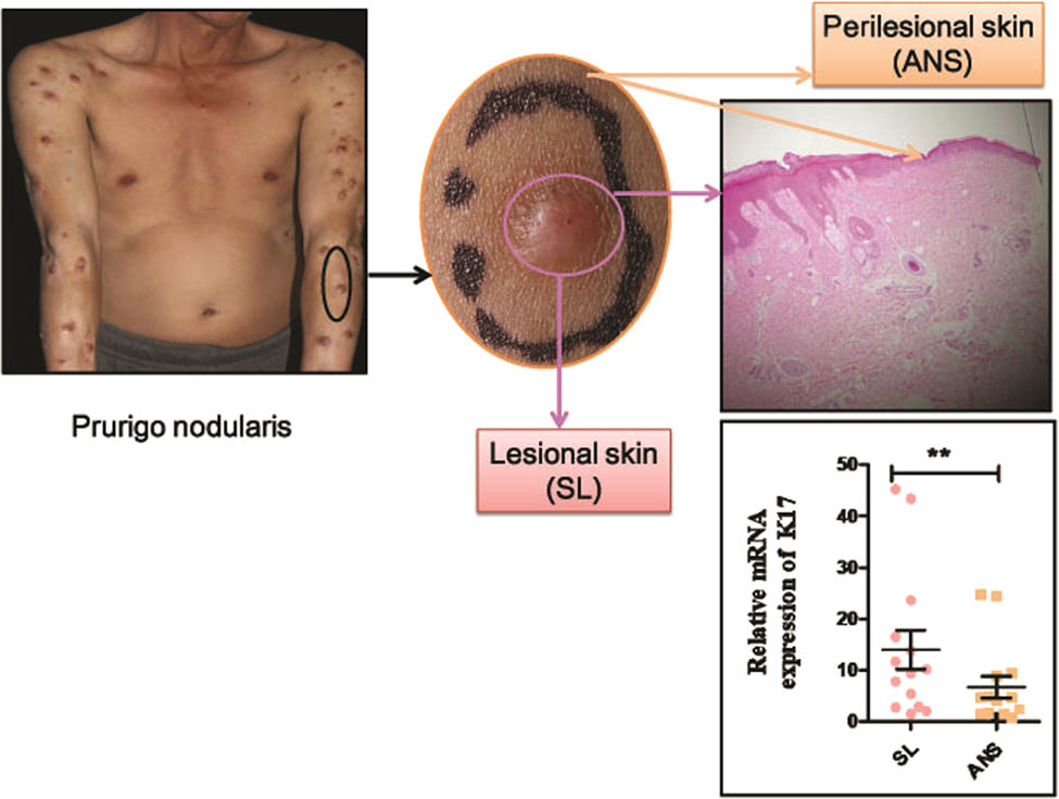
2.1 Patient population
This retrospective study was carried out on patients with PN presenting to the Department of Dermatology at Peking University Shenzhen Hospital in China. All patients were examined and diagnosed by different dermatologists according to clinical features and histopathological results. Following a 2-week washout period without active treatment (systemic immunosuppressants, corticosteroids, topical steroids, and immunomodulators), the participating dermatologist completed the questionnaire to assess patient clinical data, including demographic data, total serum immunoglobulin E (IgE) level, verbal rating scale (VRS), visual analog scale (VAS), dermatology life quality index (DLQI), and the extent of scratch lesions via PRUNOSI [26] (PRUNOSI evaluated the type [papules, nodules, lichenification excoriations, and crusts] and the percentage of lesions in the affected body area versus the whole body). Lesional skin and perilesional skin biopsy specimens from the extensor of the upper/lower extremities of patients with PN were obtained. PN lesional skin biopsies of 3–4 mm in diameter were taken from the arm, leg, or thigh, and perilesional skin biopsies were taken near the PN lesional skin. The skin specimen was divided into two parts (Figure S1): one part was used for hematoxylin and eosin staining to confirm the diagnosis, and the other part was used for mRNA analysis or protein analysis. We obtained PN specimens from 38 individuals and normal skin specimens from nine individuals (four men and five women). The normal healthy skin volunteers had no history of skin disease, including skin pruritus or atopic dermatitis (AD). Biopsies were taken from similar anatomical locations when the patient underwent surgery for some other reason. All of the experimental protocols for this research were approved by the local Ethical Committee of the Peking University Shenzhen Hospital (Figure S2). Written informed consent was obtained from all participants. The study was conducted in accordance with the ethical standards of the institutional research committee (approval no. PKUSZHEC(R)201915).
3 Methods
3.1 RNA extraction and qRT-PCR
Specimens of PN lesional skin, perilesional skin, and healthy skin were obtained. Total RNA was extracted with TRIzol (Cat. No. T9424; Sigma-Aldrich, St. Louis, MO, USA). RNA quality was detected with a NanoDrop 2000 spectrophotometer (NanoDrop Technologies, USA). The RNA product was reverse-transcribed into first-strand cDNA according to the protocol performed using a transcription kit (Cat. No. R212-02, HiScript 1st Strand cDNA Synthesis Kit; Vazyme Biotech, Nanjing, China). The expression of K17 and other keratin genes was quantified using the iTaq™ Universal SYBR®Green Supermix (Cat. No. 172-5124; Bio-Rad, Hercules, CA, USA) according to the qRT-PCR protocol. Glyceraldehyde-3-phosphate dehydrogenase (GAPDH) mRNA levels were used as an internal control to normalize the gene expression data. The qRT-PCR (40 cycles of denaturation at 98°C for 15 s and annealing at 58°C for 15 s) was performed based on the settings of the Bio-Rad system (CFX96TM Real-Time System; Bio-Rad). All reactions were performed in triplicate. The results of the difference in expression were calculated using the comparative cycle threshold (Ct) method by standardizing against GAPDH expression. Quantification of the target gene expression was performed using the 2−△△Ct method, ΔCt value = target gene Ct value − GAPDH gene CT value, ΔΔCt value = experimental group ΔCt mean value − control group ΔCt mean value. Sequence-specific primers were designed by Primer3 online as follows: keratin 5, sense: 5′-GGACAACAACCGCAACC-3′, antisense: 5′-TGCTTGGTGTTGCGGAGGT-3′; keratin 6, sense: 5′-AGGCTGAATGGCGAAGG-3′, antisense: 5′-AGGAGGTGGTGGTGTACTTGATGGT-3′; keratin 16, sense: 5′-GGTGGTGATGGGCTTCTG-3′, antisense: 5′-CGATGGTCTTGAAGTAGGGA-3′; keratin 17, sense: 5′-GGTGGGTGGTGAGATCAATGT-3′, antisense: 5′-CGCGGTTCAGTTCCTCTGTC-3′; and GAPDH, sense: 5′-GGAGTCAACGGATTTGGTCGTA-3′, antisense: 5′-GCAACAATATCCACTTTACCAGAGTTAA-3′.
3.2 Western blot and immunohistochemistry analysis
Total protein of the skin tissues was extracted according to the methods of Wang et al. (2016) using 6.5 M urea buffer with the addition of protease and phosphatase inhibitors (Cat. No. 04693132001; Roche Applied Science, Germany). Protein concentration was measured using the Pierce™ BCA protein assay kit (Cat. No. 23225; Thermo Fisher, Waltham, MA, USA). Protein (10–20 μg) was separated using 12% sodium dodecyl sulfate polyacrylamide gel electrophoresis and transferred onto a polyvinylidene fluoride (PVDF) membrane. The PVDF membrane was blocked in 5% bovine serum albumin for 45 min, followed by incubation in specific antibodies against K17 (Cat. No. 12509; Cell Signaling Technology, Beverly, MA, USA) and GAPDH (Cat. No. 2118; Cell Signaling Technology) for 2 h at room temperature or 4°C overnight. Consequently, incubation with anti-mouse IgG antibody conjugated with horseradish peroxidase (1:2,000; Cat. No. A9044; Sigma-Aldrich; secondary antibody) was performed at room temperature for 2 h. Protein bands were detected using the SuperSignal West Pico (Plus) Chemiluminescent Substrate (Cat. No. 34580; Thermo Fisher, MA, USA). The protein level of K17 was normalized against GAPDH. Immunohistochemical staining was conducted as previously described [13] with K17 rabbit polyclonal antibody (Cat. No. 103765; GeneTeX, Irvine, CA, USA).
3.3 Statistical analysis
The clinical data were expressed as mean ± standard deviation or median, while expression data were expressed as mean ± standard error of the mean (SEM). qRT-PCR expression values were normalized to the mRNA expression of GAPDH. The relative protein expression levels were quantified using Image J software (National Institutes of Health) and normalized to the controls (GAPDH).
Statistical analysis was performed with GraphPad Prism5 (GraphPad Software Inc., San Diego, CA, USA). Unpaired Student’s t test was used as an appropriate approach to compare the data between the lesional skin and healthy control groups, and paired Student’s t test was performed to determine the significant differences between lesional and perilesional skin. P < 0.05 was accepted as statistically significant. Spearman correlation was used to analyze the association among clinical variables and genes. All tests were two-tailed and considered statistically significant when P < 0.05. Statistical software SPSS version 16.0 (SPSS Inc., Chicago, IL, USA) was used for analysis.
4 Results
4.1 Demographics
We recruited 38 patients with PN in this study, including 24 men (63.2%) and 14 women (36.8%), with a mean age at first visit of 46.5 ± 14.6 years (range, 14–76 years) and average PN duration of 4.5 years (range, 0.2–40 years). Most of the patients suffered from high-intensity pruritus and accompanying sensations (e.g., pruritus, stinging, burning, or tingling). VAS (median = 5) and VRS (median = 2) scores indicated that most of the patients suffered from moderate or higher pruritus intensity. PRUNOSI scores ranged from 3 to 15, and the total serum IgE level of patients ranged from 8 to 5,500 IU/mL; 23 (60.5%) patients showed increased total IgE level (10–15 years old >200 IU/mL, adult >100 IU/mL; Table 1).
Demographic and clinical information of patients with prurigo nodularis
| Patient (n) | Gender (male:female) | Age (year) (mean ± SD) (range) | Duration (year) (mean ± SD) (range) | Atopic history | PRUNOSI (median) (range) | VRS (median) (range) | VAS (median) (range) | IgE (IU/ml) (mean ± SD) (range) | DLQI (median) (range) |
|---|---|---|---|---|---|---|---|---|---|
| 38 | 24:14 | 46.6 ± 14.6 | 4.5 ± 7.8 | 12 | 9 | 2 | 5 | 539.4 ± 967.3 | 8 |
| (14–76) | (0.2–40) | 3–15 | 0–3 | 0–10 | 8.7–5,500 | 1–26 |
DLQI: dermatology life quality index; IgE: total IgE; n: number; VAS: visual analog scale (scores from 0 to 10); and VRS: verbal rating scale (score range 0–3).
4.2 Location of lesions and histological characteristics
In this study, 32% of patients with PN had an atopic background with no signs of active dermatosis, lesions of PN manifested in circumscribed areas, and symmetrical distribution of the extensor surfaces of extremities and trunk. On the back, the untouched skin area that was free of lesions resembled a butterfly shape and is thus called the “butterfly sign” [2,27]. In addition, lesions affected patients’ face, scalp, buttock, or palms (Figure 1a–e). The biopsies from the representative lesions showed typical features, thick orthohyperkeratosis, irregular acanthosis hyperplasia, irregular elongation of the rete ridges, and focal parakeratosis in the epidermis (Figure 2a); in the dermis, mild perivascular lymphocytic infiltrate, an increased number of capillaries, and vertically oriented collagen bundles were observed (Figure 2b).
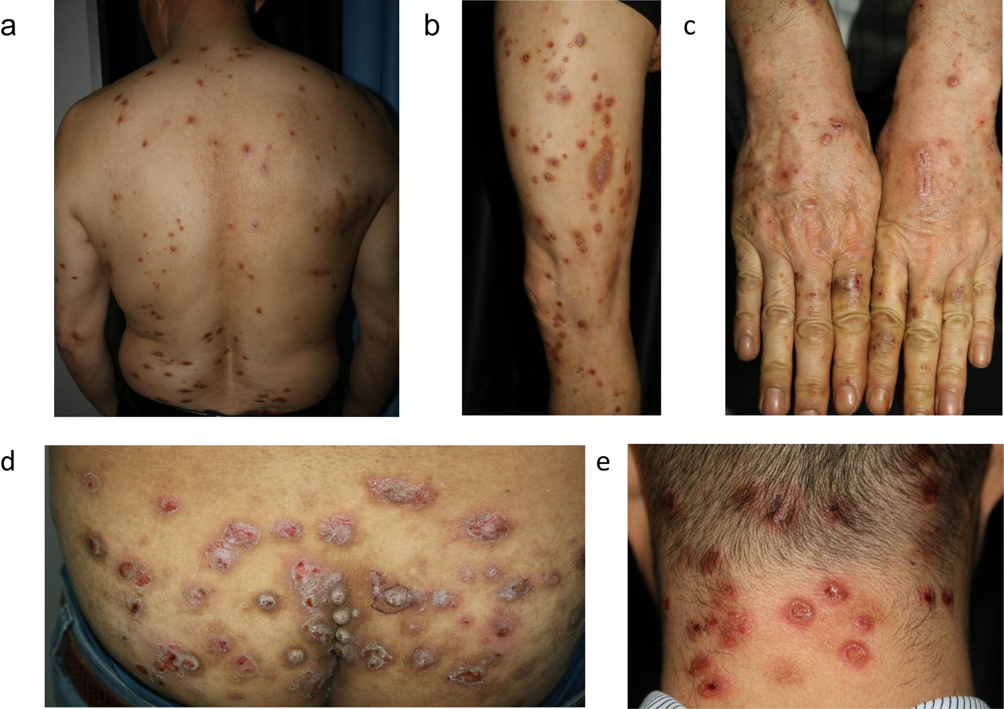
Clinicopathological features of prurigo nodularis. (a–e) Multiple excoriated papules or nodules involving typical distribution of trunk with the “butterfly sign” (no lesion on the center of the back), extensors of the extremities, palms, buttocks, and neck. These images were taken from five different patients with PN: (a) P36, (b) P23, (c) P26, (d) P18, and (e) P25.
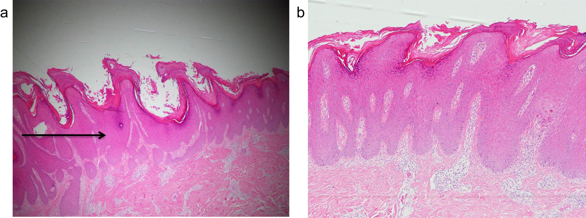
Histological observations of lesional skin biopsy from prurigo nodularis. Paraffin-embedded sections of lesional skin from PN stained with hematoxylin and eosin. Lesional skin biopsy showed orthohyperkeratosis, hypergranulosis, and epidermal hyperplasia (arrow); the epidermis mostly consisted of keratinocytes. (a) Original magnification ×40; (b) original magnification ×100.
4.3 K17 expression is induced in PN lesional skin
We first investigated the K17 transcription in the lesional and the perilesional skin area of patients with PN (eight men and six women). K17 mRNA levels were quantified by qRT-PCR analysis. In PN lesional skin, K17 transcriptions were significantly higher than those in perilesional skin (P < 0.01; Figure 3a). The average mRNA levels in the lesional skin were about 2.8-fold higher than those in perilesional skin. K17 mRNA expression was also reduced in the healthy group (n = 9) as compared with the PN group (change between the lesional skin and healthy skin was more than 40-fold, P < 0.0001; Figure 3b).
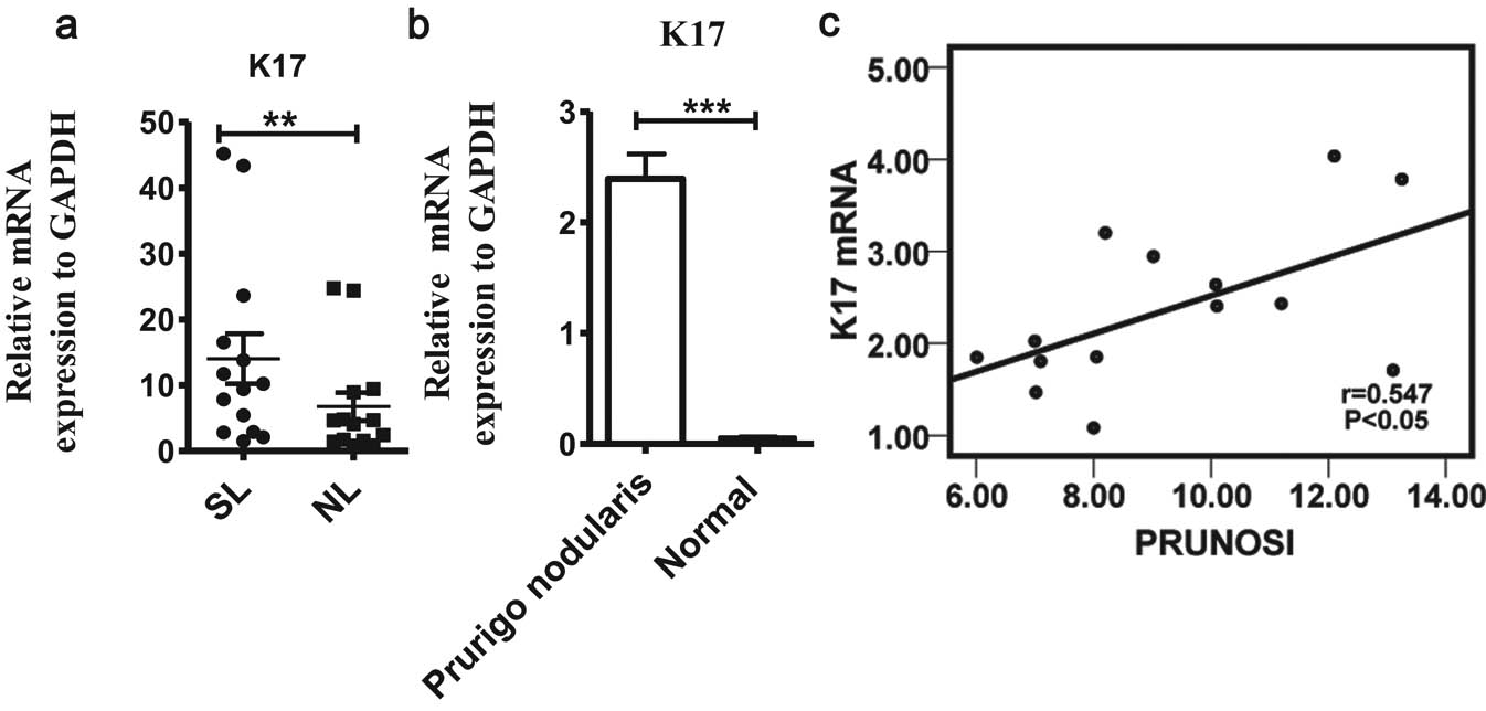
Keratin 17 (K17) mRNA expression levels in lesional and perilesional skin of patients with PN compared with those of healthy volunteers. (a) Real-time PCR analysis of K17 mRNA expression levels (n = 14). The levels are compared between the lesional skin and perilesional skin. (b) K17 mRNA expression levels are compared between the PN lesional skin and normal healthy skin. (c) Spearman analysis of PRUNOSI (x-axis) and K17 gene expression (y-axis); in the scatter plot, r = 0.547, *P < 0.05, regression line Y = 1.551x + 5.616, and regression coefficients R2 = 0.32. SL, lesional skin; NL, perilesional skin. Values show the expression/GAPDH level and are presented as mean ± SEM. Asterisks above the error bars denote comparisons between the matched lesional and perilesional skin. *P < 0.05, **P < 0.01, and ***P < 0.001, by paired Student’s t test.
To confirm the difference in K17 expression levels between the lesional and perilesional skin, we also checked the K17 protein levels in other patients with PN (five men and five women) and normal controls (n = 9) using Western blot analysis. Compared with those in the healthy controls and the perilesional skin, K17 protein levels were sharply increased in the PN lesional skin (Figure 4a), exhibiting a 3.3-fold upregulation compared with the perilesional skin by quantification of relative protein levels (Figure 4c). Among the samples, K17 protein was hardly detected in 40% of the perilesional skin samples; similarly, K17 protein levels in most of the normal skin were absent (Figure 4b). We then performed an immunohistochemical analysis of PN lesions and found that K17 was expressed in the lesional epidermis (Figure 4d). The K17 expression level in lesions was higher than in the perilesional skin.
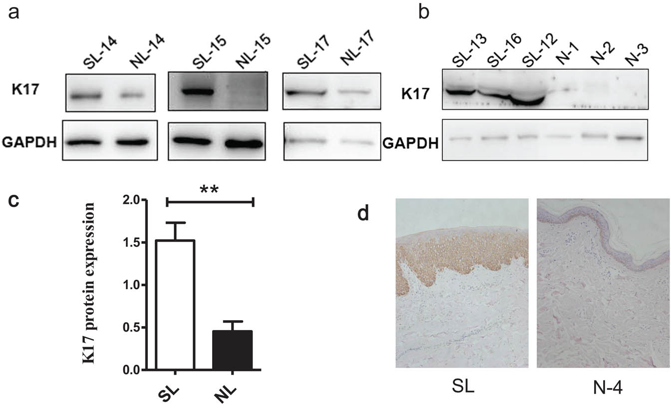
K17 expression is elevated in PN lesional skin. (a) Protein levels of K17 were detected in lesional skin and perilesional skin of patients with PN (n = 10) by Western blot and (b) protein levels of K17 from patients with PN and healthy controls (n = 9); (c) Image J analysis of K17 protein levels in lesional and perilesional skin. Band intensity was quantified by Image J. Values presented are the average of the K17 protein levels normalized to GAPDH from ten patients. (d) K17 expression in the PN lesional skin sample. Representative images of K17 expression are shown at 200 × magnification. SL, lesional skin; NL, perilesional skin; and N, normal control. **P < 0.01, by paired Student’s t test.
4.4 Higher levels of K6/K16 and K5 mRNA expression are detected in PN lesions
K17 together with K6/K16 is considered to be the marker for activated keratinocytes [28]. The mRNA expressions of K6/K16 were increased in the lesional skin (Figure S3a and b). In healthy epidermis, the expressions of specific keratin genes K5 and K14 were used as markers for the basal layer, whereas higher K5 mRNA expression levels were detected in the PN lesional skin as well (Figure S3c).
4.5 Correlations between clinical markers and K17 mRNA expression in PN lesions
We evaluated the correlations between the clinical markers and K17 relative to the mRNA expression level in PN lesions. We found that the PRUNOSI score (the extent of scratch lesions) was positively correlated with the VAS, VRS, and DLQI in a larger group of 38 patients (Figure S4). A median positive correlation was found between the K17 fold change of relative mRNA expression and PRUNOSI (r = 0.547, P < 0.05; n = 14; Figure 3c).
5 Discussion
Previous histopathological studies on PN skin lesion revealed changes in most types of skin cells, including mast cells, Merkel cells, epidermal keratinocytes, dendritic cells, and endothelial cells [15,29]. Keratinocytes are the predominant cell type in the epidermis, forming an integrated physical barrier. Epidermal keratinocytes may play an important role in the pathogenesis of PN. To date, no research has focused on the proliferation of abnormal keratinocytes in PN skin lesions, and discovery of keratin expression patterns may benefit the understanding of epidermal hyperplasia and PN pathogenesis.
K17 is a member of the type I acidic epithelial keratin family and can modulate keratinocyte growth during skin development by positively regulating protein synthesis [22,30]. K17 promotes the growth of basaloid skin tumors, oral squamous cell carcinoma, and so on [23,31]. High K17 expression is a powerful negative prognostic biomarker associated with poor outcome in patients with endocervical neoplasia [32]. In this study, we investigated the expression of K17 in PN skin lesions and its potential role in epidermal hyperplasia in PN.
In this work, comparison of K17 mRNA expression in PN skin lesions, perilesional skin, and healthy skin showed that the mRNA expression of K17 in the lesional skin was upregulated compared with the perilesional skin (Figure 3a) and healthy controls (Figure 3b). In addition, the comparison of K17 protein level in PN lesions confirmed the results of K17 mRNA expression, as the level of K17 protein was markedly increased in PN lesional skin. Compared with the healthy controls, K17 protein levels were elevated not only in the lesional skin but also in parts of the perilesional skin samples (6/10). These findings remind us that we initially selected newly developing nodules for this study, and these nodules tend to cover the healthy-appearing perilesional skin region and grow to larger nodules. These results may provide evidence for the clinical observation that the lesion frequently started as a smaller papule and rapidly processed to a larger nodule.
Overexpressed K17 in the lesion challenges the homeostasis of the skin epidermis in response to pathologic conditions. K17 together with K6/K16 is considered to be the marker for activated keratinocytes [21], and specific keratins (K6/K16) are inducible to expression in the suprabasal interfollicular epidermis [5,33], which is distinct from the keratins of healthy epidermis.
In this study, we analyzed the K6/K16 mRNA expression levels of PN skin, and the results revealed that the K6/K16 was significantly upregulated in lesional skin than in perilesional skin (P < 0.01 and P < 0.05), showing 12-fold (n = 9) and 5-fold (n = 6) increases on average, respectively (Figure S1a and b). Furthermore, positive trends of the relative mRNA expression of K6 correlated with K17 (r = 0.617, P = 0.077; n = 9; Figure S1d). K17 was mainly expressed in the epidermal keratinocytes of PN lesional skin (Figure 4d). Taken together, as the activated keratinocyte markers [21], the increased expression of K17 and K6/K16 implied that keratinocytes in PN lesions may be in the activated state. In basaloid skin tumors, co-polymerization of K17 with K5 was induced, where K17 as an immunomodulator accelerated epithelial proliferation and tumor formation in situations with an activated Hedgehog signaling pathway [23]. We also checked the K5 mRNA levels in both the lesional and perilesional skin and observed 19-fold higher K5 mRNA expression levels in the lesional skin (P = 0.089; n = 9; Figure S1c). This finding reflected the preferential activation and proliferation of the basal keratinocytes. We proposed that the activated basal keratinocytes in lesions expressed the K6/K16; consequently, some cytokines induced K17 expression [21]. These activated keratinocytes may lead to the hyperkeratotic epidermis of the PN.
From the correlation analysis, the K17 relative mRNA expression levels (fold change) in the 14 samples were positively correlated with the PRUNOSI score (Figure 3c), suggesting that K17 may play a role in the severity of PN skin lesions. During follow-up detection in a larger group of 38 patients, we found that the PRUNOSI score was positively correlated with the VAS, VRS, and DLQI (Figure S4), which indicates that severe itching leads to scratch-induced skin lesions [13], and damaged lesional skin negatively affects a patient’s quality of life. Therefore, we hypothesize that K17 could be critical to PN pathogenesis and a potential therapy target.
In this research, 60.5% of patients showed increased total serum IgE levels (Table 1). Spearman analysis showed that no correlations were detected between the total serum IgE levels and disease severity (PRUNOSI, VAS, and VRS). Specific IgE reactivities against a variety of bacterial antigens were observed in a subgroup comprising a third of AD patients [28], as an elevated serum IgE level is one of the clinical criteria of AD [20,34,35,36], and significantly high total serum IgE is also a hallmark of visceral infections by parasites [37]. Fifty percent of the patients with PN showed an atopic predisposition [4], and infection (hepatitis C and HIV) has been considered to be linked to PN. In this research, 31.6% of patients with PN had an atopic history; half of these patients with PN showed insect-bite dermatitis, and the IgE levels of the AD patients were greater than 100 IU/mL. Because increased levels of total IgE may be partly associated with PN pathogenesis, we will investigate the details of this in the future.
Therapies for PN generally focus on interrupting the itch–scratch cycle. In addition to addressing the cause of chronic pruritus [38], PN lesion healing should be considered as the goal of PN therapy. As far as we are concerned, there has been no standard criterion of treatment for itch until now. Treatment typically relies on the use of topical or intralesional steroids, although more severe or recalcitrant cases require novel therapeutic agents for treatment according to the underlying factors of PN [39]. Calcipotriol ointment, a synthetic form of vitamin D, acts to inhibit skin keratinocyte cell proliferation and enhance cell differentiation in the skin of patients with psoriasis, and downregulation of K17 is vital for its effect [40]. In a small randomized controlled trial comparing calcipotriol ointment with betamethasone valerate 0.1% ointment, calcipotriol ointment showed greater efficacy and more rapid clearance of the PN lesions [41].
In summary, we preliminarily identified that K17 was upregulated in PN lesions. Along with an aberrant expression of K6/K16 and K5, we speculated that keratinocytes may be in the activated cycles, and K17 might contribute to the proliferation of keratinocytes in PN epidermis and lesion progression. However, all of these results were obtained from a small sample size; in the future, we will use immunochemistry staining studies of keratins in the lesional epidermis to understand the dysregulated patterns of keratinocytes. The K17 regulation mechanism and its exact biological role in PN remains to be elucidated in future studies.
Acknowledgments
The authors thank Professor Gang Li for his kindness and patient guidance in the academic study. The authors also thank all of the patients as well as Ning Xu, Shi-hong Chen, Jian-jun Li, Wen-juan Yu, Xia Hong, Bo Lin, and Zhao-xue Ma for generously supporting this study.
Funding source: This work was supported by grants from the National Natural Science Foundation in China (81803139), the Science and Technology Program of Shenzhen Foundation of China (JCYJ 20160427185241351), Shenzhen San-ming Project (SZSM201812059).
Conflict of interest: There are no conflicts of interest.
References
[1] Zeidler C, Tsianakas A, Pereira M, Ständer H, Yosipovitch G, Ständer S. Chronic prurigo of nodular type: a review. Acta Derm Venereol. 2018 Feb;98(2):173–9.10.2340/00015555-2774Search in Google Scholar PubMed
[2] Schedel F, Schürmann C, Metze D, Ständer S. Prurigo. Clinical definition and classification. Hautarzt. 2014 Aug;65(8):684–90.10.1007/s00105-014-2753-zSearch in Google Scholar PubMed
[3] Puxeddu I, Pratesi F, Ribatti D, Migliorini P. Mediators of inflammation and angiogenesis in chronic spontaneous urticaria: are they potential biomarkers of the disease? Mediators Inflamm. 2017:4123694.10.1155/2017/4123694Search in Google Scholar PubMed PubMed Central
[4] Tanaka M, Aiba S, Matsumura N, Aoyama H, Tagami H. Prurigo nodularis consists of two distinct forms: early-onset atopic and late-onset non-atopic. Dermatology. 1995;190(4):269–76.10.1159/000246715Search in Google Scholar PubMed
[5] Boozalis E, Tang O, Patel S, Semenov YR, Pereira MP, Stander S, et al. Ethnic differences and comorbidities of 909 prurigo nodularis patients. J Am Acad Dermatology. 2018;79(4):714–9 e713.10.1016/j.jaad.2018.04.047Search in Google Scholar PubMed PubMed Central
[6] Tseng HW, Ger LP, Liang CK, Liou HH, Lam HC. High prevalence of cutaneous manifestations in the elderly with diabetes mellitus: an institution-based cross-sectional study in Taiwan. J Eur Acad Dermatol Venereol. 2015 Aug;29(8):1631–5.10.1111/jdv.12664Search in Google Scholar PubMed
[7] Kestner RI, Ständer S, Osada N, Ziegler D, Metze D. Acquired reactive perforating dermatosis is a variant of prurigo nodularis. Acta Derm Venereol. 2017 Feb;97(2):249–54.10.2340/00015555-2492Search in Google Scholar PubMed
[8] Mettang T, Vonend A, Raap U. Prurigo nodularis: its association with dermatoses and systemic disorders. Hautarzt. 2014 Aug;65(8):697–703.10.1007/s00105-014-2755-xSearch in Google Scholar PubMed
[9] Gieler U, Consoli SG, Tomás-Aragones L, Linder DM, Jemec GB, Poot F, et al. Self-inflicted lesions in dermatology: terminology and classification—a position paper from the European Society for Dermatology and Psychiatry (ESDaP). Acta Derm Venereol. 2013 Jan;93(1):4–12.10.2340/00015555-1506Search in Google Scholar PubMed
[10] Akarsu S, Ozbagcivan O, Ilknur T, Semiz F, Inci BB, Fetil E. Xerosis cutis and associated co-factors in women with prurigo nodularis. An Bras Dermatol. 2018 Sep–Oct;93(5):671–9.10.1590/abd1806-4841.20187127Search in Google Scholar PubMed PubMed Central
[11] Zeidler C, Ständer S. The pathogenesis of Prurigo nodularis ‘Super-Itch’ in exploration. Eur J Pain. 2016 Jan;20(1):37–40.10.1002/ejp.767Search in Google Scholar PubMed
[12] Stanisic M, Lyngstadaas SP, Pripp AH, Aasen AO, Lindegaard KF, Ivanovic J, et al. Chemokines as markers of local inflammation and angiogenesis in patients with chronic subdural hematoma: a prospective study. Acta Neurochir. 2012 Jan;154(1):113–20.10.1007/s00701-011-1203-2Search in Google Scholar PubMed
[13] Zhong W, Wu X, Zhang W, Zhang J, Chen X, Chen S, et al. Aberrant expression of histamine-independent pruritogenic mediators in keratinocytes may be involved in the pathogenesis of prurigo nodularis. Acta Derm Venereol. 2019 May;99(6):579–86.10.2340/00015555-3150Search in Google Scholar PubMed
[14] Knöbel M, O’Toole EA, Smith FJ. Keratins and skin disease. Cell Tissue Res. 2015 Jun;360(3):583–9.10.1007/s00441-014-2105-4Search in Google Scholar PubMed
[15] Weigelt N, Metze D, Ständer S. Prurigo nodularis: systematic analysis of 58 histological criteria in 136 patients. J Cutan Pathol. 2010 May;37(5):578–86.10.1111/j.1600-0560.2009.01484.xSearch in Google Scholar PubMed
[16] Moll R, Divo M, Langbein L. The human keratins: biology and pathology. Histochem Cell Biol. 2008 Jun;129(6):705–33.10.1007/s00418-008-0435-6Search in Google Scholar PubMed PubMed Central
[17] Paladini RD, Takahashi K, Bravo NS, Coulombe PA. Onset of re-epithelialization after skin injury correlates with a reorganization of keratin filaments in wound edge keratinocytes: defining a potential role for keratin 16. J Cell Biol. 1996 Feb;132(3):381–97.10.1083/jcb.132.3.381Search in Google Scholar PubMed PubMed Central
[18] Toivola DM, Boor P, Alam C, Strnad P. Keratins in health and disease. Curr Opin Cell Biol. 2015 Feb;32:73–81.10.1016/j.ceb.2014.12.008Search in Google Scholar PubMed
[19] Moll R, Franke WW, Volc-Platzer B, Krepler R. Different keratin polypeptides in epidermis and other epithelia of human skin: a specific cytokeratin of molecular weight 46,000 in epithelia of the pilosebaceous tract and basal cell epitheliomas. J Cell Biol. 1982 Oct;95(1):285–95.10.1083/jcb.95.1.285Search in Google Scholar PubMed PubMed Central
[20] Mevorah B, Frenk E, Wietlisbach V, Carrel CF. Minor clinical features of atopic dermatitis. Evaluation their diagnostic significance. Dermatologica. 1988;177(6):360–4.10.1159/000248607Search in Google Scholar PubMed
[21] Freedberg IM, Tomic-Canic M, Komine M, Blumenberg M. Keratins and the keratinocyte activation cycle. J Invest Dermatol. 2001 May;116(5):633–40.10.1046/j.1523-1747.2001.01327.xSearch in Google Scholar PubMed
[22] Kim S, Wong P, Coulombe PA. A keratin cytoskeletal protein regulates protein synthesis and epithelial cell growth. Nature. 2006 May;441(7091):362–5.10.1038/nature04659Search in Google Scholar PubMed
[23] Depianto D, Kerns ML, Dlugosz AA, Coulombe PA. Keratin 17 promotes epithelial proliferation and tumor growth by polarizing the immune response in skin. Nat Genet. 2010 Oct;42(10):910–4.10.1038/ng.665Search in Google Scholar PubMed PubMed Central
[24] Fu M, Wang G. Keratin 17 as a therapeutic target for the treatment of psoriasis. J Dermatol Sci. 2012 Sep;67(3):161–5.10.1016/j.jdermsci.2012.06.008Search in Google Scholar PubMed
[25] Gudmundsdottir AS, Sigmundsdottir H, Sigurgeirsson B, Good MF, Valdimarsson H, Jonsdottir I. Is an epitope on keratin 17 a major target for autoreactive T lymphocytes in psoriasis? Clin Exp Immunol. 1999 Sep;117(3):580–6.10.1046/j.1365-2249.1999.01013.xSearch in Google Scholar PubMed PubMed Central
[26] Siepmann D, Lotts T, Blome C, Braeutigam M, Phan NQ, Butterfass-Bahloul T, et al. Evaluation of the antipruritic effects of topical pimecrolimus in non-atopic prurigo nodularis: results of a randomized, hydrocortisone-controlled, double-blind phase II trial. Dermatology. 2013;227(4):353–60.10.1159/000355671Search in Google Scholar PubMed
[27] Pölking J, Zeidler C, Schedel F, Osada N, Augustin M, Metze D, et al. Prurigo activity score (PAS): validity and reliability of a new instrument to monitor chronic prurigo. J Eur Acad Dermatol Venereol. 2018 Oct;32(10):1754–60.10.1111/jdv.15040Search in Google Scholar PubMed
[28] Reginald K, Westritschnig K, Werfel T, Heratizadeh A, Novak N, Focke-Tejkl M, et al. Immunoglobulin E antibody reactivity to bacterial antigens in atopic dermatitis patients. Clin Exp Allergy. 2011 Mar;41(3):357–69.10.1111/j.1365-2222.2010.03655.xSearch in Google Scholar PubMed PubMed Central
[29] Schuhknecht B, Marziniak M, Wissel A, Phan NQ, Pappai D, Dangelmaier J, et al. Reduced intraepidermal nerve fibre density in lesional and nonlesional prurigo nodularis skin as a potential sign of subclinical cutaneous neuropathy. Br J Dermatol. 2011 Jul;165(1):85–91.10.1111/j.1365-2133.2011.10306.xSearch in Google Scholar PubMed
[30] Kim S, Coulombe PA. Emerging role for the cytoskeleton as an organizer and regulator of translation. Nat Rev Mol Cell Biol. 2010 Jan;11(1):75–81.10.1038/nrm2818Search in Google Scholar PubMed
[31] Khanom R, Nguyen CT, Kayamori K, Zhao X, Morita K, Miki Y, et al. Keratin 17 is induced in oral cancer and facilitates tumor growth. PLoS One 2016 Aug;11(8):e0161163.10.1371/journal.pone.0161163Search in Google Scholar PubMed PubMed Central
[32] Mockler D, Escobar-Hoyos LF, Akalin A, Romeiser J, Shroyer AL, Shroyer KR. Keratin 17 is a prognostic biomarker in endocervical glandular neoplasia. Am J Clin Pathol. 2017 Sep;148(3):264–73.10.1093/ajcp/aqx077Search in Google Scholar PubMed
[33] Schneider G, Driesch G, Heuft G, Evers S, Luger TA, Ständer S. Psychosomatic cofactors and psychiatric comorbidity in patients with chronic itch. Clin Exp Dermatol. 2006 Nov;31(6):762–7.10.1111/j.1365-2230.2006.02211.xSearch in Google Scholar PubMed
[34] Lodén M, Andersson AC, Lindberg M. The number of diagnostic features in patients with atopic dermatitis correlates with dryness severity. Acta Derm Venereol. 1998 Sep;78(5):387–8.10.1080/000155598443187Search in Google Scholar PubMed
[35] Eigenmann PA. Clinical features and diagnostic criteria of atopic dermatitis in relation to age. Pediatr Allergy Immunol. 2001;12(Suppl 14):69–74.10.1034/j.1399-3038.2001.121416.xSearch in Google Scholar PubMed
[36] Sharma L. Diagnostic clinical features of atopic dermatitis. Indian J Dermatol Venereol Leprol. 2001 Jan–Feb;67(1):25–7.Search in Google Scholar
[37] Humbert P, Buchet S, Barde T. Toxocariasis. A cosmopolitan parasitic zoonosis. Allerg Immunol. 1995 Oct;27(8):284–91.Search in Google Scholar
[38] Zeidler C, Yosipovitch G, Ständer S. Prurigo nodularis and its management. Dermatol Clin. 2018 Jul;36(3):189–97.10.1016/j.det.2018.02.003Search in Google Scholar PubMed
[39] Kowalski EH, Kneiber D, Valdebran M, Patel U, Amber KT. Treatment-resistant prurigo nodularis: challenges and solutions. Clin Cosmet Investig Dermatol. 2019 Feb;12:163–72.10.2147/CCID.S188070Search in Google Scholar PubMed PubMed Central
[40] Zhang J, Fang H, Wang R, Dang E, Jiang M, Wang G. Effect of calcipotriol on IFN-gamma-induced keratin 17 expression in immortalized human epidermal keratinocyte cells. Med Sci Monit. 2017 Dec;23:6049–56.10.12659/MSM.904850Search in Google Scholar
[41] Wong SS, Goh CL. Double-blind, right/left comparison of calcipotriol ointment and betamethasone ointment in the treatment of Prurigo nodularis. Arch Dermatol. 2000 Jun;136(6):807–8.10.1001/archderm.136.6.807Search in Google Scholar PubMed
© 2020 Li-Li Yang et al., published by De Gruyter
This work is licensed under the Creative Commons Attribution 4.0 International License.
Articles in the same Issue
- Regular Articles
- Electrochemical antioxidant screening and evaluation based on guanine and chitosan immobilized MoS2 nanosheet modified glassy carbon electrode (guanine/CS/MoS2/GCE)
- Kinetic models of the extraction of vanillic acid from pumpkin seeds
- On the maximum ABC index of bipartite graphs without pendent vertices
- Estimation of the total antioxidant potential in the meat samples using thin-layer chromatography
- Molecular dynamics simulation of sI methane hydrate under compression and tension
- Spatial distribution and potential ecological risk assessment of some trace elements in sediments and grey mangrove (Avicennia marina) along the Arabian Gulf coast, Saudi Arabia
- Amino-functionalized graphene oxide for Cr(VI), Cu(II), Pb(II) and Cd(II) removal from industrial wastewater
- Chemical composition and in vitro activity of Origanum vulgare L., Satureja hortensis L., Thymus serpyllum L. and Thymus vulgaris L. essential oils towards oral isolates of Candida albicans and Candida glabrata
- Effect of excess Fluoride consumption on Urine-Serum Fluorides, Dental state and Thyroid Hormones among children in “Talab Sarai” Punjab Pakistan
- Design, Synthesis and Characterization of Novel Isoxazole Tagged Indole Hybrid Compounds
- Comparison of kinetic and enzymatic properties of intracellular phosphoserine aminotransferases from alkaliphilic and neutralophilic bacteria
- Green Organic Solvent-Free Oxidation of Alkylarenes with tert-Butyl Hydroperoxide Catalyzed by Water-Soluble Copper Complex
- Ducrosia ismaelis Asch. essential oil: chemical composition profile and anticancer, antimicrobial and antioxidant potential assessment
- DFT calculations as an efficient tool for prediction of Raman and infra-red spectra and activities of newly synthesized cathinones
- Influence of Chemical Osmosis on Solute Transport and Fluid Velocity in Clay Soils
- A New fatty acid and some triterpenoids from propolis of Nkambe (North-West Region, Cameroon) and evaluation of the antiradical scavenging activity of their extracts
- Antiplasmodial Activity of Stigmastane Steroids from Dryobalanops oblongifolia Stem Bark
- Rapid identification of direct-acting pancreatic protectants from Cyclocarya paliurus leaves tea by the method of serum pharmacochemistry combined with target cell extraction
- Immobilization of Pseudomonas aeruginosa static biomass on eggshell powder for on-line preconcentration and determination of Cr (VI)
- Assessment of methyl 2-({[(4,6-dimethoxypyrimidin-2-yl)carbamoyl] sulfamoyl}methyl)benzoate through biotic and abiotic degradation modes
- Stability of natural polyphenol fisetin in eye drops Stability of fisetin in eye drops
- Production of a bioflocculant by using activated sludge and its application in Pb(II) removal from aqueous solution
- Molecular Properties of Carbon Crystal Cubic Structures
- Synthesis and characterization of calcium carbonate whisker from yellow phosphorus slag
- Study on the interaction between catechin and cholesterol by the density functional theory
- Analysis of some pharmaceuticals in the presence of their synthetic impurities by applying hybrid micelle liquid chromatography
- Two mixed-ligand coordination polymers based on 2,5-thiophenedicarboxylic acid and flexible N-donor ligands: the protective effect on periodontitis via reducing the release of IL-1β and TNF-α
- Incorporation of silver stearate nanoparticles in methacrylate polymeric monoliths for hemeprotein isolation
- Development of ultrasound-assisted dispersive solid-phase microextraction based on mesoporous carbon coated with silica@iron oxide nanocomposite for preconcentration of Te and Tl in natural water systems
- N,N′-Bis[2-hydroxynaphthylidene]/[2-methoxybenzylidene]amino]oxamides and their divalent manganese complexes: Isolation, spectral characterization, morphology, antibacterial and cytotoxicity against leukemia cells
- Determination of the content of selected trace elements in Polish commercial fruit juices and health risk assessment
- Diorganotin(iv) benzyldithiocarbamate complexes: synthesis, characterization, and thermal and cytotoxicity study
- Keratin 17 is induced in prurigo nodularis lesions
- Anticancer, antioxidant, and acute toxicity studies of a Saudi polyherbal formulation, PHF5
- LaCoO3 perovskite-type catalysts in syngas conversion
- Comparative studies of two vegetal extracts from Stokesia laevis and Geranium pratense: polyphenol profile, cytotoxic effect and antiproliferative activity
- Fragmentation pattern of certain isatin–indole antiproliferative conjugates with application to identify their in vitro metabolic profiles in rat liver microsomes by liquid chromatography tandem mass spectrometry
- Investigation of polyphenol profile, antioxidant activity and hepatoprotective potential of Aconogonon alpinum (All.) Schur roots
- Lead discovery of a guanidinyl tryptophan derivative on amyloid cascade inhibition
- Physicochemical evaluation of the fruit pulp of Opuntia spp growing in the Mediterranean area under hard climate conditions
- Electronic structural properties of amino/hydroxyl functionalized imidazolium-based bromide ionic liquids
- New Schiff bases of 2-(quinolin-8-yloxy)acetohydrazide and their Cu(ii), and Zn(ii) metal complexes: their in vitro antimicrobial potentials and in silico physicochemical and pharmacokinetics properties
- Treatment of adhesions after Achilles tendon injury using focused ultrasound with targeted bFGF plasmid-loaded cationic microbubbles
- Synthesis of orotic acid derivatives and their effects on stem cell proliferation
- Chirality of β2-agonists. An overview of pharmacological activity, stereoselective analysis, and synthesis
- Fe3O4@urea/HITh-SO3H as an efficient and reusable catalyst for the solvent-free synthesis of 7-aryl-8H-benzo[h]indeno[1,2-b]quinoline-8-one and indeno[2′,1′:5,6]pyrido[2,3-d]pyrimidine derivatives
- Adsorption kinetic characteristics of molybdenum in yellow-brown soil in response to pH and phosphate
- Enhancement of thermal properties of bio-based microcapsules intended for textile applications
- Exploring the effect of khat (Catha edulis) chewing on the pharmacokinetics of the antiplatelet drug clopidogrel in rats using the newly developed LC-MS/MS technique
- A green strategy for obtaining anthraquinones from Rheum tanguticum by subcritical water
- Cadmium (Cd) chloride affects the nutrient uptake and Cd-resistant bacterium reduces the adsorption of Cd in muskmelon plants
- Removal of H2S by vermicompost biofilter and analysis on bacterial community
- Structural cytotoxicity relationship of 2-phenoxy(thiomethyl)pyridotriazolopyrimidines: Quantum chemical calculations and statistical analysis
- A self-breaking supramolecular plugging system as lost circulation material in oilfield
- Synthesis, characterization, and pharmacological evaluation of thiourea derivatives
- Application of drug–metal ion interaction principle in conductometric determination of imatinib, sorafenib, gefitinib and bosutinib
- Synthesis and characterization of a novel chitosan-grafted-polyorthoethylaniline biocomposite and utilization for dye removal from water
- Optimisation of urine sample preparation for shotgun proteomics
- DFT investigations on arylsulphonyl pyrazole derivatives as potential ligands of selected kinases
- Treatment of Parkinson’s disease using focused ultrasound with GDNF retrovirus-loaded microbubbles to open the blood–brain barrier
- New derivatives of a natural nordentatin
- Fluorescence biomarkers of malignant melanoma detectable in urine
- Study of the remediation effects of passivation materials on Pb-contaminated soil
- Saliva proteomic analysis reveals possible biomarkers of renal cell carcinoma
- Withania frutescens: Chemical characterization, analgesic, anti-inflammatory, and healing activities
- Design, synthesis and pharmacological profile of (−)-verbenone hydrazones
- Synthesis of magnesium carbonate hydrate from natural talc
- Stability-indicating HPLC-DAD assay for simultaneous quantification of hydrocortisone 21 acetate, dexamethasone, and fluocinolone acetonide in cosmetics
- A novel lactose biosensor based on electrochemically synthesized 3,4-ethylenedioxythiophene/thiophene (EDOT/Th) copolymer
- Citrullus colocynthis (L.) Schrad: Chemical characterization, scavenging and cytotoxic activities
- Development and validation of a high performance liquid chromatography/diode array detection method for estrogen determination: Application to residual analysis in meat products
- PCSK9 concentrations in different stages of subclinical atherosclerosis and their relationship with inflammation
- Development of trace analysis for alkyl methanesulfonates in the delgocitinib drug substance using GC-FID and liquid–liquid extraction with ionic liquid
- Electrochemical evaluation of the antioxidant capacity of natural compounds on glassy carbon electrode modified with guanine-, polythionine-, and nitrogen-doped graphene
- A Dy(iii)–organic framework as a fluorescent probe for highly selective detection of picric acid and treatment activity on human lung cancer cells
- A Zn(ii)–organic cage with semirigid ligand for solvent-free cyanosilylation and inhibitory effect on ovarian cancer cell migration and invasion ability via regulating mi-RNA16 expression
- Polyphenol content and antioxidant activities of Prunus padus L. and Prunus serotina L. leaves: Electrochemical and spectrophotometric approach and their antimicrobial properties
- The combined use of GC, PDSC and FT-IR techniques to characterize fat extracted from commercial complete dry pet food for adult cats
- MALDI-TOF MS profiling in the discovery and identification of salivary proteomic patterns of temporomandibular joint disorders
- Concentrations of dioxins, furans and dioxin-like PCBs in natural animal feed additives
- Structure and some physicochemical and functional properties of water treated under ammonia with low-temperature low-pressure glow plasma of low frequency
- Mesoscale nanoparticles encapsulated with emodin for targeting antifibrosis in animal models
- Amine-functionalized magnetic activated carbon as an adsorbent for preconcentration and determination of acidic drugs in environmental water samples using HPLC-DAD
- Antioxidant activity as a response to cadmium pollution in three durum wheat genotypes differing in salt-tolerance
- A promising naphthoquinone [8-hydroxy-2-(2-thienylcarbonyl)naphtho[2,3-b]thiophene-4,9-dione] exerts anti-colorectal cancer activity through ferroptosis and inhibition of MAPK signaling pathway based on RNA sequencing
- Synthesis and efficacy of herbicidal ionic liquids with chlorsulfuron as the anion
- Effect of isovalent substitution on the crystal structure and properties of two-slab indates BaLa2−xSmxIn2O7
- Synthesis, spectral and thermo-kinetics explorations of Schiff-base derived metal complexes
- An improved reduction method for phase stability testing in the single-phase region
- Comparative analysis of chemical composition of some commercially important fishes with an emphasis on various Malaysian diets
- Development of a solventless stir bar sorptive extraction/thermal desorption large volume injection capillary gas chromatographic-mass spectrometric method for ultra-trace determination of pyrethroids pesticides in river and tap water samples
- A turbidity sensor development based on NL-PI observers: Experimental application to the control of a Sinaloa’s River Spirulina maxima cultivation
- Deep desulfurization of sintering flue gas in iron and steel works based on low-temperature oxidation
- Investigations of metallic elements and phenolics in Chinese medicinal plants
- Influence of site-classification approach on geochemical background values
- Effects of ageing on the surface characteristics and Cu(ii) adsorption behaviour of rice husk biochar in soil
- Adsorption and sugarcane-bagasse-derived activated carbon-based mitigation of 1-[2-(2-chloroethoxy)phenyl]sulfonyl-3-(4-methoxy-6-methyl-1,3,5-triazin-2-yl) urea-contaminated soils
- Antimicrobial and antifungal activities of bifunctional cooper(ii) complexes with non-steroidal anti-inflammatory drugs, flufenamic, mefenamic and tolfenamic acids and 1,10-phenanthroline
- Application of selenium and silicon to alleviate short-term drought stress in French marigold (Tagetes patula L.) as a model plant species
- Screening and analysis of xanthine oxidase inhibitors in jute leaves and their protective effects against hydrogen peroxide-induced oxidative stress in cells
- Synthesis and physicochemical studies of a series of mixed-ligand transition metal complexes and their molecular docking investigations against Coronavirus main protease
- A study of in vitro metabolism and cytotoxicity of mephedrone and methoxetamine in human and pig liver models using GC/MS and LC/MS analyses
- A new phenyl alkyl ester and a new combretin triterpene derivative from Combretum fragrans F. Hoffm (Combretaceae) and antiproliferative activity
- Erratum
- Erratum to: A one-step incubation ELISA kit for rapid determination of dibutyl phthalate in water, beverage and liquor
- Review Articles
- Sinoporphyrin sodium, a novel sensitizer for photodynamic and sonodynamic therapy
- Natural products isolated from Casimiroa
- Plant description, phytochemical constituents and bioactivities of Syzygium genus: A review
- Evaluation of elastomeric heat shielding materials as insulators for solid propellant rocket motors: A short review
- Special Issue on Applied Biochemistry and Biotechnology 2019
- An overview of Monascus fermentation processes for monacolin K production
- Study on online soft sensor method of total sugar content in chlorotetracycline fermentation tank
- Studies on the Anti-Gouty Arthritis and Anti-hyperuricemia Properties of Astilbin in Animal Models
- Effects of organic fertilizer on water use, photosynthetic characteristics, and fruit quality of pear jujube in northern Shaanxi
- Characteristics of the root exudate release system of typical plants in plateau lakeside wetland under phosphorus stress conditions
- Characterization of soil water by the means of hydrogen and oxygen isotope ratio at dry-wet season under different soil layers in the dry-hot valley of Jinsha River
- Composition and diurnal variation of floral scent emission in Rosa rugosa Thunb. and Tulipa gesneriana L.
- Preparation of a novel ginkgolide B niosomal composite drug
- The degradation, biodegradability and toxicity evaluation of sulfamethazine antibiotics by gamma radiation
- Special issue on Monitoring, Risk Assessment and Sustainable Management for the Exposure to Environmental Toxins
- Insight into the cadmium and zinc binding potential of humic acids derived from composts by EEM spectra combined with PARAFAC analysis
- Source apportionment of soil contamination based on multivariate receptor and robust geostatistics in a typical rural–urban area, Wuhan city, middle China
- Special Issue on 13th JCC 2018
- The Role of H2C2O4 and Na2CO3 as Precipitating Agents on The Physichochemical Properties and Photocatalytic Activity of Bismuth Oxide
- Preparation of magnetite-silica–cetyltrimethylammonium for phenol removal based on adsolubilization
- Topical Issue on Agriculture
- Size-dependent growth kinetics of struvite crystals in wastewater with calcium ions
- The effect of silica-calcite sedimentary rock contained in the chicken broiler diet on the overall quality of chicken muscles
- Physicochemical properties of selected herbicidal products containing nicosulfuron as an active ingredient
- Lycopene in tomatoes and tomato products
- Fluorescence in the assessment of the share of a key component in the mixing of feed
- Sulfur application alleviates chromium stress in maize and wheat
- Effectiveness of removal of sulphur compounds from the air after 3 years of biofiltration with a mixture of compost soil, peat, coconut fibre and oak bark
- Special Issue on the 4th Green Chemistry 2018
- Study and fire test of banana fibre reinforced composites with flame retardance properties
- Special Issue on the International conference CosCI 2018
- Disintegration, In vitro Dissolution, and Drug Release Kinetics Profiles of k-Carrageenan-based Nutraceutical Hard-shell Capsules Containing Salicylamide
- Synthesis of amorphous aluminosilicate from impure Indonesian kaolin
- Special Issue on the International Conf on Science, Applied Science, Teaching and Education 2019
- Functionalization of Congo red dye as a light harvester on solar cell
- The effect of nitrite food preservatives added to se’i meat on the expression of wild-type p53 protein
- Biocompatibility and osteoconductivity of scaffold porous composite collagen–hydroxyapatite based coral for bone regeneration
- Special Issue on the Joint Science Congress of Materials and Polymers (ISCMP 2019)
- Effect of natural boron mineral use on the essential oil ratio and components of Musk Sage (Salvia sclarea L.)
- A theoretical and experimental study of the adsorptive removal of hexavalent chromium ions using graphene oxide as an adsorbent
- A study on the bacterial adhesion of Streptococcus mutans in various dental ceramics: In vitro study
- Corrosion study of copper in aqueous sulfuric acid solution in the presence of (2E,5E)-2,5-dibenzylidenecyclopentanone and (2E,5E)-bis[(4-dimethylamino)benzylidene]cyclopentanone: Experimental and theoretical study
- Special Issue on Chemistry Today for Tomorrow 2019
- Diabetes mellitus type 2: Exploratory data analysis based on clinical reading
- Multivariate analysis for the classification of copper–lead and copper–zinc glasses
- Special Issue on Advances in Chemistry and Polymers
- The spatial and temporal distribution of cationic and anionic radicals in early embryo implantation
- Special Issue on 3rd IC3PE 2020
- Magnetic iron oxide/clay nanocomposites for adsorption and catalytic oxidation in water treatment applications
- Special Issue on IC3PE 2018/2019 Conference
- Exergy analysis of conventional and hydrothermal liquefaction–esterification processes of microalgae for biodiesel production
- Advancing biodiesel production from microalgae Spirulina sp. by a simultaneous extraction–transesterification process using palm oil as a co-solvent of methanol
- Topical Issue on Applications of Mathematics in Chemistry
- Omega and the related counting polynomials of some chemical structures
- M-polynomial and topological indices of zigzag edge coronoid fused by starphene